Abstract
A recombinant capripoxvirus vaccine containing a cDNA of the peste-des-petits-ruminants virus (PPRV) fusion protein gene was constructed. A quick and efficient method was used to select a highly purified recombinant virus clone. A trial showed that a dose of this recombinant as low as 0.1 PFU protected goats against challenge with a virulent PPRV strain.
Sheeppox and goatpox are contagious diseases of sheep and goats, respectively, characterized by fever, lacrymation, and secondary bronchopneumonia with nasal discharges. The characteristic lesions are skin macules that evolve into papules, which then progress in some cases to vesicles or nodules. The causal agents are classified in the Capripoxvirus genus. Members of this virus group have a host range specific to sheep (sheeppox), goats (goatpox), or cattle (lumpy skin disease), and attenuated vaccines are already available to control capripoxvirus infections (12). Capripoxvirus is therefore an ideal poxvirus vector for the development of recombinant multivalent vaccines to enable delivery of immunogenic genes from other ruminant pathogens that share the same geographical distribution. With that in mind, the attenuated capripoxvirus strain KS-1 was used to develop an effective recombinant rinderpest vaccine expressing the fusion (F) and hemagglutinin (H) proteins of the rinderpest virus (22, 23). This vaccine has now been tested and shown to be effective in long-term trials (19, 20). Capripoxviruses are present in Asia, the Middle East, and Africa. In many of these areas of endemicity, the most important contagious disease of small ruminants is peste des petits ruminants (PPR). It is generally characterized by erosive stomatitis, catarrhal inflammation of the ocular and nasal mucous membranes, diarrhea, and death in 50 to 80% of the acute cases (14). The causal agent, the peste-des-petits-ruminants virus (PPRV), is a member of the Morbillivirus genus within the family Paramyxoviridae (11). An effective live vaccine is currently in use. It was attenuated by serial passage of the Nigeria 75/1 strain of PPRV in Vero cells (7). As is the case for other morbilliviruses, this vaccine is thermolabile, and it is necessary to maintain it in an effective cold chain, a condition that is sometimes difficult to achieve in many of the developing countries where the disease is endemic. Thus a more heat-stable vaccine would be beneficial for use in countries with hot climates.
PPRV, like other viruses in the family Paramyxoviridae, is an enveloped RNA virus with two external glycoproteins, F and H, associated with the envelope. The F protein enables the virus to penetrate the cell membrane and enter the cytoplasm by effecting fusion of the virus and host cell membranes. This phenomenon is also responsible for virus spread from cell to cell without the formation of free viral particles, and this protein is critical for the induction of an effective protective immune response (4, 13, 16, 17). The F proteins of several morbilliviruses have been expressed in different poxviruses by using recombinant DNA technology, and the resultant viruses have proven to be effective vaccines (1, 2, 8, 21, 22, 25, 26, 28-30). Here we report the insertion of the F gene of PPRV into the genome of the attenuated capripoxvirus strain KS-1.
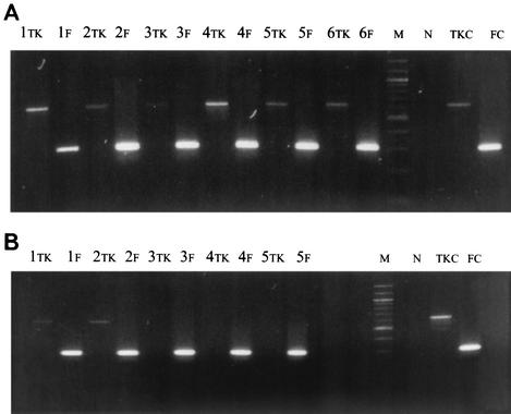
The cDNA corresponding to the PPRV F gene was released by enzymatic digestion from plasmid FNYZ12 as previously described (18). It was subcloned into plasmid JC-35, which contains the vaccinia virus early/late p7.5 promoter. The insertion was made so that, in the new plasmid, FPJ2, expression of the PPRV F protein gene was driven by this promoter. The Escherichia coli xanthine-guanine -phosphoribosyltransferase gene (gpt) was used as a dominant-selectable marker to isolate the recombinant (3, 9). This gene is under the control of a p7.5 promoter in the plasmid GPT. It was released from that plasmid and inserted into FPJ2 in a way to place the two promoters back to back. From this final construct, the E. coli gpt-p7.5-p7.5-PPR F cassette was released by enzymatic digestion and inserted into the plasmid pKH66T at the KpnI site of the gene coding for capripoxvirus thymidine kinase (TK). Indeed, pKH66T contains the HindIII S fragment of KS-1 DNA, which encompasses the complete TK gene of capripoxvirus KS-1 (10). The final construct was used to generate recombinant viruses with the capripoxvirus KS-1 in lamb testis (LT) cells as described previously (22). The virus suspension obtained following transfection of capripoxvirus-infected cells with the insertion plasmid was used to select the recombinants. This was done in the presence of the E. coli gpt selection medium containing mycophenolic acid (MPA) (22). However, while the previous authors were confirming the nature of their clones by DNA probes, we adopted a faster PCR-based method that could be performed directly on the samples without the need for DNA extraction. For that, we used both capripoxvirus TK and PPRV F gene-specific primers (CPTK7 and CPTK8 and F1ab and F2ab, respectively) (Fig. 1). Five microliters of each sample to be tested was used directly in the PCR, which consisted of 10 μl of 10× Pfu buffer, deoxynucleoside triphosphates (dNTPs [250 μM each]), 25 pmol of each primer, and 2.5 U of Pfu polymerase (Stratagene) in a total volume of 100 μl. The PCR was carried out under the following conditions: a first step at 94°C for 5 min; 30 cycles of amplification, each consisting of 94°C for 1 min, 50°C for 1 min, and 72°C for 3 min; and a final step at 4°C to keep the samples. Fifteen microliters of the amplified products was analyzed by electrophoresis on agarose gels. The primers CPTK7 and CPTK8, located in the TK gene of the capripoxvirus strain KS-1 at each side of the insertion site, amplify a fragment about 600 nucleotides long in the absence of a foreign gene insert. The PPRV F gene insert is about 2,400 nucleotides long and is very GC rich (18); therefore, the expected size of the target to be amplified by the primers CPTK7 and CPTK8 in the recombinant would be about 3,000 nucleotides. The conditions used for the PCR allow amplification of the 600-nucleotide target, but not a target of 3,000 nucleotides. Thus, in the case of a recombinant, the PCR with primers CPTK7 and CPTK8 will be negative, while it should give a product of 300 nucleotides amplified on the PPRV F gene with primers F1ab and F2ab. Figure 1A shows the DNA amplified from samples collected at different steps in the procedures of recombinant selection. As can been seen (lanes 1 to 6), a clone that was obtained was still contaminated with parental virus even after three plaque purifications and two rounds of growth in selection medium. The PCR test was positive with both pairs of primers (lanes TK and F). Similar results were obtained with five other selected clones. We presumed that capripoxviruses have a high tendency to form clumps, and by this means, the recombinant viruses, which can grow in the selection medium MPA, could act as a helper virus and allow replication of parental virus in the same clump. We therefore modified the selection method to include a sonication step. One milliliter of the stock virus suspension was submitted to sonication for 30 s in a water bath sonicator with the power set at 15 W (Ultrasonic Processor, Bioblock Scientific). After sonication, 10-fold serial dilutions of the sample were made from 10−1 to 10−7. Confluent LT cells in 96-well plates were preincubated in selection medium for 16 h and then infected with 200 μl of diluted virus suspension (2 wells per dilution). After 10 days of incubation at 37°C, the supernatants from wells from the most dilute suspensions showing a virus cytopathic effect were harvested. These were then subjected to another round of limiting dilution selection and plaque purification. The virus material collected at each step of the purification process was always sonicated to ensure that the virus was not clumped, and its purity was checked by PCR. No DNA amplification was obtained with the TK primers after the second limiting dilution selection (Fig. 1B, lanes 3Tk, 4Tk, and 5Tk), while the presence of the PPRV F protein gene was still detected by primers F1ab and F2ab (Fig. 1B, lanes 3F, 4F, and 5F).
FIG. 1.
Screening of the recombinant virus clones by PCR. Primers CPTK7 (5′ ACTTATCAGATTTTGTTACGACATT) and CPTK8 (5′ CGATGAGTTCTATTTCCTTTTCTTTAG) were located in the TK gene of capripoxvirus strain KS-1 at each side of the insertion site KpnI. Primers F1ab (5′ ATGCTCTGTCAGTGATAACC) and F2ab (5′ TTATGGACAG AAGGGACAAG) were located in the PPRV F gene. The PCR products obtained with these primers from samples collected during the different steps of the recombinant virus selection process were analyzed by agarose gel electrophoresis (see the text). The samples amplified with the capripoxvirus TK primers are indicated by TK, while those amplified with the PPRV fusion protein gene primers are in lanes F. M, molecular size markers (100-bp ladders); TKc, positive control capripoxvirus DNA; Fc, positive control PPRV F cDNA; N, negative control (no DNA template). (A) Samples from the first method. Sample 1 is the starting virus stock suspension to be screened for the recombinants. Samples 2, 3, and 4 are from three successive plaque purifications. Samples 5 and 6 are from two successive growth rounds of sample 4 in selection medium. (B) Samples from the second method. Sample 1 is the starting virus stock suspension to be screened for the recombinant. Samples 2 and 3 are from two successive limiting dilution purifications. Sample 4 is a clone collected by plaque purification of sample 4. Sample 5 is aliquot of the virus suspension obtained by one growth round of sample 4 in the selection medium.
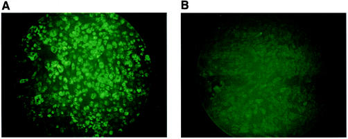
The expression of the PPRV F protein by the pure recombinant virus clone (Fig. 1B, lane 5), was analyzed by immunofluorescence in paraformaldehyde (PFA)-fixed infected LT cells (22), but without treating the cells with Triton. Cells expressing the PPR F protein were detected with a PPRV F-specific monoclonal antibody (MAb) (15) and fluorescein isothiocyanate (FITC)-conjugated rabbit anti-mouse immunoglobulin G (IgG). As can be seen in Fig. 2A, cell surface fluorescence was detected on LT cells infected with recCapPPR/F, but not on those infected with the parental capripoxvirus (Fig. 2B).
FIG. 2.
Detection of the PPRV F protein by immunofluorescence staining. LT cells were infected with the recombinant reCapPPR/F (A) or the parental capripoxvirus KS1 (B). Two days after infection, the cells were submitted to indirect immunofluorescence staining with the anti-PPRV F MAb as described by Romero et al. (22), but without Triton treatment.
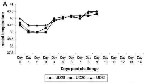
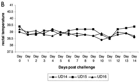
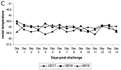
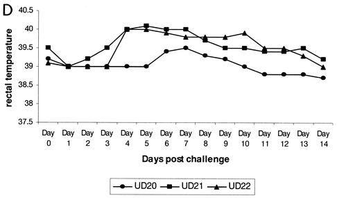
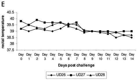
The recombinant clone recCapPPR/F was tested as a dual vaccine to see if it could protect goats against both PPR and capripoxvirus challenge. For that test, 18 British goats were purchased and housed in six groups of three (groups I to VI). Animals in groups I, II, III, and IV were inoculated with 104, 102, 10, and 0.1 PFU of recCapPPR/F, respectively. Animals in group V were vaccinated with 103 PFU of KS-1 vaccine, while those in group VI were used as unvaccinated controls. One month after vaccination, the animals, with the exception of those of group V, were challenged with virulent PPRV (Guinée Bissau/89 isolate) at 104 50% tissue culture infective doses (TCID50) per animal by subcutaneous injection. The animals were examined daily for clinical signs of infection, and their rectal temperatures were recorded. All unvaccinated animals developed high fever 4 days after challenge (Fig. 3A). They also developed necrotic mouth lesions, nasal and ocular discharges, and profuse diarrhea. They were euthanized on day 9 because of the severity of the disease. Among the animals that had received the recombinant vaccine, only two developed a transient fever (Fig. 3D). They were in the group vaccinated with a dose of 100 PFU. No other clinical signs were apparent.
FIG. 3.
Determination of the minimum effective dose of the recombinant vaccine represented by daily rectal temperatures of goats following challenge with virulent PPRV. Goats were vaccinated with different doses of the recombinant virus reCapPPR/F and challenged 1 month later with the virulent PPRV Guinée Bissau/89 isolate as indicated in the text. Shown are results for unvaccinated animals (A) and animals vaccinated with 0.1 (B), 10 (C), 100 (D), and 10,000 (E) PFU of the recombinant virus reCapPPR/F.
Morbilliviruses are lymphotropic viruses and virulent strains induce a severe leukopenia in infected hosts. In the present experiment, leukocytes (WBC) were collected from animals on days 2, 5, 7, 9, 12, and 14 days following challenge and counted. A moderate and transient reduction in the WBC number was observed for a couple of days following challenge in the case of the vaccinated animals (Table 1); however, the unvaccinated animals showed a dramatic leukopenia (reduction of ≥50%). Total RNA extracted from these cells was assayed with a reverse transcription-PCR to detect nucleocapsid protein gene-specific RNA of the challenge virus (5). The quality of the RNA was determined by amplifying a fragment of the cellular β-actin gene by using actin-specific primers. As can be seen in Table 2, PPRV nucleic acid was detected in the WBC from all three unvaccinated animals from day 5 or 7 after challenge. However, in the case of 6 of the 12 vaccinated goats, no DNA amplification could be obtained with the PPRV primers, while the same samples were positive for the β-actin gene RNA, indicating the integrity of the RNA extracts. For the remaining six vaccinated goats, PPRV nucleic acid was detected in only one or two postchallenge samples. Blood for serum antibody analysis was collected weekly following vaccination and challenge. The sera were analyzed for PPRV-specific antibodies by using an F protein-based competitive enzyme-linked immunosorbent assay (cELISA) (Table 3). All were negative before challenge. However, following challenge, an anamnestic response was observed after 1 week, indicating that replication of the challenge virus had occurred. One month after PPRV challenge, the surviving animals and those in group V, in addition to three new control goats, were submitted to capripoxvirus challenge by intradermal inoculation of 0.2 ml of virulent capripoxvirus (Yemen isolate). The animals were examined daily for 2 weeks to observe their clinical reactions. The control goats, as expected, developed capripox disease, while all of the others resisted.
TABLE 1.
Percentage reduction in WBC counts in goats following challenge with virulent PPRV
| Goat | Vaccine dose (PFU/goat) | % Decrease in cell count on postchallenge daya:
|
|||
|---|---|---|---|---|---|
| 5 | 7 | 9 | 14 | ||
| UD 29 | 0 | 30.6 | 47.5 | 48 | Dead |
| UD 30 | 0 | 22.8 | 30.4 | 55.6 | Dead |
| UD 31 | 0 | 43.4 | 49.1 | 71.8 | Dead |
| UD 14 | 0.1 | 39 | 15.4 | 0 | 44.5 |
| UD 15 | 0.1 | 21 | 25 | 15 | 6 |
| UD 16 | 0.1 | 55 | 39 | 33 | 18 |
| UD 17 | 10 | 0 | 0 | 0 | 7.8 |
| UD 18 | 10 | 0 | 6.5 | 23 | 5 |
| UD 19 | 10 | 19.2 | 31 | 32 | 2 |
| UD 20 | 100 | 0 | 6.5 | 23 | 5 |
| UD 21 | 100 | 13 | 31.9 | 10 | 0 |
| UD 22 | 100 | 32.8 | 44.5 | 46 | 0 |
| UD 26 | 10,000 | 28.7 | 0 | 0 | 0 |
| UD 27 | 10,000 | 37.9 | 37.3 | 0 | 37 |
| UD 28 | 10,000 | 0 | 0 | 0 | 13.3 |
The values are expressed as percent decrease in the cell count from that found on day 2 postchallenge. A decrease of a 50% (boldface) is considered an indication of severe leukopenia.
TABLE 2.
Detection of PPRV N gene-specific RNA by RT-PCR in WBC collected from goats after challenge with virulent PPRVa
| Goat | Vaccine dose (PFU/goat) | Result on day postchallengeb:
|
|||||||||
|---|---|---|---|---|---|---|---|---|---|---|---|
| 2
|
5
|
7
|
9
|
14
|
|||||||
| Actin | N gene | Actin | N gene | Actin | N gene | Actin | N gene | Actin | N gene | ||
| UD 29 | 0 | + | − | + | + | + | + | + | + | Dead | |
| UD 30 | 0 | + | − | + | + | + | + | + | + | Dead | |
| UD 31 | 0 | + | − | + | − | + | + | + | + | Dead | |
| UD 14 | 0.1 | + | − | + | − | + | − | + | − | + | − |
| UD 15 | 0.1 | + | − | + | − | + | − | + | − | + | − |
| UD 16 | 0.1 | + | − | + | − | + | − | + | − | + | − |
| UD 17 | 10 | + | − | + | − | + | − | + | − | + | − |
| UD 18 | 10 | + | − | + | − | + | − | + | − | + | − |
| UD 19 | 10 | + | − | + | − | + | + | + | − | + | − |
| UD 20 | 100 | + | − | + | − | + | + | + | + | + | − |
| UD 21 | 100 | + | − | + | + | + | + | + | − | + | − |
| UD 22 | 100 | + | − | + | − | + | + | + | − | + | − |
| UD 26 | 10,000 | + | − | + | − | + | + | + | − | + | − |
| UD 27 | 10,000 | + | − | + | − | + | − | + | − | + | − |
| UD 28 | 10,000 | + | − | + | + | + | − | + | − | + | − |
RNA was detected as described by Couacy et al. (5).
A β-actin-specific primer set, bAct1 (5′ ACCAACTGGGACGACATGGAGA) and bAct2 (5′ AGCCATCTCC TGCTCGAAGTC), was used to amplify the β-actin mRNA as a control for the quality of the extracted RNA.
TABLE 3.
Serological responses in goats following challenge with virulent PPRVa
| Goat | Vaccine dose (PFU/goat) | % Inhibition (status) on day:
|
||
|---|---|---|---|---|
| 0 postinfection | 7 postinfection | 14 postinfection | ||
| UD 29 | 0 | 19 (−) | 16 (−) | Dead |
| UD 30 | 0 | 21 (−) | 15 (−) | Dead |
| UD 31 | 0 | 16 (−) | 17 (−) | Dead |
| UD 14 | 0.1 | 5 (−) | 31 (−) | 74 (+) |
| UD 15 | 0.1 | −5 (−) | 17 (−) | 37 (−) |
| UD 16 | 0.1 | 8 (−) | 20 (−) | 71 (+) |
| UD 17 | 10 | 5 (−) | 44 (−) | 52 (+) |
| UD 18 | 10 | −14 (−) | 31 (−) | 69 (+) |
| UD 19 | 10 | 4 (−) | 46 (−) | 56 (+) |
| UD 20 | 100 | −9 (−) | 39 (−) | 76 (+) |
| UD 21 | 100 | −10 (−) | 15 (−) | 47 (−) |
| UD 22 | 100 | −6 (−) | 37 (−) | 74 (+) |
| UD 26 | 10,000 | 22 (−) | 51 (+) | 71 (+) |
| UD 27 | 10,000 | 14 (−) | 56 (+) | 75 (+) |
| UD 28 | 10,000 | 18 (−) | 53 (+) | 65 (+) |
Serological responses were measured by cELISA based on the use of an anti-PPRV F protein MAb with the attenuated PPRV as an antigen for coating the ELISA plates. Both the test serum sample and the MAb were used at the final concentration of 1/20 in phosphate-buffered saline diluted 1/5 and containing 0.05% Tween and 0.5% lamb serum (total volume, 100 μl per well). Four wells were used as controls and contained only the MAb in the dilution buffer. For the interpretation of the results, inhibition of 50% or greater was considered a positive sample (shown in boldface).
The F protein of rinderpest virus has been expressed in both vaccinia virus and capripoxvirus vector systems (1, 22, 30). Here we report the construction of a recombinant capripoxvirus expressing the PPRV F protein. The modifications to the previous selection methodology (22), a sonication step carried out to disrupt potential clumps of virus before the limit dilution followed by PCR screening, allowed a recombinant clone free of parental virus to be selected easily. This recombinant was able to protect goats against both PPR and capripox at a dose as low as 0.1 PFU. With the original capripoxvirus-rinderpest virus F recombinant, a dose of 1.5 × 10³ PFU was unable to protect cattle against virulent rinderpest virus challenge (24); apparently it was 104 times less effective than the recombinant (reCaPPR/F) reported here. This difference between the two recombinants may be due to the type of promoter used: vaccinia virus p11 in the case of the rinderpest virus F protein and vaccinia virus p7.5 in the case of the PPRV F protein. Tsukiyama et al. (27) developed two rinderpest vaccinia virus (RRV) recombinant vaccines in which the H protein gene was expressed under the control of either the vaccinia virus early/late promoter p7.5 or the cowpox virus A-type inclusion body (ATI) promoter. They reported the induction of a higher immune response with p7.5/RVV than with ATI/RVV in vaccinated rabbits. Both ATI and p11 are strong late promoters. Proteins expressed under the control of such promoters may induce less cellular immunity than those that start synthesis early in the viral replication cycle (6). Another possibility may explain the lower dose requirement of reCaPPR/F than that reported by Romero et al. (24) for rinderpest: that is, the degrees of purity of the recombinant clones may have differed and the original capripoxvirus-rinderpest virus recombinant clone may have been contaminated by the parental virus, since its purity was not checked by PCR, a very sensitive technique able to detect extremely small amounts of parental virus contaminant.
Capripoxvirus is a highly host-specific virus, the host range being limited to cattle and small ruminants. It is nonpathogenic to humans, and so it is an ideal vector for the development of recombinant vaccines for use against ruminant diseases. The results reported here indicate that a capripoxvirus recombinant that expresses the PPR F protein can protect goats against two diseases that are of great economic importance in many developing countries: PPR and capripox. The possibility of dual vaccines made by such recombinants and providing efficacy at doses as low as 0.1 PFU would dramatically cut the cost of controlling these diseases by vaccination. However, more work is needed to establish the duration of immunity provided by this vaccine and to test its efficacy in presence of antibodies against capripoxvirus or PPRV. This work is now in progress.
Acknowledgments
This work was supported by a grant from the Ministère des Affaires Etrangères (projet Ravira no. 9502220002307501). G.B. was supported by a fellowship from the Ministère des Affaires Etrangères.
We thank Gerrit Viljoen (OVI, South Africa) for helpful discussions during the course of this work.
REFERENCES
- 1.Barrett, T., G. J. Belsham, S. M. Subbarao, and S. A. Evans. 1989. Immunization with a vaccinia recombinant expressing the F protein protects rabbits from challenge with a lethal dose of rinderpest virus. Virology 170:11-18. [DOI] [PubMed] [Google Scholar]
- 2.Belsham, G. J., E. C. Anderson, P. K. Murray, J. Anderson, and T. Barrett. 1989. Immune response and protection of cattle and pigs generated by a vaccinia virus recombinant expressing the F protein of rinderpest virus. Vet. Rec. 124:655-658. [DOI] [PubMed] [Google Scholar]
- 3.Boyle, D. B., and B. E. H. Coupar. 1988. A dominant selectable marker for the construction of recombinant poxviruses. Gene 65:123-128. [DOI] [PubMed] [Google Scholar]
- 4.Choppin, P. W., and A. Scheid. 1980. The role of viral glycoproteins in adsorption, penetration, and pathogenicity of viruses. Rev. Infect. Dis. 2:40-61. [DOI] [PubMed] [Google Scholar]
- 5.Couacy-Hymann, R. F., C. Hurard, J. P. Guillou, G. Libeau, and A. Diallo. 2002. Rapid and sensitive detection of peste des petits ruminants virus by a polymerase chain reaction assay. J. Virol. Methods 100:17-25. [DOI] [PubMed] [Google Scholar]
- 6.Coupar, B. E., M. E. Andrew, G. W. Both, and D. B. Boyle. 1986. Temporal regulation of influenza haemagglutin expression in vaccinia virus recombinants and effects of the immune response. Eur. J. Immunol. 16:1479-1487. [DOI] [PubMed] [Google Scholar]
- 7.Diallo, A., W. P. Taylor, P. C. Lefèvre, and A. Provost. 1989. Atténuation d'une souche de virus de la peste des petits ruminants: candidat pour un vaccin homologue. Rev. Elev. Med. Vet. Pays Trop. 42:311-317. [PubMed] [Google Scholar]
- 8.Drillien, R., D. Spehner, A. Kirn, P. Giraudon, R. Buckland, F. Wild, and J. P. Lecocq. 1988. Protection of mice from fatal measles encephalitis by vaccination with vaccinia virus recombinants encoding either the hemagglutinin or the fusion protein. Proc. Natl. Acad. Sci. USA 85:1252-1256. [DOI] [PMC free article] [PubMed] [Google Scholar]
- 9.Falkner, F. G., and B. Moss. 1988. Escherichia coli gpt gene provides dominant selection for vaccinia virus open reading frame expression vectors. J. Virol. 62:1849-1854. [DOI] [PMC free article] [PubMed] [Google Scholar]
- 10.Gershon, P. D., and D. N. Black. 1989. The nucleotide sequence around the capripoxvirus thymidine kinase gene reveals a gene shared specifically with leporipoxvirus. J. Gen. Virol. 70:525-533. [DOI] [PubMed] [Google Scholar]
- 11.Gibbs, P. J. E., W. P. Taylor, M. P. J. Lawman, and J. Bryant. 1979. Classification of the peste-des-petits-ruminants virus as the fourth member of the genus Morbillivirus. Intervirology 11:268-274. [DOI] [PubMed] [Google Scholar]
- 12.Kitching, R. P., J. M. Hammond, and W. P. Taylor. 1987. A single vaccine for the control of capripox infection in sheep and goats. Res. Vet. Sci. 42:53-60. [PubMed] [Google Scholar]
- 13.Lamb, R. A. 1993. Paramyxovirus fusion protein: a hypothesis for changes. Virology 197:1-11. [DOI] [PubMed] [Google Scholar]
- 14.Lefèvre, P. C., and A. Diallo. 1990. Peste des petits ruminants. Rev. Sci. Tech. Off. Int. Epizoot. 9:951-965. [DOI] [PubMed] [Google Scholar]
- 15.Libeau, G., J. T. Saliki, and A. Diallo. 1997. Caractérisation d'anticorps monoclonaux dirigés contre les virus de la peste bovine et de la peste des petits ruminants: identifications d'épitopes conservés ou de spécificité stricte sur la nucléoprotéine. Rev. Elev. Méd. Vét. Pays Trop. 50:109-118. [Google Scholar]
- 16.Merz, D. C., A. Scheid, and P. W. Choppin. 1980. Importance of antibodies to the fusion glycoprotein of paramyxoviruses in the prevention of spread of infection. J. Exp. Med. 151:275-288. [DOI] [PMC free article] [PubMed] [Google Scholar]
- 17.Merz, D. C., A. Scheid, and P. W. Choppin. 1981. Immunological studies of the functions of paramyxovirus glycoproteins. Virology 109:94-105. [DOI] [PubMed] [Google Scholar]
- 18.Meyer, G., and A. Diallo. 1995. The nucleotide sequence of the fusion protein gene of the peste des petits ruminants virus: the long untranslated region in the 5′ end of the F protein gene of morbilliviruses seems to be specific to each virus. Virus Res. 37:23-38. [DOI] [PubMed] [Google Scholar]
- 19.Ngichabe, C. K., H. M. Wamwayi, T. Barrett, E. K. Ndungu, D. N. Black, and C. J. Bostock. 1997. Trial of a capripoxvirus-rinderpest recombinant vaccine in African cattle. Epidemiol. Infect. 118:63-70. [DOI] [PMC free article] [PubMed] [Google Scholar]
- 20.Ngichabe, C. K., H. M. Wamwayi, E. K. Ndungu, P. K. Mirangi, C. J. Bostock, D. N. Black, and T. Barrett. 2002. Long term immunity in African cattle vaccinated with a recombinant capripox-rinderpest virus vaccine. Epidemiol. Infect. 128:343-346. [DOI] [PMC free article] [PubMed] [Google Scholar]
- 21.Ohishi, K., K. Inui, T. Barrett, and K. Yamanouchi. 2000. Long-term protective immunity to rinderpest in cattle following a single vaccination with a recombinant vaccinia virus expressing the virus haemagglutinin protein. J. Gen. Virol. 81:1439-1446. [DOI] [PubMed] [Google Scholar]
- 22.Romero, C. H., T. Barrett, S. A. Evans, R. P. Kitching, P. D. Gershon, C. Bostock, and D. N. Black. 1993. Single capripoxvirus recombinant vaccine for the protection of cattle against rinderpest and lumpy skin disease. Vaccine 11:737-742. [DOI] [PubMed] [Google Scholar]
- 23.Romero, C. H., T. Barrett, R. W. Chamberlain, R. P. Kitching, M. Fleming, and D. N. Black. 1994. Recombinant capripoxvirus expressing the hemagglutinin protein gene of rinderpest virus: protection of cattle against rinderpest and lumpy skin disease viruses. Virology 204:425-429. [DOI] [PubMed] [Google Scholar]
- 24.Romero, C. H., T. Barrett, R. P. Kitching, V. M. Carn, and D. N. Black. 1994. Protection of cattle against rinderpest and lumpy skin disease with a recombinant capripoxvirus expressing the fusion protein gene of rinderpest virus. Vet. Rec. 135:152-154. [DOI] [PubMed] [Google Scholar]
- 25.Stephensen, C. B., J. Welter, S. R. Thaker, J. Taylor, J. Tartaglia, and E. Paoletti. 1997. Canine distemper virus (CDV) infection of ferrets as a model for testing Morbillivirus vaccine strategies: NYVAC- and ALVAC-based CDV recombinants protect against symptomatic infection. J. Virol. 71:1506-1513. [DOI] [PMC free article] [PubMed] [Google Scholar]
- 26.Taylor, J., R. Weinberg, J. Tartaglia, C. Richardson, G. Alkhatib, D. Briedis, M. Appel, E. Norton, and E. Paoletti. 1992. Nonreplicating viral vectors as potential vaccines: recombinant canarypox virus expressing measles virus fusion (F) and hemagglutinin (HA) glycoproteins. Virology 187:321-328. [DOI] [PubMed] [Google Scholar]
- 27.Tsukiyama, T., Y. Yoshikawa, H. Kamata, H. Imaoka, K. Asano, S. Funahashi, T. Maruyama, H. Shida, M. Sugimoto, and K. Yamanouchi. 1989. Development of heat-stable recombinant rinderpest vaccine. Arch. Virol. 107:225-235. [DOI] [PubMed] [Google Scholar]
- 28.Wild, F., P. Giraudon, D. Spehner, R. Drillien, and J. P. Lecocq. 1990. Fowlpox virus recombinant encoding the measles virus fusion protein: protection of mice against fatal measles encephalitis. Vaccine 8:441-442. [DOI] [PubMed] [Google Scholar]
- 29.Wild, T. F., A. Bernard, D. Spehner, D. Villeval, and R. Drillien. 1993. Vaccination of mice against canine distemper virus-induced encephalitis with vaccinia virus recombinants encoding measles or canine distemper virus antigens. Vaccine 11:438-444. [DOI] [PubMed] [Google Scholar]
- 30.Yilma, T., D. Hsu, L. Jones, S. Owens, M. Grubman, C. Mebus, M. Yamanaka, and B. Dale. 1988. Protection of cattle against rinderpest with vaccinia virus recombinants expressing the HA or F gene. Science 242:1058-1061. [DOI] [PubMed] [Google Scholar]


