Abstract
We have generated a recombinant Newcastle disease virus (NDV) that expresses the green fluorescence protein (GFP) in infected chicken embryo fibroblasts (CEFs). This virus is interferon (IFN) sensitive, and pretreatment of cells with chicken alpha/beta IFN (IFN-α/β) completely blocks viral GFP expression. Prior transfection of plasmid DNA induces an IFN response in CEFs and blocks NDV-GFP replication. However, transfection of known inhibitors of the IFN-α/β system, including the influenza A virus NS1 protein and the Ebola virus VP35 protein, restores NDV-GFP replication. We therefore conclude that the NDV-GFP virus could be used to screen proteins expressed from plasmids for the ability to counteract the host cell IFN response. Using this system, we show that expression of the NDV V protein or the Nipah virus V, W, or C proteins rescues NDV-GFP replication in the face of the transfection-induced IFN response. The V and W proteins of Nipah virus, a highly lethal pathogen in humans, also block activation of an IFN-inducible promoter in primate cells. Interestingly, the amino-terminal region of the Nipah virus V protein, which is identical to the amino terminus of Nipah virus W, is sufficient to exert the IFN-antagonist activity. In contrast, the anti-IFN activity of the NDV V protein appears to be located in the carboxy-terminal region of the protein, a region implicated in the IFN-antagonist activity exhibited by the V proteins of mumps virus and human parainfluenza virus type 2.
The alpha/beta interferon (IFN-α/β) system is a major component of the host innate immune response to viral infection (reviewed in reference 1). IFN (i.e., IFN-β and several IFN-α types) is synthesized in response to viral infection due to the activation of several factors, including IFN regulatory factor proteins, NF-κB, and AP-1 family members. As a consequence, viral infection induces the transcriptional upregulation of IFN genes. Secreted IFNs signal through a common receptor activating a JAK/STAT signaling pathway which leads to the transcriptional upregulation of numerous IFN-responsive genes, a number of which encode antiviral proteins, and leads to the induction in cells of an antiviral state. Among the antiviral proteins induced in response to IFN are PKR, 2′,5′-oligoadenylate synthetase (OAS), and the Mx proteins (10, 15, 23).
Many viruses have evolved mechanisms to counteract the host IFN response and, in some viruses, including vaccinia virus, adenovirus, and hepatitis C virus, multiple IFN-antagonist activities have been reported (3, 6, 12, 16, 17, 28, 35, 57, 58). Among negative-strand RNA viruses, several different IFN-subverting strategies have been identified that target a variety of components of the IFN system. The influenza virus NS1 protein, for example, prevents production of IFN by inhibiting the activation of the transcription factors IFN regulatory factor 3 and NF-κB and blocks the activation of the IFN-induced antiviral proteins PKR and OAS (4, 18, 55, 59; N. Donelan, X. Wang, and A. García-Sastre, unpublished data). Among the paramyxoviruses, different mechanisms are employed by different viruses (60). For example, the “V” proteins of several paramyxoviruses have previously been shown to inhibit IFN signaling, but the targets of different V proteins vary (32, 47). In the case of Sendai virus, the “C” proteins, a set of four carboxy-coterminal proteins, have been reported to block IFN signaling both in infected cells and when expressed alone (19, 21, 22, 27, 30). In contrast, respiratory syncytial virus, which encodes neither a C nor a V protein, produces two nonstructural proteins, NS1 and NS2, that are reported to cooperatively counteract the antiviral effects of IFN (5, 54). Ebola virus, a nonsegmented, negative-strand RNA virus of the family Filoviridae that possesses a genome structure similar to that of the paramyxoviruses (29), also encodes at least one protein, VP35, that counteracts the host IFN response (2).
Viral IFN antagonists have been shown to be important virulence factors in several viruses, including herpes simplex virus type 1, vaccinia virus, influenza virus, and Sendai virus. Analysis of viruses with mutations in genes encoding herpes simplex virus type 1 ICP34.5 (8, 38), vaccinia virus E3L (6), influenza virus NS1 (18, 56), and Sendai virus C (13, 20) proteins has demonstrated an important role for each of these IFN antagonists in viral pathogenicity in mice. Because IFN antagonists are important virulence factors, their identification and characterization should provide important insights into viral pathogenesis.
Infectious cDNAs for Newcastle disease virus (NDV) have recently been developed (31, 42, 49, 51) and permit the introduction of foreign genes into the NDV genome (31, 42, 53). We constructed a recombinant NDV expressing the green fluorescence protein (GFP), NDV-GFP, and show that this virus is sensitive to the antiviral effects of IFN. We have taken advantage of this IFN-sensitive property and developed an NDV-GFP-based assay to identify proteins that exhibit IFN-antagonist activity. Using this system, we provide evidence that the NDV V protein possesses IFN-antagonist activity. We further use this assay to show that the V, W, and C proteins of Nipah virus, an important emerging pathogen that is highly lethal in humans (9, 14, 34), also exhibit IFN-antagonist activity.
MATERIALS AND METHODS
Cells and plasmids.
Chicken embryo fibroblasts (CEFs) were prepared from 10-day-old specific-pathogen-free embryos (Charles River SPAFAS, North Franklin, Conn.). CEFs and Vero cells were maintained in minimal essential medium (MEM) containing 10% fetal bovine serum (FBS) and Dulbecco modified Eagle medium with 10% FBS, respectively. The influenza virus NS1 and the Ebola virus VP35 protein expression plasmids have been described previously (2, 55).
Construction and growth of a GFP-expressing NDV.
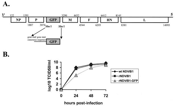
The enhanced GFP open reading frame (ORF) from the plasmid pEGFP-c1 (Clontech) was cloned between the P and M genes of the previously described NDV Hitchner B1 cDNA (42) (Fig. 1). This virus, NDV-GFP, was then rescued from cDNA by using previously described methods (42), and the presence in the viral genome of the inserted GFP gene was confirmed by reverse transcription-PCR and sequencing (data not shown). Virus stocks were prepared in 10-day-old embryonated chicken eggs. Virus titers were determined as TCID50/ml on CEFs. Briefly, 96-well plates containing CEFs were infected with 10-fold serial dilutions of virus. At 2 days postinfection, cells were fixed with 2.5% formaldehyde containing 0.1% Triton X-100. Infection of individual wells was determined by indirect immunofluorescence with polyclonal, anti-NDV rabbit antiserum as the primary antibody and fluorescein isothiocyanate-conjugated anti-rabbit immunoglobulin (Dako Corp.).
FIG. 1.
Construction and growth of an NDV expressing GFP. (A) Diagram of the NDV-GFP virus genome. (B) Comparison of growth of recombinant NDV Hitchner B1 and NDV-GFP viruses in CEFs (MOI = 0.001).
Cloning of the NDV V ORF and the Nipah virus V, W, and C ORFs.
The NDV V ORF was constructed by PCR amplification from the wild-type NDV Hitchner B1 P protein expression plasmid pCAGGS-NDV P (42). The NDV V ORF was amplified as two PCR fragments by using the following primers. For the amino-terminal fragment, the sense primer 5′-CC GAA TTC ATGGCC ACC TTT ACA GAT, containing an EcoRI site (underlined), and the antisense primer 5′-GGC TCG ACC ATG GGC CCC TT, containing an NcoI restriction site (underlined), were used. For the carboxy-terminal fragment, the sense primer 5′-AAG GGG CCC ATG GTC GAG CC and the antisense primer 5′-CG CTC GAG TTA CTT ACT CTC TGT GAT ATC, each containing an XhoI restriction site (underlined), were used. For the NDV VN ORF, the sense primer 5′-CC GAA TTC ATGGCC ACC TTT ACA GAT and the antisense primer 5′-CG CTC GAG TCA TTT AGC ATT GGA CGA TTT were used. For the NDV VC ORF, the sense primer 5′-CC GAA TTC CCC ATG GTC GAG CCC CCA and the antisense primer 5′-CG CTC GAG TTA CTT ACT CTC TGT GAT ATC were used.
The Nipah virus cDNAs were constructed by PCR without template by using overlapping deoxyoligonucleotides corresponding to GenBank accession number NC_002728. The V ORF corresponds to nucleotides 2406 to 3775 but contains a single non-template-encoded G residue inserted after position 3624. The W ORF corresponds to nucleotides 2406 to 3756 but contains two non-template-encoded G residues inserted after position 3624. The C ORF corresponds to nucleotides 2428 to 2928. All ORFs were cloned into the mammalian expression plasmid pCAGGS (44). Because the C ORF lies within the V and W ORFs, it could conceivably be produced from the Nipah virus V and W expression plasmids if the first ATG in these plasmids were bypassed. Therefore, all plasmids expressing V or W proteins contained mutations in which the two tandem ATGs at the predicted start of the C protein were mutated to ACG. These mutations would be expected to eliminate or significantly reduce the level of full-length C protein that might be expressed. Additionally, a V protein expression plasmid was generated in which the two tandem ATGs at the predicted start of the C protein were mutated to ACG and a stop codon was introduced at the fourth codon of the C ORF. The changes did not affect the translation of the V protein. Primer sequences and PCR conditions are available upon request.
Transfection and infection of CEF cells.
One day prior to transfection, CEFs were seeded onto 24-well plates such that they would be 80% confluent the following morning. The medium was replaced with 0.3 ml of Dulbecco modified Eagle medium with 5% FBS. Transfection mixtures were prepared in polystyrene tubes as follows. First, 2 μg of plasmid DNA was diluted to 50 μl in OptiMEM (Gibco). Then, 6 μl of Fugene 6 (Roche) previously diluted to 50 μl in OptiMEM was added to the polystyrene tube. The transfection mixtures were incubated for 20 min at room temperature and added to the cells. After 20 h of incubation at 37°C in 5% CO2, the transfected cells were washed briefly with phosphate-buffered saline and infected with NDV-GFP (multiplicity of infection [MOI] of 1 to 2) at room temperature for 30 to 60 min. The inoculum was then aspirated and 0.5 ml of MEM-10% FBS was added to the cells. The infected cells were incubated for 20 h at 37°C in 5% CO2 prior to detection of green fluorescence.
Neutralization of chicken IFN.
Chicken IFN-α/β (1,000 or 500 U) was added to cells 8 h after transfection or to nontransfected cells where specified. Neutralizing mouse antibody against chicken IFN was added together with IFN or immediately after transfection where specified. The chicken IFN and the anti-chicken IFN antibodies were kindly provided by Peter Staeheli (University of Freiburg) and Bernd Kaspers (University of Munich).
FACS analysis.
Cells either mock-infected or infected with NDV-GFP were detached from dishes by treating the cells with trypsin-EDTA and resuspending them in 1 ml of MEM-10% FBS. The relative mean intensity of green fluorescence of the cells was determined by fluorescence-activated cell-sorting (FACS) analysis with a Beckman-Coulter Epics XL-MCL fluorescence-activated cell sorter.
Reporter gene assays in Vero cells.
Vero cells were transfected with three plasmids: a pCAGGS construct of the gene of interest, a plasmid containing the chloramphenicol acetyltransferase (CAT) gene downstream of an IFN-stimulated response element (ISRE) (pHISG-54-CAT), and a construct encoding the Renilla luciferase protein (pRL-tk). The cells were transfected with 1 μg of each reporter plasmid and 5 μg of the expression plasmid by using the transfection reagent Lipofectamine 2000 (Invitrogen). The following day the medium was replaced with medium containing 1,000 IU of IFN-β/ml, and incubation was continued overnight. The cells were harvested, and the cell pellet was resuspended in phosphate-buffered saline and divided into two aliquots for analysis of the CAT and luciferase activities. CAT assays were performed as previously described (50a). Luciferase assays were performed by using the Renilla luciferase assay system (Promega) according to the manufacturer's instructions.
RESULTS
Construction of an NDV that expresses GFP.
The GFP ORF was cloned between the P and M genes of the previously described NDV Hitchner B1 cDNA (42) (Fig. 1A). This virus, NDV-GFP, was then rescued from cDNA by previously described methods, and the presence of the inserted GFP gene in the viral genome was confirmed by reverse transcription-PCR and sequencing (data not shown). The growth of this virus on CEFs was compared to that of the parental recombinant Hitchner B1 strain and to the original NDV Hitchner B1 virus used to construct the infectious cDNA (Fig. 1B). Growth of the GFP-expressing virus was similar to that of the viruses without the GFP insert. As expected, infected CEFs displayed strong green fluorescence at 1 day postinfection (Fig. 2A).
FIG. 2.
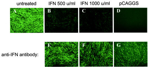
Transfection of CEFs with plasmid DNA induces production of IFN-α/β and represses NDV-GFP replication. CEFs were left untreated (A), were treated with chicken IFN-α/β at 500 U (B) or 1,000 U (C)/ml, or were transfected with the empty expression plasmid pCAGGS (D). IFN treatment was performed in the absence (A to C) or presence (E and F) of neutralizing antibody against chicken IFN. Transfection of cells with empty vector was performed in the absence of anti-IFN antibody (D), or else the antibody was added immediately after the transfection (G). Cells were infected with NDV-GFP at an MOI of 1 at 20 h posttreatment or posttransfection and then examined for green fluorescence at 24 h postinfection.
Transfection of CEFs with plasmid DNA induces production of IFN and represses NDV-GFP replication.
We examined the effect of treating CEFs with chicken IFN prior to NDV-GFP infection. Infection of untreated CEFs resulted in GFP expression (Fig. 2A). When cells were treated with 500 or 1,000 U of chicken IFN-α/β per ml 1 day prior to infection, viral GFP expression was greatly reduced, suggesting that viral replication was significantly impaired (Fig. 2B and C). However, viral GFP expression was restored when a neutralizing anti-chicken IFN antibody was added 8 h after addition of IFN (Fig. 2E and F). We also found that prior transfection of cells with plasmid DNA blocked GFP expression, and this inhibition was even more pronounced than when cells were pretreated with 1,000 U of IFN/ml (Fig. 2D). GFP expression could be restored in transfected and infected cells by addition of anti-IFN antibody to the cell culture supernatant, suggesting that DNA transfection inhibits NDV-GFP replication largely, if not exclusively, through the induction of IFN production (Fig. 2G). We have also established that cell supernatants taken from transfected cells and transferred to fresh CEFs inhibit NDV-GFP replication (data not shown). Interestingly, the induction of an IFN response in CEFs appears to require both the transfection reagent and DNA. Inhibition of NDV-GFP expression was not seen when only transfection reagent or only DNA was added to cells (data not shown). The requirement for both the transfection reagent and the plasmid DNA to induce IFN production suggests that transcription from the transfected plasmids is the actual trigger of IFN production. Transcription from the mammalian expression plasmids can lead to the production of double-stranded RNA, a factor known to induce IFN production (37).
Expression of the IFN-antagonist influenza virus NS1 or Ebola virus VP35 proteins prevents plasmid transfection-induced inhibition of NDV-GFP replication.
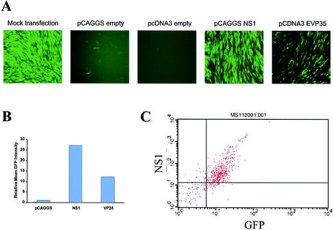
The influenza virus NS1 protein and the Ebola virus VP35 protein have previously been shown to inhibit cellular IFN responses (2, 18, 55, 59). Transfection of plasmids encoding either of these IFN antagonists could overcome the transfection-mediated inhibition of NDV-GFP replication. As seen previously, transfection of empty vector (either pCAGGS or pcDNA3) 1 day before infection prevented GFP expression from NDV-GFP-infected cells (Fig. 3A). However, when either NS1 or VP35 expression plasmids were transfected, GFP expression could be readily detected 1 day postinfection (Fig. 3A). When the intensity of GFP expression was analyzed by FACS, the enhancement of green fluorescence in NS1- or VP35-expressing cells was clearly demonstrated (Fig. 3B). When NS1-expressing, NDV-GFP-infected cells were fixed and stained with anti-NS1 antiserum and then analyzed by FACS, the levels of GFP expression were found to correlate with the levels of NS1 expression (Fig. 3C). These data demonstrate that expression of IFN antagonists prevents transfection-induced inhibition of NDV-GFP replication and provide further evidence that the transfection-mediated inhibition of NDV-GFP growth is related to activation of the cellular IFN response. These experiments also suggest that the NDV-GFP assay can be used as a screen for proteins with IFN-antagonist activity.
FIG. 3.
Expression of the influenza virus NS1 or Ebola virus VP35 proteins prevents plasmid transfection-induced inhibition of NDV-GFP replication. (A) GFP expression in NDV-GFP-infected cells transfected 24 h prior to infection with various plasmids. Cells were mock transfected, transfected with empty pCAGGS or pcDNA3 expression plasmids, or transfected with expression plasmids for the influenza A virus NS1 protein or the Ebola virus VP35 protein as indicated. (B) Mean relative green fluorescence intensity of CEFs transfected with the indicated plasmids (pCAGGS, pCAGGS-NS1, or pcDNA3-VP35) and subsequently infected with NDV-GFP. The graph shows the mean relative intensity of green fluorescence for 104 cells from each culture as determined by FACS. The results from panel A and panel B are from the same experiment and are representative of typical results with the indicated plasmids. (C) Expression of NS1 in green fluorescent cells. CEFs were transfected with NS1 expression plasmid and infected with NDV-GFP 24 h posttransfection. At 24 h postinfection, the cells were fixed, permeabilized, and stained with rabbit antiserum against the influenza A virus NS1 protein. FACS analysis was then performed to detect both NS1 (y axis) and GFP (x axis).
Expression of the NDV V protein prevents transfection-induced inhibition of NDV-GFP replication.
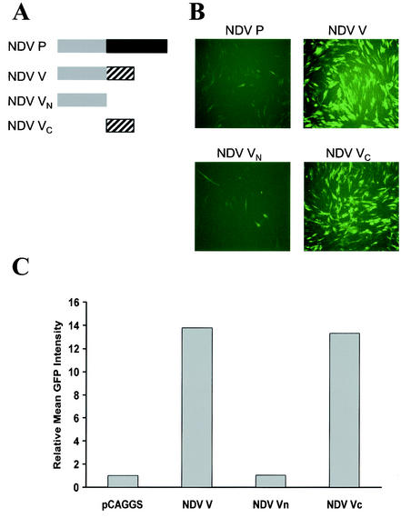
Because NDV encodes a V protein but not a C protein and because the V proteins of several paramyxoviruses display IFN-antagonist activity, the V protein of NDV was tested for its ability to rescue the growth of NDV-GFP virus. Transfection of an empty plasmid once again inhibited NDV-GFP replication. In contrast, transfection of CEF cells with an NDV V protein expression plasmid restored the ability of NDV-GFP to grow (Fig. 4A and B). It should be noted that although the NDV-GFP virus possesses an intact V ORF, the IFN-induced inhibition of viral growth occurs prior to infection, allowing an antiviral state to be established before V protein is expressed from virus.
FIG. 4.
Expression of the NDV V protein prevents transfection-induced inhibition of NDV-GFP replication. (A) Diagram of the four NDV P and V constructs used. The gray boxes on the left indicate shared amino-terminal domains. The V protein possesses a cysteine-rich carboxy-terminal region distinct from the P protein. This V-specific domain arises due to the insertion of a single non-template-encoded G residue and is indicated by the hatched box. VN possesses only the shared amino-terminal region. VC possesses the V carboxy terminus to which an initiator ATG has been added. (B) GFP expression in NDV-GFP-infected cells transfected 24 h prior to infection with NDV P, NDV V, NDV VN, or NDV VC expression plasmids. (C) Relative mean green fluorescence of CEFs transfected with empty vector (pCAGGS) or transfected with NDV P, NDV V, NDV VN, or NDV VC expression plasmids as indicated and subsequently infected with NDV-GFP. Shown is the relative mean intensity of green fluorescence for 104 cells from each culture as determined by FACS. The results from panels B and C are from the same experiment and are representative of typical results with the indicated plasmids.
The NDV V protein is produced in infected cells from an edited transcript in which the viral polymerase has added a single non-template-encoded G residue. As a result, the amino-terminal region of V is identical to the amino terminus of P. However, after the editing site, the proteins are different (Fig. 4A). In order to determine which region of V was responsible for the restoration of GFP expression, three additional constructs were tested. These plasmids encode the full-length P protein, the amino-terminal region of V which is shared in common with the P protein or the carboxy-terminal domain of V which is distinct from P (Fig. 4A). Whereas the carboxy-terminal domain of V and the full-length V were able to rescue GFP expression, neither the VN nor the P constructs rescued NDV-GFP growth (Fig. 4B). Analysis of the relative intensity of green fluorescence from this experiment demonstrated that cells expressing either full-length NDV V or the carboxy-terminal domain resulted in an average GFP fluorescence intensity approximately 13 times that of mock-transfected, infected cells. (Fig. 4C). These data indicate that the observed IFN-antagonist activity is encoded by the carboxy terminus of NDV V.
The Nipah virus V, W, and C proteins display IFN-antagonist activity in the NDV-GFP system.
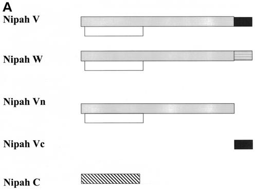
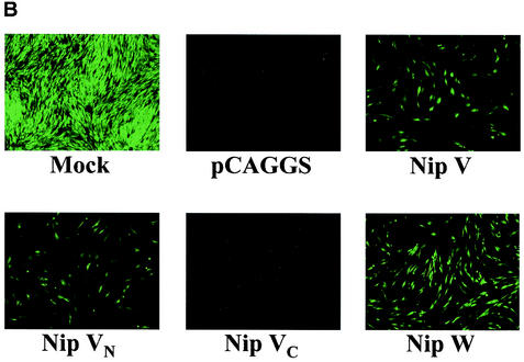
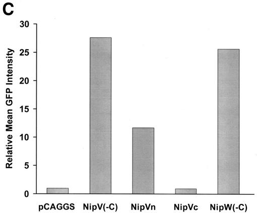
The NDV-GFP assay was then used to screen several Nipah virus protein expression plasmids for IFN-antagonist activity. Nipah virus was chosen because of its importance as an emerging paramyxovirus that causes severe disease in humans (9). Because this virus is expected to produce V, W, and C proteins from its P gene (7; B. H. Harcourt, A. Tamin, B. Newton, A. Sanchez, T. G. Ksiazek, P. E. Rollin, W. J. Bellini, and P. A. Rota, Abstr. 11th Int. Conf. Negative Strand Viruses, abstr. 170, 2000), we tested the V, W, and C ORFs in the NDV-GFP assay (Fig. 5A). Expression of the Nipah virus V protein rescued the growth of NDV-GFP (Fig. 5B and C). Likewise, a W protein expression plasmid was also found to enhance NDV-GFP replication (Fig. 5B and C). The V and W plasmids used for these experiments were altered to decrease the possibility that they would also express the C protein, which is encoded entirely within the amino terminus of the V or W proteins. The two tandem ATGs predicted to begin the C ORF were mutated to ACG. These mutations were expected to reduce or eliminate C protein expression. Although ACG is used as a start codon to initiate translation of the Sendai virus C′ protein, it is an inefficient start codon (11). We have also tested an additional V protein expression plasmid in which the C gene was mutated not only at these two ATGs but also by introduction of a stop codon at position four of the C ORF. These changes did not affect V protein translation, and this C knockout construct also rescued NDV-GFP growth (data not shown). These data suggest that the enhancement of NDV-GFP replication seen with V and W expression plasmids occurs independent of C protein expression.
FIG. 5.
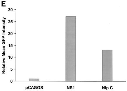
(A) Diagram of the five Nipah P, V, and W constructs used. The shaded boxes indicate the shared amino-terminal domains. The V protein possesses a cysteine-rich carboxy-terminal region distinct from the P protein. This V-specific domain arises due to the insertion of a single non-template-encoded G residue and is indicated by the solid box. The W protein possesses a carboxy-terminal region distinct from both the P and the V proteins. This W-specific domain arises due to the insertion of two non-template-encoded G residues and is indicated by the box with horizontal lines. VN possesses only the shared amino-terminal region. VC possesses the V carboxy terminus, to which an initiator ATG has been added. The C ORF is indicated by the hatched box. Because the ATGs at the beginning of the C ORF have been mutated in the V, W, and VN constructs, the C ORF in these constructs is indicated by the open box. (B) GFP expression in NDV-GFP-infected cells that were, 24 h prior to infection, mock transfected or transfected with the empty pCAGGS plasmid or expression plasmids for Nipah virus V (Nip VN), the amino-terminal domain of Nipah virus V (Nip VN), the carboxy-terminal domain of V (Nip VC), or Nipah virus W (Nip W). (C) Relative mean green fluorescence of CEFs transfected with empty vector (pCAGGS) or transfected with the plasmids encoding Nip V, Nip VN, Nip VC, or Nip W. These plasmids were engineered so as not to express the C protein [indicated by (−C)]. Shown is the relative mean intensity of green fluorescence for 104 cells from each culture as determined by FACS. (D) GFP expression in NDV-GFP-infected cells that were, 24 h prior to infection, mock transfected or transfected with the empty pCAGGS plasmid or expression plasmids for influenza virus NS1 or Nipah virus C proteins. (E) Relative mean green fluorescence of CEFs transfected with empty vector (pCAGGS) or transfected with the plasmids encoding influenza virus NS1 or Nipah virus C. Shown is the relative mean intensity of green fluorescence for 104 cells from each culture as determined by FACS. The results from panels B and C are from the same experiment, and the results from panels D and E are from the same experiment and are representative of typical results with the indicated plasmids.
It was striking that both V and W exerted an anti-IFN effect in this system. Because these proteins possess the same 407 amino-terminal amino acids, this common region was also tested and found to also enhance green fluorescence in NDV-GFP-infected cells (Fig. 5B and C). Interestingly and in contrast to the data obtained with the NDV V, the carboxy-terminal region of Nipah virus V, which possesses the relatively conserved cysteine-rich region, did not show significant IFN-antagonist activity in this assay (Fig. 5B and C).
Because the C proteins of Sendai virus have been reported to counter the host IFN response, we also tested the Nipah virus C protein in the NDV-GFP assay. When a Nipah virus C protein expression plasmid was transfected, GFP expression was also detected, although the intensity of the green fluorescence in C-transfected cells was typically lower than that seen with the V or W constructs (Fig. 5D and E).
The Nipah virus V and W proteins inhibit IFN-β-mediated activation of an IFN responsive promoter.
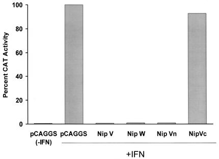
The V proteins of human parainfluenza virus type 2 (hPIV-2), simian virus 5 (SV5), and mumps virus reportedly inhibit the IFN signaling pathways (32, 47). We therefore determined whether expression plasmids encoding the Nipah virus V or W or truncated forms of Nipah virus V inhibited, in Vero cells, the activation by IFN-β of an IFN-inducible promoter (Fig. 6). Transfection of plasmids possessing the entire V ORF, the entire V ORF in which the C ORF was disrupted, or the entire W ORF resulted in a near-complete block in ISRE reporter gene expression in response to IFN (Fig. 6). Further, a plasmid containing only the amino-terminal domain common to V and W also blocked reporter gene activation, whereas a plasmid containing only the unique carboxy-terminal region of V failed to block reporter gene activation (Fig. 6). These data are consistent with the IFN-antagonist activity seen with these proteins in the NDV-GFP assay and suggest that the common amino-terminal domain of Nipah virus V and W can function to block the IFN signaling pathway. When the Nipah virus C expression plasmid which enhanced NDV-GFP replication in CEFs was tested in the reporter gene assay in Vero cells, it had a much less dramatic effect on IFN-induced reporter gene activation. Nipah virus C was found to reproducibly decrease reporter gene activity to a modest degree (data not shown). It is unclear whether this modest inhibition of IFN signaling is sufficient to permit NDV-GFP replication or whether Nipah virus C may enhance NDV-GFP replication by an alternative mechanism.
FIG. 6.
Inhibition in Vero cells of IFN-induced activation of an ISRE promoter by Nipah virus proteins. Effect of Nipah virus V, W, and V truncation mutant expression plasmids on IFN-β-induced activation of an IFN-inducible promoter. Vero cells were transfected with the indicated plasmids and either mock treated (−IFN) or treated with human IFN-β (+IFN). Τhe expression plasmids encoded Nipah virus V (Nip V), Nipah virus W (Nip W), the amino-terminal region common to Nipah virus V and W (Nip Vn), or the carboxy-terminal region of Nipah virus V specific to V (Nip Vc). Shown is the relative activity of an IFN-inducible CAT reporter gene under the control of the IFN-inducible HISG54 promoter normalized to an internal control consisting of a constitutively expressed Renilla luciferase expression plasmid. The cells were treated with 1,000 U of human IFN-β/ml at 1 day posttransfection, and CAT and luciferase assays were performed 1 day later.
DISCUSSION
Using an NDV-GFP-based assay, we found that expression of the NDV V protein or of the (putative) Nipah virus V, W, and C proteins can prevent establishment of an IFN-induced antiviral state. The NDV-GFP assay provides a straightforward system by which cloned viral genes can be screened for IFN-antagonist activity. NDV-GFP is a virus that is susceptible to the antiviral effects of IFN, although it encodes a functional IFN antagonist, the V protein. It thus appears that the presence of a functional NDV V gene may not be sufficient to overcome the previously established antiviral state within the time frame of the assay. This contention is supported by the following observation: when CEF cells are treated once with IFN-α/β at 20 h prior to infection, NDV-GFP replication is undetectable, although green fluorescence becomes detectable at 48 h postinfection under these conditions (data not shown). Likewise, prior transfection of CEF cells with plasmid DNA results in the secretion of IFN, and this IFN response suppresses, for at least 24 h, the replication of subsequently added NDV-GFP. It is likely that the transfection produces sufficient IFN that an antiviral state is well established before NDV-GFP infection. However, when cells are transfected with plasmids expressing IFN antagonists, including the influenza virus NS1 protein or the Ebola virus VP35 protein, the induction of an antiviral state can be reduced or prevented, and GFP expression is easily detected less than 24 h postinfection.
One advantage of the NDV-GFP assay is the simple and rapid readout. It should be noted that the absolute number and intensity of green cells seen from experiment to experiment are variable, but the relative activity of the various transfected proteins is consistent between experiments. Additionally, the levels of NDV-GFP replication vary depending on which IFN antagonist is expressed. The reason(s) that one protein enhances NDV-GFP replication to a different degree than another is not clear but might be related to different levels of expression or to different mechanisms of action (e.g., one may target the JAK/STAT signaling pathway, whereas another might prevent the production of IFN). Another helpful aspect of the system is that the test virus (NDV-GFP) readily grows to high titers in 10-day-old embryonated chicken eggs. It remains to be seen whether the use of chicken cells limits the usefulness of the assay. In this respect, it is encouraging that the influenza virus NS1 protein, the Ebola virus VP35 protein, and the Nipah virus proteins all appear to function as IFN antagonists in both CEFs and mammalian systems. This is despite the fact that some paramyxovirus V proteins appear to function in a species-specific manner (46). We also have preliminary data indicating that the NDV-GFP assay can be adapted to at least some mammalian cells. The use of different cell lines may help in the identification of IFN antagonists that function in a cell type-dependent fashion.
Paramyxovirus V, W, and C proteins are encoded by the viral phosphoprotein (P) gene. The P protein and the V protein always share a common amino terminus but possess unique carboxy termini due to the insertion of a non-template-encoded G residue(s) at a precise point during transcription of the P gene, a process called “editing.” In some paramyxoviruses, including all members of the Rubulavirus genus except for NDV, the V protein is encoded by the unedited mRNA (33). For the remaining paramyxoviruses that encode V proteins, V is encoded by an edited transcript in which one non-template-encoded G has been inserted (33). In addition, some paramyxoviruses produce additional edited P-gene transcripts that encode proteins with amino-terminal sequences identical to that of the P protein. These include “W” proteins, such as that predicted for Nipah virus (Harcourt et al., 11th Int. Conf. Negative Strand Viruses), which would arise from edited mRNAs in which two G residues are inserted at the editing site into the P gene mRNA. The C proteins are encoded by the P transcripts as well but arise due to the use of alternate start codons and do not possess amino acid identity with the P protein (33). As with V proteins, not all paramyxoviruses encode C proteins, although some, such a Nipah virus, encode C proteins, V proteins, and additional P gene-derived proteins (e.g., W proteins) (33).
It will be important to determine the specific mechanisms by which the NDV V and the Nipah virus V, W, and C proteins counter the IFN response. In this regard, the Nipah virus V protein has recently been reported to cause cytoplasmic retention of STAT1 and STAT2 (50a). Among other paramyxoviruses, different viruses have been shown to employ different mechanisms to overcome the host IFN response (5, 19, 21, 22, 27, 30, 32, 46, 47, 54, 60). Several paramyxovirus V proteins have previously been shown to inhibit IFN signaling. For example, the mumps virus V protein has been reported to decrease cellular STAT1 levels, possibly by targeting STAT1 for proteosome-mediated degradation (32). Similarly, the “V” proteins of SV5 and hPIV-2 also appear to promote STAT protein degradation although, whereas SV5 V promotes STAT1 degradation, hPIV-2 V promotes the degradation of STAT2 (47). Interestingly, V-mediated degradation of one specific STAT (STAT1 or STAT2) protein required the presence of the second STAT (47), and the inability of SV5 V to promote the degradation of STAT1 in mouse cells was related to the absence of a compatible (to the SV5 V protein) STAT2 protein (46). Our data indicate that the NDV V also blocks the host cell IFN response, although its mechanism of action has yet to be defined. The ability of the Nipah virus V or W protein expression plasmids to not only rescue NDV-GFP growth but also prevent activation of the IFN-inducible reporter genes in response to IFN-β treatment suggests that the Nipah virus V and W proteins will also affect some component of the IFN-α/β signaling pathway. It is interesting that the Nipah virus C expression plasmid also showed an ability to rescue NDV-GFP replication but displayed only a weak inhibition in the IFN-induced reporter gene system. In the case of Sendai virus, the “C” proteins, a set of four carboxy-coterminal proteins, have been reported to block IFN signaling both in infected cells and when expressed individually (19, 21, 22, 27, 30, 52). It is therefore unclear whether the IFN-antagonist activity of the Nipah virus C seen in the NDV-GFP assay can be fully accounted for by a weak block in the IFN signaling pathway or whether Nipah virus C has additional biological functions.
It was striking that different regions of the NDV V and the Nipah virus V proteins were required for anti-IFN activity. The carboxy-terminal domain of the various paramyxovirus V proteins is relatively conserved and includes seven cysteine residues that together form a zinc finger (24, 36, 45, 48). As was the case for the NDV V in the present study, the conserved, carboxy-terminal domains of either the mumps virus V protein or the hPIV-2 V protein were capable of blocking the host cell IFN signaling pathway (32, 43). In contrast, in both the NDV-GFP assay (Fig. 5) and the reporter gene assay (Fig. 6), IFN-antagonist activity was clearly associated with the amino-terminal region common to the Nipah virus P, V, and W proteins. In contrast, little to no IFN-antagonist activity was detected with the cysteine-rich carboxy terminus of the Nipah virus V protein. However, although it is clear that the amino terminus of V is sufficient for IFN-antagonist activity in these assays, it remains possible that the carboxy-terminal domain of Nipah virus V contributes to the IFN-antagonist function of the full-length Nipah virus V protein or that the carboxy terminus of V will display a species-specific IFN-antagonist activity. It is interesting that the V, W, and P proteins of Nipah virus and Hendra virus possess longer amino termini than do other paramyxoviruses (7). Perhaps the unique 210 amino-terminal amino acids common to the Nipah virus V and W proteins possess the IFN-antagonist activity. It is also interesting that the Ebola virus VP35 protein is functionally analogous to the paramyxovirus P proteins and also counteracts the host IFN response (Fig. 2) (2, 40, 41). However, filovirus VP35 proteins do not appear to encode C, V, or W protein equivalents. Thus, it is clear that P genes (or their equivalents) of negative-strand RNA viruses have frequently evolved IFN-antagonist functions. In cases such as Ebola virus, the P protein itself may be sufficient to carry out this function. In other viruses, such “auxiliary” functions may have been shifted exclusively to C or V proteins during the course of evolution. In the case of Nipah virus, although V and W proteins are produced and exert an anti-IFN function, it remains possible that the P protein also blocks the host IFN system.
Recently, data were presented indicating that the amino-terminal domain of the measles virus (a morbilivirus) P protein is a “natively unfolded protein” (25). It was also predicted that the amino-terminal region of the P proteins of other morbilliviruses, of the paramyxovirus Sendai virus, and of the rhabdovirus vesicular stomatitis virus are also natively unfolded (25). This finding is in contrast to findings for the amino-terminal domains of the P proteins of the rubulaviruses, a group that includes NDV, which are predicted to be folded (25). When the amino-terminal domain common to the Nipah virus P, V, and W is analyzed in the same way, it is also predicted to be a natively unfolded protein (data not shown). Any connection between this property and the ability to counteract the IFN response remains to be determined.
The identification of the NDV V and the Nipah virus C, V, and W proteins as having IFN-antagonist activity suggests that they are important virulence factors. A role for the NDV V protein in virulence has already been demonstrated. A recombinant NDV with an editing site mutation such that the virus produces 20-fold less V protein than wild-type NDV was highly attenuated in chicken embryos (39). An NDV completely unable to produce V, but presumably able to produce a truncated “amino terminus only” form of V, was highly impaired in tissue culture and unable to replicate in 10-day-old embryonated chicken eggs (39). Other studies provide additional evidence for the importance of the V protein in the virulence of several other paramyxoviruses. Mutations truncating the V protein before the unique carboxy terminus or mutations affecting the ability of V protein to bind zinc attenuated Sendai virus in mice (24, 26). Mutations preventing expression of the unique domains of either V or D (produced from a +2G transcript) had little effect on hPIV-3 replication, either in tissue culture or in vivo. However, mutation of both the V and the D ORFs did yield a modest attenuation phenotype in vivo in a hamster model and in a monkey model (13). The relationship between the IFN system and the various attenuation phenotypes seen with these particular mutant viruses remains to be determined. In contrast, there is a clear correlation between virulence and the anti-IFN function of the Sendai virus C proteins (13, 20).
The NDV-GFP-based assay used to identify IFN-antagonist functions for NDV and Nipah virus proteins is similar to our previously described assay which used a mutant influenza virus, influenza delNS1 virus, which lacks the influenza virus IFN-antagonist NS1 protein (2). In this previously described assay, we found that transfection of MDCK cells with plasmids encoding IFN antagonists greatly enhanced growth of the mutant influenza virus (2). The NDV-GFP-based assay should be complementary to the influenza delNS1 virus-based assay. For example, the use of different viruses with different host ranges may allow a wider range of cell lines to be used when screening for IFN antagonists. Such assays will likely provide new insights into viral pathogenesis. Previous studies on NDV and other paramyxoviruses suggest that these anti-IFN functions play important roles in viral pathogenesis. The observations made in the present report regarding Nipah virus may therefore be of particular interest because Nipah virus is a highly lethal, emerging virus of concern as a potential agent of bioterrorism (9, 14, 34).
Acknowledgments
This work was supported by NIH grants to C.F.B., A.G.-S., and P.P. C.F.B. is an Ellison Medical Foundation New Scholar in Global Infectious Diseases.
We thank Neva Morales and Estanis Nistal-Villan for expert technical assistance. We thank Peter Staeheli (University of Freiburg) and Bernd Kaspers (University of Munich) for providing chicken IFN and anti-chicken IFN antibodies.
REFERENCES
- 1.Basler, C. F., and A. García-Sastre. 2002. Viruses and the type I interferon antiviral system: induction and evasion. Int. Rev. Immunol. 21:305-338. [DOI] [PubMed] [Google Scholar]
- 2.Basler, C. F., X. Wang, E. Mühlberger, V. Volchkov, J. Paragas, H. D. Klenk, A. Garcia-Sastre, and P. Palese. 2000. The Ebola virus VP35 protein functions as a type I IFN antagonist. Proc. Natl. Acad. Sci. USA 97:12289-12294. [DOI] [PMC free article] [PubMed] [Google Scholar]
- 3.Beattie, E., K. L. Denzler, J. Tartaglia, M. E. Perkus, E. Paoletti, and B. L. Jacobs. 1995. Reversal of the interferon-sensitive phenotype of a vaccinia virus lacking E3L by expression of the reovirus S4 gene. J. Virol. 69:499-505. [DOI] [PMC free article] [PubMed] [Google Scholar]
- 4.Bergmann, M., A. García-Sastre, E. Carnero, H. Pehamberger, K. Wolff, P. Palese, and T. Muster. 2000. Influenza virus NS1 protein counteracts PKR-mediated inhibition of replication. J. Virol. 74:6203-6206. [DOI] [PMC free article] [PubMed] [Google Scholar]
- 5.Bossert, B., and K. K. Conzelmann. 2002. Respiratory syncytial virus (RSV) nonstructural (NS) proteins as host range determinants: a chimeric bovine RSV with NS genes from human RSV is attenuated in interferon-competent bovine cells. J. Virol. 76:4287-4293. [DOI] [PMC free article] [PubMed] [Google Scholar]
- 6.Brandt, T. A., and B. L. Jacobs. 2001. Both carboxy- and amino-terminal domains of the vaccinia virus interferon resistance gene, E3L, are required for pathogenesis in a mouse model. J. Virol. 75:850-856. [DOI] [PMC free article] [PubMed] [Google Scholar]
- 7.Chan, Y. P., K. B. Chua, C. L. Koh, M. E. Lim, and S. K. Lam. 2001. Complete nucleotide sequences of Nipah virus isolates from Malaysia. J. Gen. Virol. 82:2151-2155. [DOI] [PubMed] [Google Scholar]
- 8.Chou, J., E. R. Kern, R. J. Whitley, and B. Roizman. 1990. Mapping of herpes simplex virus-1 neurovirulence to gamma 134.5, a gene nonessential for growth in culture. Science 250:1262-1266. [DOI] [PubMed] [Google Scholar]
- 9.Chua, K. B., W. J. Bellini, P. A. Rota, B. H. Harcourt, A. Tamin, S. K. Lam, T. G. Ksiazek, P. E. Rollin, S. R. Zaki, W. Shieh, C. S. Goldsmith, D. J. Gubler, J. T. Roehrig, B. Eaton, A. R. Gould, J. Olson, H. Field, P. Daniels, A. E. Ling, C. J. Peters, L. J. Anderson, and B. W. Mahy. 2000. Nipah virus: a recently emergent deadly paramyxovirus. Science 288:1432-1435. [DOI] [PubMed] [Google Scholar]
- 10.Clemens, M. J. 1997. PKR: a protein kinase regulated by double-stranded RNA. Int. J. Biochem. Cell Biol. 29:945-949. [DOI] [PubMed] [Google Scholar]
- 11.Curran, J., and D. Kolakofsky. 1988. Ribosomal initiation from an ACG codon in the Sendai virus P/C mRNA. EMBO J. 7:245-251. [DOI] [PMC free article] [PubMed] [Google Scholar]
- 12.Davies, M. V., H. W. Chang, B. L. Jacobs, and R. J. Kaufman. 1993. The E3L and K3L vaccinia virus gene products stimulate translation through inhibition of the double-stranded RNA-dependent protein kinase by different mechanisms. J. Virol. 67:1688-1692. [DOI] [PMC free article] [PubMed] [Google Scholar]
- 13.Durbin, A. P., J. M. McAuliffe, P. L. Collins, and B. R. Murphy. 1999. Mutations in the C, D, and V open reading frames of human parainfluenza virus type 3 attenuate replication in rodents and primates. Virology 261:319-330. [DOI] [PubMed] [Google Scholar]
- 14.Field, H., P. Young, J. M. Yob, J. Mills, L. Hall, and J. Mackenzie. 2001. The natural history of Hendra and Nipah viruses. Microbes Infect. 3:307-314. [DOI] [PubMed] [Google Scholar]
- 15.Floyd-Smith, G., E. Slattery, and P. Lengyel. 1981. Interferon action: RNA cleavage pattern of a (2′-5′)oligoadenylate-dependent endonuclease. Science 212:1030-1032. [DOI] [PubMed] [Google Scholar]
- 16.Francois, C., G. Duverlie, D. Rebouillat, H. Khorsi, S. Castelain, H. E. Blum, A. Gatignol, C. Wychowski, D. Moradpour, and E. F. Meurs. 2000. Expression of hepatitis C virus proteins interferes with the antiviral action of interferon independently of PKR-mediated control of protein synthesis. J. Virol. 74:5587-5596. [DOI] [PMC free article] [PubMed] [Google Scholar]
- 17.Gale, M. J., Jr., M. J. Korth, N. M. Tang, S. L. Tan, D. A. Hopkins, T. E. Dever, S. J. Polyak, D. R. Gretch, and M. G. Katze. 1997. Evidence that hepatitis C virus resistance to interferon is mediated through repression of the PKR protein kinase by the nonstructural 5A protein. Virology 230:217-227. [DOI] [PubMed] [Google Scholar]
- 18.García-Sastre, A., A. Egorov, D. Matassov, S. Brandt, D. E. Levy, J. E. Durbin, P. Palese, and T. Muster. 1998. Influenza A virus lacking the NS1 gene replicates in interferon-deficient systems. Virology 252:324-330. [DOI] [PubMed] [Google Scholar]
- 19.Garcin, D., J. Curran, and D. Kolakofsky. 2000. Sendai virus C proteins must interact directly with cellular components to interfere with interferon action. J. Virol. 74:8823-8830. [DOI] [PMC free article] [PubMed] [Google Scholar]
- 20.Garcin, D., M. Itoh, and D. Kolakofsky. 1997. A point mutation in the Sendai virus accessory C proteins attenuates virulence for mice, but not virus growth in cell culture. Virology 238:424-431. [DOI] [PubMed] [Google Scholar]
- 21.Garcin, D., P. Latorre, and D. Kolakofsky. 1999. Sendai virus C proteins counteract the interferon-mediated induction of an antiviral state. J. Virol. 73:6559-6565. [DOI] [PMC free article] [PubMed] [Google Scholar]
- 22.Gotoh, B., K. Takeuchi, T. Komatsu, J. Yokoo, Y. Kimura, A. Kurotani, A. Kato, and Y. Nagai. 1999. Knockout of the Sendai virus C gene eliminates the viral ability to prevent the interferon-α/β-mediated responses. FEBS Lett. 459:205-210. [DOI] [PubMed] [Google Scholar]
- 23.Haller, O., M. Frese, and G. Kochs. 1998. Mx proteins: mediators of innate resistance to RNA viruses. Rev. Sci. Technol. 17:220-230. [DOI] [PubMed] [Google Scholar]
- 24.Huang, C., K. Kiyotani, Y. Fujii, N. Fukuhara, A. Kato, Y. Nagai, T. Yoshida, and T. Sakaguchi. 2000. Involvement of the zinc-binding capacity of Sendai virus V protein in viral pathogenesis. J. Virol. 74:7834-7841. [DOI] [PMC free article] [PubMed] [Google Scholar]
- 25.Karlin, D., S. Longhi, V. Receveur, and B. Canard. 2002. The N-terminal domain of the phosphoprotein of morbilliviruses belongs to the natively unfolded class of proteins. Virology 296:251-262. [DOI] [PubMed] [Google Scholar]
- 26.Kato, A., K. Kiyotani, Y. Sakai, T. Yoshida, T. Shioda, and Y. Nagai. 1997. Importance of the cysteine-rich carboxyl-terminal half of V protein for Sendai virus pathogenesis. J. Virol. 71:7266-7272. [DOI] [PMC free article] [PubMed] [Google Scholar]
- 27.Kato, A., Y. Ohnishi, M. Kohase, S. Saito, M. Tashiro, and Y. Nagai. 2001. Y2, the smallest of the Sendai virus C proteins, is fully capable of both counteracting the antiviral action of interferons and inhibiting viral RNA synthesis. J. Virol. 75:3802-3810. [DOI] [PMC free article] [PubMed] [Google Scholar]
- 28.Kitajewski, J., R. J. Schneider, B. Safer, S. M. Munemitsu, C. E. Samuel, B. Thimmappaya, and T. Shenk. 1986. Adenovirus VAI RNA antagonizes the antiviral action of interferon by preventing activation of the interferon-induced eIF-2 alpha kinase. Cell 45:195-200. [DOI] [PubMed] [Google Scholar]
- 29.Klenk, H.-D., W. Slenczka, and H. Feldmann. 1994. Marburg and Ebola viruses, p. 827-831. In R. G. Webster and A. Granoff (ed.), Encyclopedia of virology, vol. 2. Academic Press, New York, N.Y.
- 30.Komatsu, T., K. Takeuchi, J. Yokoo, Y. Tanaka, and B. Gotoh. 2000. Sendai virus blocks alpha interferon signaling to signal transducers and activators of transcription. J. Virol. 74:2477-2480. [DOI] [PMC free article] [PubMed] [Google Scholar]
- 31.Krishnamurthy, S., Z. Huang, and S. K. Samal. 2000. Recovery of a virulent strain of Newcastle disease virus from cloned cDNA: expression of a foreign gene results in growth retardation and attenuation. Virology 278:168-182. [DOI] [PubMed] [Google Scholar]
- 32.Kubota, T., N. Yokosawa, S. Yokota, and N. Fujii. 2001. C terminal CYS-RICH region of mumps virus structural V protein correlates with block of interferon alpha and gamma signal transduction pathway through decrease of STAT 1-alpha. Biochem. Biophys. Res. Commun. 283:255-259. [DOI] [PubMed] [Google Scholar]
- 33.Lamb, R. A., and D. Kolakofsky. 2001. Paramyxoviridae: the viruses and their replication, p. 1305-1340. In D. M. Knipe and P. M. Howley (ed.), Fields virology, 4th ed., vol. 1. Lippincott, Williams & Wilkins Co., Philadelphia, Pa.
- 34.Lane, H. C., J. L. Montagne, and A. S. Fauci. 2001. Bioterrorism: a clear and present danger. Nat. Med. 7:1271-1273. [DOI] [PubMed] [Google Scholar]
- 35.Leonard, G. T., and G. C. Sen. 1997. Restoration of interferon responses of adenovirus E1A-expressing HT1080 cell lines by overexpression of p48 protein. J. Virol. 71:5095-5101. [DOI] [PMC free article] [PubMed] [Google Scholar]
- 36.Lin, G. Y., R. G. Paterson, C. D. Richardson, and R. A. Lamb. 1998. The V protein of the paramyxovirus SV5 interacts with damage-specific DNA binding protein. Virology 249:189-200. [DOI] [PubMed] [Google Scholar]
- 37.Mabrouk, T., C. Danis, and G. Lemay. 1995. Two basic motifs of reovirus sigma 3 protein are involved in double-stranded RNA binding. Biochem. Cell Biol. 73:137-145. [DOI] [PubMed] [Google Scholar]
- 38.Markovitz, N. S., D. Baunoch, and B. Roizman. 1997. The range and distribution of murine central nervous system cells infected with the γ134.5− mutant of herpes simplex virus 1. J. Virol. 71:5560-5569. [DOI] [PMC free article] [PubMed] [Google Scholar]
- 39.Mebatsion, T., S. Verstegen, L. T. De Vaan, A. Römer-Oberdörfer, and C. C. Schrier. 2001. A recombinant Newcastle disease virus with low-level V protein expression is immunogenic and lacks pathogenicity for chicken embryos. J. Virol. 75:420-428. [DOI] [PMC free article] [PubMed] [Google Scholar]
- 40.Mühlberger, E., B. Lotfering, H. D. Klenk, and S. Becker. 1998. Three of the four nucleocapsid proteins of Marburg virus, NP, VP35, and L, are sufficient to mediate replication and transcription of Marburg virus-specific monocistronic minigenomes. J. Virol. 72:8756-8764. [DOI] [PMC free article] [PubMed] [Google Scholar]
- 41.Mühlberger, E., M. Weik, V. E. Volchkov, H. D. Klenk, and S. Becker. 1999. Comparison of the transcription and replication strategies of Marburg virus and Ebola virus by using artificial replication systems. J. Virol. 73:2333-2342. [DOI] [PMC free article] [PubMed] [Google Scholar]
- 42.Nakaya, T., J. Cros, M. S. Park, Y. Nakaya, H. Zheng, A. Sagrera, E. Villar, A. García-Sastre, and P. Palese. 2001. Recombinant Newcastle disease virus as a vaccine vector. J. Virol. 75:11868-11873. [DOI] [PMC free article] [PubMed] [Google Scholar]
- 43.Nishio, M., M. Tsurudome, M. Ito, M. Kawano, H. Komada, and Y. Ito. 2001. High resistance of human parainfluenza type 2 virus protein-expressing cells to the antiviral and anti-cell proliferative activities of alpha/beta interferons: cysteine-rich V-specific domain is required for high resistance to the interferons. J. Virol. 75:9165-9176. [DOI] [PMC free article] [PubMed] [Google Scholar]
- 44.Niwa, H., K. Yamamura, and J. Miyazaki. 1991. Efficient selection for high-expression transfectants with a novel eukaryotic vector. Gene 108:193-199. [DOI] [PubMed] [Google Scholar]
- 45.Ohgimoto, S., H. Bando, M. Kawano, K. Okamoto, K. Kondo, M. Tsurudome, M. Nishio, and Y. Ito. 1990. Sequence analysis of P gene of human parainfluenza type 2 virus: P and cysteine-rich proteins are translated by two mRNAs that differ by two nontemplated G residues. Virology 177:116-123. [DOI] [PubMed] [Google Scholar]
- 46.Parisien, J. P., J. F. Lau, and C. M. Horvath. 2002. STAT2 acts as a host range determinant for species-specific paramyxovirus interferon antagonism and simian virus 5 replication. J. Virol. 76:6435-6441. [DOI] [PMC free article] [PubMed] [Google Scholar]
- 47.Parisien, J. P., J. F. Lau, J. J. Rodriguez, B. M. Sullivan, A. Moscona, G. D. Parks, R. A. Lamb, and C. M. Horvath. 2001. The V protein of human parainfluenza virus 2 antagonizes type I interferon responses by destabilizing signal transducer and activator of transcription 2. Virology 283:230-239. [DOI] [PubMed] [Google Scholar]
- 48.Paterson, R. G., G. P. Leser, M. A. Shaughnessy, and R. A. Lamb. 1995. The paramyxovirus SV5 V protein binds two atoms of zinc and is a structural component of virions. Virology 208:121-131. [DOI] [PubMed] [Google Scholar]
- 49.Peeters, B. P., O. S. de Leeuw, G. Koch, and A. L. Gielkens. 1999. Rescue of Newcastle disease virus from cloned cDNA: evidence that cleavability of the fusion protein is a major determinant for virulence. J. Virol. 73:5001-5009. [DOI] [PMC free article] [PubMed] [Google Scholar]
- 50.Percy, N., W. S. Barclay, A. García-Sastre, and P. Palese. 1994. Expression of a foreign protein by influenza A virus. J. Virol. 68:4486-4492. [DOI] [PMC free article] [PubMed] [Google Scholar]
- 50a.Rodriguez, J. J., J. F. Lau, and C. M. Horvath. 2002. Nipah virus V protein evades alpha and gamma interferons by preventing STAT1 and STAT2 activation and nuclear accumulation. J. Virol. 76:11476-11483. [DOI] [PMC free article] [PubMed] [Google Scholar]
- 51.Römer-Oberdörfer, A., E. Mundt, T. Mebatsion, U. J. Buchholz, and T. C. Mettenleiter. 1999. Generation of recombinant lentogenic Newcastle disease virus from cDNA. J. Gen. Virol. 80:2987-2995. [DOI] [PubMed] [Google Scholar]
- 52.Saito, S., T. Ogino, N. Miyajima, A. Kato, and M. Kohase. 2002. Dephosphorylation failure of tyrosine-phosphorylated STAT1 in IFN-stimulated Sendai virus C protein-expressing cells. Virology 293:205-209. [DOI] [PubMed] [Google Scholar]
- 53.Schickli, J. H., A. Flandorfer, T. Nakaya, L. Martinez-Sobrido, A. García-Sastre, and P. Palese. 2001. Plasmid-only rescue of influenza A virus vaccine candidates. Philos. Trans. R. Soc. London B Biol. Sci. 356:1965-1973. [DOI] [PMC free article] [PubMed] [Google Scholar]
- 54.Schlender, J., B. Bossert, U. Buchholz, and K. K. Conzelmann. 2000. Bovine respiratory syncytial virus nonstructural proteins NS1 and NS2 cooperatively antagonize alpha/beta interferon-induced antiviral response. J. Virol. 74:8234-8242. [DOI] [PMC free article] [PubMed] [Google Scholar]
- 55.Talon, J., C. M. Horvath, R. Polley, C. F. Basler, T. Muster, P. Palese, and A. García-Sastre. 2000. Activation of interferon regulatory factor 3 is inhibited by the influenza A virus NS1 protein. J. Virol. 74:7989-7996. [DOI] [PMC free article] [PubMed] [Google Scholar]
- 56.Talon, J., M. Salvatore, R. E. O'Neill, Y. Nakaya, H. Zheng, T. Muster, A. Garcia-Sastre, and P. Palese. 2000. Influenza A and B viruses expressing altered NS1 proteins: a vaccine approach. Proc. Natl. Acad. Sci. USA 97:4309-4314. [DOI] [PMC free article] [PubMed] [Google Scholar]
- 57.Taylor, D. R., S. T. Shi, P. R. Romano, G. N. Barber, and M. M. Lai. 1999. Inhibition of the interferon-inducible protein kinase PKR by HCV E2 protein. Science 285:107-110. [DOI] [PubMed] [Google Scholar]
- 58.Taylor, D. R., B. Tian, P. R. Romano, A. G. Hinnebusch, M. M. Lai, and M. B. Mathews. 2001. Hepatitis C virus envelope protein E2 does not inhibit PKR by simple competition with autophosphorylation sites in the RNA-binding domain. J. Virol. 75:1265-1273. [DOI] [PMC free article] [PubMed] [Google Scholar]
- 59.Wang, X., M. Li, H. Zheng, T. Muster, P. Palese, A. A. Beg, and A. García-Sastre. 2000. Influenza A virus NS1 protein prevents activation of NF-κB and induction of alpha/beta interferon. J. Virol. 74:11566-11573. [DOI] [PMC free article] [PubMed] [Google Scholar]
- 60.Young, D. F., L. Didcock, S. Goodbourn, and R. E. Randall. 2000. Paramyxoviridae use distinct virus-specific mechanisms to circumvent the interferon response. Virology 269:383-390. [DOI] [PubMed] [Google Scholar]


