Abstract
We have previously shown that replication of foot-and-mouth disease virus (FMDV) is highly sensitive to alpha/beta interferon (IFN-α/β). In the present study, we constructed recombinant, replication-defective human adenovirus type 5 vectors containing either porcine IFN-α or IFN-β (Ad5-pIFNα or Ad5-pIFNβ). We demonstrated that cells infected with these viruses express high levels of biologically active IFN. Swine inoculated with 109 PFU of a control Ad5 virus lacking the IFN gene and challenged 24 h later with FMDV developed typical signs of foot-and-mouth disease (FMD), including fever, vesicular lesions, and viremia. In contrast, swine inoculated with 109 PFU of Ad5-pIFNα were completely protected when challenged 24 h later with FMDV. These animals showed no clinical signs of FMD and no viremia and did not develop antibodies against viral nonstructural proteins, suggesting that complete protection from infection was achieved.
Foot-and-mouth disease virus (FMDV) causes a highly contagious vesicular disease of cloven-hoofed animals. The virus spreads rapidly via aerosol, by contact with infected animals, or by movement of contaminated farm equipment, humans, and other nonsusceptible animals (2). The economic and social impact of foot-and-mouth disease (FMD) can be catastrophic when an outbreak occurs in FMD-free countries populated with immunologically naive animals. The FMD outbreaks in Taiwan in 1997 and the 2001 outbreak in the United Kingdom that spread to France and The Netherlands resulted in the culling of millions of infected and in-contact susceptible animals and billions of dollars (U.S.) in direct and indirect costs (8, 12, 22).
Current protocols to contain an FMD outbreak in disease-free countries include control of animal movement, slaughter of infected and in-contact animals, disinfection, and occasionally ring vaccination followed by slaughter of vaccinated animals. The current vaccine is a chemically inactivated preparation of concentrated infected cell culture supernatant. Depending on the manufacturer, the vaccine contains various amounts of contaminating viral nonstructural (NS) proteins. Thus, although vaccination can be effective in control and elimination of the disease, FMD-free countries generally prohibit its use because of the lack of an approved diagnostic test that can reliably distinguish vaccinated from infected animals and the possibility that vaccinated animals can become disease carriers following contact with FMDV (3, 18). The FMD outbreaks in previously disease-free countries have also emphasized the importance of rapid control in preventing spread of the disease. However, current vaccines can induce a protective response only by about 7 days postvaccination; thus, there is a need to develop disease control strategies that more rapidly induce protection.
Type I alpha/beta interferon (IFN-α/β) is the first line of host cell defense against virus infection (21). Virus-infected cells are induced to express and secrete IFN-α/β, which binds to specific receptors on neighboring cells, priming them to a virus-resistant state via a series of events leading to activation of IFN-α/β-stimulated genes (9, 21, 23). Members of our group and others have demonstrated that FMDV is highly sensitive to IFN-α/β (1, 4, 5, 20) and inhibition of virus replication involves two IFN-α/β-stimulated-gene products: double-stranded RNA-dependent protein kinase and 2′-5′A synthetase/RNase L (4). These results suggest that IFN-α/β may be useful in vivo as an anti-FMDV agent. However, IFN-α/β protein is rapidly cleared; therefore, clinical use requires multiple inoculations of high doses for a prolonged time (13, 16, 19), which can lead to adverse systemic effects (16).
We selected recombinant, replication-defective human adenovirus type 5 (Ad5) as an alternative way to deliver IFN-α/β, thus allowing animals to produce IFN-α/β endogenously for a period of time. Because IFN-α/β is continuously expressed, this protocol can overcome rapid clearance of IFN from the body, and the amount of IFN-α/β delivered can be controlled by the dosage of the recombinant virus.
Construction of Ad5-pIFN viruses and expression in cell culture.
Porcine IFN-α or IFN-β genes (pIFN), including the signal sequence, were amplified from PK cells by PCR (primer sets were designed based on GenBank accession numbers X57191 [pIFN-α gene 1] and S41178 [pIFN-β]), sequenced, and inserted into the vector pAd5-Blue at the unique ClaI and XbaI sites (14). Viruses were produced by PacI digestion of the plasmids and transfection into 293 cells (10, 11, 14, 15) and examined for expression of the IFN genes by infection of IBRS2 (swine kidney) cells, a cell line susceptible to Ad5 infection but not productive replication. IBRS2 cells do not produce IFN-α/β mRNAs after virus infection (4), and therefore, IFN that is detected is a result of Ad5-pIFN infection and expression.
IBRS2 cells were infected at a multiplicity of infection of 20 with Ad5-pIFNα or Ad5-pIFNβ, and supernatant fluids were removed at various times postinfection (p.i.). IFN-α and -β were expressed as early as 4.5 to at least 30 h p.i. (data not shown). Infection of IBRS2 cells by a replication-defective adenovirus lacking the IFN gene (Ad5-A24; a recombinant virus containing the FMDV serotype A24 capsid coding region [15]) did not produce a band comigrating with IFN (data not shown).
To examine the biological activity of the pIFN-α and -β released from Ad5-pIFNα and Ad5-pIFNβ-infected IBRS2 cells, supernatant fluids were harvested at various times after infection and examined for antiviral activity by a plaque reduction assay carried out with IBRS2 cells with FMDV (5). Activity was first detected at 4 h p.i. and was as high as 256,000 U at 25 h p.i. for pIFN-α or 64,000 U for pIFN-β (Table 1). Supernatant fluids from Ad5-A24-infected IBRS2 cells showed no detectable activity (data not shown). Porcine IFN-α and -β had essentially the same level of antiviral activity in Madin-Darby bovine kidney (MDBK) cells, a bovine cell line (Table 1).
TABLE 1.
Antiviral activity of supernatantsa from Ad5-pIFNα- and Ad5-pIFNβ-infected IBRS2 cells in swine and bovine cells
| Time p.i. (h) | Activity in cell line for vector:
|
|||
|---|---|---|---|---|
| Ad5-pIFNα
|
Ad5-pIFNβ
|
|||
| IBRS2b | MDBKc | IBRS2b | MDBKc | |
| 1 | <50 | <50 | <50 | <50 |
| 2 | <50 | <50 | <50 | <50 |
| 4 | 800 | 400 | 200 | 100 |
| 6 | 3,200 | 3,200 | 1,600 | 400 |
| 25 | 256,000 | 128,000 | 64,000 | 32,000 |
Supernatants were centrifuged through a Centricon 100 membrane, pH2 treated and neutralized, and assayed for antiviral activity.
Highest dilution that reduced FMDV A12 plaque number by 50%.
Highest dilution that reduced VSV-NJ plaque number by 50%.
Effect of Ad5-pIFNα on swine.
In a preliminary dose-response study in swine (one animal per dose), animals were inoculated intramuscularly with 109 PFU of an Ad5 virus lacking IFN-α and 107, 108, or 109 PFU of Ad5-pIFNα. Only animals inoculated with 108 or 109 PFU of Ad5-pIFNα had detectable antiviral activity in their plasma samples. Antiviral activity was detectable at 16 h p.i. (400 U in the animal inoculated with 109 PFU), and activity was still detectable at 4 days p.i. in the animal inoculated with the highest dose. None of the inoculated animals developed a fever or any other adverse effects after inoculation.
Based on this preliminary experiment, we designed a two-part study to measure antiviral activity and examine the protective effect against FMDV challenge 24 h after inoculation with Ad5-pIFNα. Groups of Yorkshire gilts, weighing 35 to 40 lb, were housed in separate rooms, and experimental parameters used in the study are shown in Tables 2 and 3.
TABLE 2.
Antiviral response of swine inoculated with Ad5-pIFNαa
| Group | Animal no. | Inoculum | Antiviral activity for day p.i.b
|
|||||
|---|---|---|---|---|---|---|---|---|
| 0 | 1 | 2 | 3 | 4 | 5 | |||
| 1 | 367-1, 368-1, 402-1 | 109 PFU of Ad5-Blue | <25 | <25 | <25 | <25 | <25 | <25 |
| 2 | 204-1, 208-6, 365-4 | 108 PFU of Ad5-pIFNα | <25 | 133 | 58 | 25 | <25 | <25 |
| 3 | 202-4, 202-6, 203-3 | 109 PFU of Ad5-pIFNα | <25 | 800 | 400 | 267 | 25 | 25 |
All animal experiments were performed in the disease-secure isolation facilities at the Plum Island Animal Disease Center.
Highest dilution that reduced FMDV A12 plaque number by 50%. The antiviral activity is the average for the three animals in each group.
TABLE 3.
Efficacy of inoculation of swine with Ad5-pIFNα
| Group | Animal no. | Inoculum | Viremia/FMDV Neut. Aba | Lesion scoreb |
|---|---|---|---|---|
| 4 | 304-12, 305-2, 305-3 | 109 PFU of Ad5-Blue | +, +, +/16,000, 32,000, 16,000 | 9, 13, 11 |
| 5 | 186-1, 199-2, 501-3 | 108 PFU of Ad5-pIFNα | +, +, +/6,400, 6,400, 3,200 | 1, 11, 16 |
| 6 | 3-1, 3-2, 7-7 | 109 PFU of Ad5-pIFNα | −, −, −/32, <8, 8 | 0, 0, 0 |
The FMDV-specific neutralizing antibody titer (highest dilution that resulted in a 70% reduction in the number of plaques) was determined with 14-dpc sera (12-dpc serum for animal 7-7). Viremia and FMDV-specific neutralizing antibody titer were reported, for each animal, in the same order as shown in the Animal no. column.
Lesion score was determined at 14 dpc by the number of digits plus snout with vesicles and is reported, for each animal, in the same order as shown in the Animal no. column.
In the first part of the study, groups of animals (three per group) were inoculated with 109 PFU of Ad5-Blue, a virus containing the α gene fragment of β-galactosidase (14), or 108 or 109 PFU of Ad5-pIFNα but were not challenged. Plasma antiviral activity was detected by 1 day p.i. only in animals inoculated with Ad5-pIFNα (Table 2). Animals inoculated with the high dose of Ad5-pIFNα (group 3) showed higher titers (800 U) and a longer duration (5 days) of antiviral activity than the low-dose-inoculated animals (up to 200 U with detectable activity for 3 days). Most of the Ad5-inoculated animals showed a transient increase in temperature in the first 7 days (reaching 40°C for two to three consecutive days) but otherwise appeared healthy throughout the study.
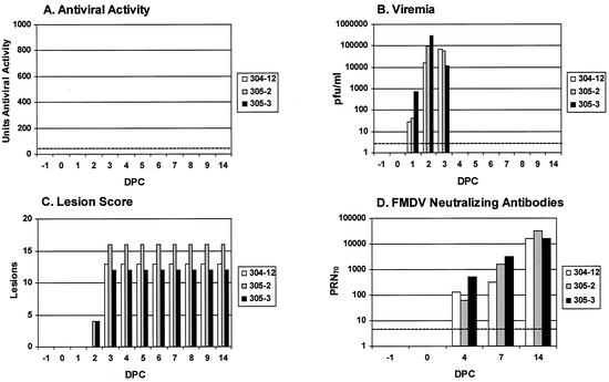
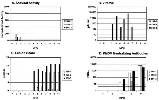
In the second part of the study, three groups of animals were inoculated with the same Ad5 viruses (Table 3, groups 4 to 6) and challenged 1 day p.i. with 105 50% bovine infectious doses of animal-derived FMDV A24 in the heel bulb of the left front foot. Among the FMDV-challenged groups, all the Ad5-Blue-inoculated animals (group 4) developed signs of FMD, including viremia between 1 to 3 days postchallenge (dpc), vesicles by 3 dpc, an FMDV-specific neutralizing antibody response by 4 dpc, and a fever (temperature, >40°C) for 1 to 3 days (Fig. 1). In group 5 (inoculated with the low dose of Ad5-pIFNα) (Fig. 2), animal #501-3 had detectable antiviral activity, 100 U, only at 1 day p.i. (0 dpc) and developed viremia at 2 dpc and vesicles at 4 dpc. Animal #199-2 had detectable antiviral activity at 1 day p.i. (0 dpc) that lasted for an additional day. This animal had viremia at 5 dpc, which continued for 2 days, and developed two lesions at 5 dpc, and disease became more severe by 7 dpc (Fig. 2). Both of these animals developed fever for several days. Animal #186-1 also had detectable antiviral activity by 1 day p.i. (0 dpc) that lasted for an additional day (Fig. 2). This animal did not develop viremia or fever but had one lesion on the left rear foot at 8 dpc and had a low FMDV-specific neutralizing antibody response by 4 dpc (Fig. 2). Animal #186-1 most likely developed disease by contact or aerosol transmission from animal #501-3, 199-2, or both, since they were all housed in the same room. Clearly the FMDV-challenged low-Ad5-pIFNα-dose group showed delayed disease and milder disease than the control animals.
FIG. 1.
Effect of FMDV challenge on animals inoculated with Ad5-Blue. Animals were challenged 1 day p.i. with FMDV A24 (17). Plasma, blood, and serum samples were taken from swine 304-12, 305-2, and 305-3, and the animals were physically monitored for fever and lesion score as described in Table 3. The dashed lines in panels A, B, and D represent the lowest detectable levels in each assay, i.e., <25 U, 5 PFU, and <8 PRN70 (serum dilution yielding a 70% reduction in the number of plaques), respectively.
FIG. 2.
Effect of FMDV challenge on animals inoculated with a low dose of Ad5-pIFNα. Animals were challenged 1 day p.i. with FMDV A24. Plasma, blood, and serum samples were taken from swine 186-1, 199-2, and 501-3, and the animals were physically monitored for fever and lesion score as described in Table 3. The dashed lines in panels A, B, and D represent the lowest detectable levels in each assay, i.e., <25 U, 5 PFU, and <8 PRN70, respectively.
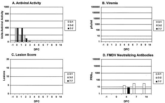
Animals in group 6, given the high dose of Ad5-pIFNα (Fig. 3), had high levels of antiviral activity at 1 day p.i. (0 dpc) (400 U) that was detectable for two to three additional days. None of the animals in this group developed viremia, and all were completely protected from virus challenge. Two animals (#3-1 and 7-7) developed a very low FMDV-specific neutralizing antibody response, while animal #3-2 had no detectable neutralizing antibody (Table 3; Fig. 3). Animal #7-7 died at 12 dpc of causes unrelated to FMD and showed massive peritonitis, probably from ileal perforation.
FIG. 3.
Effect of FMDV challenge on animals inoculated with a high dose of Ad5-pIFNα. Animals were challenged 1 day p.i. with FMDV A24. Plasma, blood, and serum samples were taken from swine 3-1, 3-2, and 7-7, and the animals were physically monitored for fever and lesion score as described in Table 3. The dashed lines in panels A, B, and D represent the lowest detectable levels in each assay, i.e., <25 U, 5 PFU, and <8 PRN70, respectively.
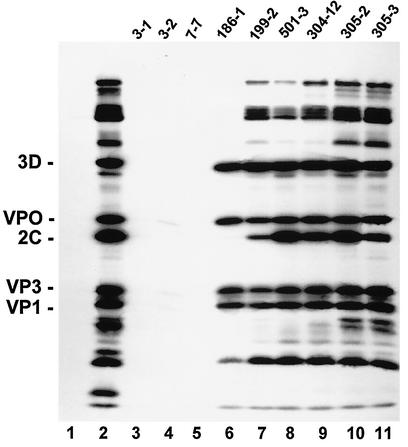
We also examined 14-dpc sera by radioimmunoprecipitation for the presence of antibodies against FMDV NS proteins as a more sensitive measure of challenge virus replication. All animals in groups 4 and 5 developed antibodies against FMDV structural and NS proteins, while all animals given the high dose of Ad5-pIFNα (group 6) had no detectable antibodies against NS proteins, and only one animal had low-level but detectable antibodies against the viral structural proteins (Fig. 4, lane 4). Identical results were obtained using a 3ABC enzyme-linked immunosorbent assay, an assay currently used to differentiate vaccinated animals from infected or convalescent animals (data not shown) (6).
FIG. 4.
Examination of immune response in 14-dpc serum from swine inoculated with Ad5-vectors and challenged with FMDV A24. [35S]Methionine-labeled cell lysates from A24-infected IBRS2 cells were immunoprecipitated with the following: lane 1, 0-dpc serum from swine 3-1; lane 2, bovine convalescent-phase serum as a positive control; lanes 3 to 5, 14-dpc serum from swine 3-1 and 3-2 and 12-dpc serum from swine 7-7 (group 6); lanes 6 to 8, 14-dpc serum from swine 186-1, 199-2, and 501-3 (group 5); lanes 9 to 11, 14-dpc serum from swine 304-12, 305-2, and 305-3 (group 4). Samples were examined by sodium dodecyl sulfate-polyacrylamide gel electrophoresis on a 15% gel.
These results demonstrate that swine can be protected from FMD 1 day after inoculation with Ad5-pIFNα and presumably could be protected earlier, since significant antiviral activity (400 U) was detected as early as 16 h p.i. in the dose-response experiment. Animals inoculated with the high dose of Ad5-pIFNα appeared to clear the challenge virus rapidly, prior to virus replication, preventing detectable viremia and the induction of an antibody response against viral NS proteins. The induction of a low FMDV-specific neutralizing antibody titer in this group suggested that these animals were exposed to a very small amount of antigen for a short time. The presence of plasma antiviral activity for several days following administration of the high dose of Ad5-pIFNα indicates that animals may be protected for at least this time period (Table 2; Fig. 3), and more recent experiments directly support this (M. P. Moraes et al., unpublished data).
IFN-α/β is an ideal candidate for rapid induction of FMDV resistance in animals because it requires a short period of time to function, while vaccination can require 7 to 14 days for induction of protective immunity. Administration of IFN-α/β should also provide protection against all serotypes and subtypes of FMDV, an inherent strategic problem for FMD vaccination during an outbreak where effective vaccination requires the vaccine to be matched to the outbreak strain (7).
To our knowledge, this is the first demonstration that pretreatment with IFN-α successfully protects economically important animals against virus challenge. We are currently exploring a strategy that combines IFN-α/β administration with vaccination so as to provide both immediate nonspecific as well as long-lasting protection against the virus serotype(s) present in the vaccine. These experiments indicate that dual inoculation induces protection when the animals are challenged 5 days later (Moraes et al., unpublished data). Thus, this combined protocol may enable previously disease-free countries to control an FMD outbreak and avoid large-scale slaughter of animals and the subsequent environmental and social concerns that arise. These strategies could also be used as a prophylactic treatment against other acute infectious viral diseases.
Acknowledgments
We thank Tracy DeMeola and Mario Brum for laboratory assistance and the animal care staff for assistance with the experimental animals. We also thank Barry Baxt, Peter Mason, and Luis Rodriguez for critical reading of the manuscript.
REFERENCES
- 1.Ahl, R., and A. Rump. 1976. Assay of bovine interferons in cultures of the porcine cell line IB-RS-2. Infect. Immun. 14:603-606. [DOI] [PMC free article] [PubMed] [Google Scholar]
- 2.Bachrach, H. L. 1978. Foot-and-mouth disease: world-wide impact and control measures, p. 299-310. In E. Kurstak and K. Maramorosch (ed.), Viruses and environment. Academic Press, Inc., New York, N.Y.
- 3.Barteling, S. J., and J. Vreeswijk. 1991. Developments in foot-and-mouth disease vaccines. Vaccine 9:75-88. [DOI] [PubMed] [Google Scholar]
- 4.Chinsangaram, J., M. Koster, and M. J. Grubman. 2001. Inhibition of L-deleted foot-and-mouth disease virus replication by alpha/beta interferon involves double-stranded RNA-dependent protein kinase. J. Virol. 12:5498-5503. [DOI] [PMC free article] [PubMed] [Google Scholar]
- 5.Chinsangaram, J., M. E. Piccone, and M. J. Grubman. 1999. Ability of foot-and mouth disease virus to form plaques in cell culture is associated with suppression of alpha/beta interferon. J. Virol. 73:9891-9898. [DOI] [PMC free article] [PubMed] [Google Scholar]
- 6.De Diego, M., E. Brocchi, D. Mackay, and F. De Simone. 1997. The non-structural polyprotein 3ABC of foot-and-mouth disease virus as a diagnostic antigen in ELISA to differentiate infected from vaccinated cattle. Arch. Virol. 142:2021-2033. [DOI] [PubMed] [Google Scholar]
- 7.Doel, T., M. Lombard, D. Fawthrop, C. Schermbrucker, and P. Dubourget. 1998. Testing of FMD vaccines. Session of the Research Group of the Standing Technical Committee of the European Commission for the Control of Foot-and-Mouth Disease, appendix 32, p. 227-237. European Commission for the Control of Foot-and-Mouth Disease, Food and Agriculture Organization, Aldershot, United Kingdom.
- 8.Gibbens, J. C., C. E. Sharp, J. W. Wilesmith, L. M. Mansley, E. Michalopoulou, J. B. M. Ryan, and M. Hudson. 2001. Descriptive epidemiology of the 2001 foot-and-mouth disease epidemic in Great Britain: the first five months. Vet. Rec. 149:729-743. [PubMed] [Google Scholar]
- 9.Goodbourn, S., L. Didcock, and R. E. Randall. 2000. Interferons: cell signalling, immune modulation, antiviral response and virus countermeasures. J. Gen. Virol. 81:2341-2364. [DOI] [PubMed] [Google Scholar]
- 10.Graham, F. L., and L. Prevec. 1991. Gene transfer and expression protocols: manipulation of adenovirus vectors, p. 109-128. In E. J. Murray (ed.), Methods in molecular biology. Humana Press, Totowa, New Jersey. [DOI] [PubMed]
- 11.Graham, F. L., J. Smiley, W. C. Russell, and R. Nairn. 1977. Characteristics of a human cell line transformed by DNA from human adenovirus 5. J. Gen. Virol. 36:59-72. [DOI] [PubMed] [Google Scholar]
- 12.Knowles, N. J., A. R. Samuel, P. R. Davies, R. P. Kitching, and A. I. Donaldson. 2001. Outbreak of foot-and-mouth disease virus serotype O in the UK caused by a pandemic strain. Vet. Rec. 148:258-259. [PubMed] [Google Scholar]
- 13.Lukaszewski, R. A., and T. J. Brooks. 2000. Pegylated alpha interferon is an effective treatment for virulent Venezuelan equine encephalitis virus and has profound effects on the host immune response to infection. J. Virol. 74:5006-5015. [DOI] [PMC free article] [PubMed] [Google Scholar]
- 14.Moraes, M. P., G. A. Mayr, and M. J. Grubman. 2001. pAd5-Blue: an easy to use, efficient, direct ligation system for engineering recombinant adenovirus constructs. BioTechniques 31:1050-1056. [DOI] [PubMed] [Google Scholar]
- 15.Moraes, M. P., G. A. Mayr, P. W. Mason, and M. J. Grubman. 2002. Early protection against homologous challenge after a single dose of replication-defective human adenovirus type 5 expressing capsid proteins of foot-and-mouth disease virus (FMDV) strain A24. Vaccine 20:1631-1639. [DOI] [PubMed] [Google Scholar]
- 16.Qin, X. Q., N. Tao, A. Dergay, P. Moy, S. Fawell, A. Davis, J. M. Wilson, and J. Barsoum. 1998. Interferon-beta gene therapy inhibits tumor formation and causes regression of established tumors in immune-deficient mice. Proc. Natl. Acad. Sci. USA 95:14411-14416. [DOI] [PMC free article] [PubMed] [Google Scholar]
- 17.Sa-Carvalho, D., E. Rieder, B. Baxt, R. Rodarte, A. Tanuri, and P. W. Mason. 1997. Tissue culture adaptation of foot-and-mouth disease virus selects viruses that bind to heparin and are attenuated in cattle. J. Virol. 71:5115-5123. [DOI] [PMC free article] [PubMed] [Google Scholar]
- 18.Salt, J. S. 1993. The carrier state in foot and mouth disease—an immunological review. Br. Vet. J. 149:207-223. [DOI] [PubMed] [Google Scholar]
- 19.Santodonato, L., M. Ferrantini, F. Palombo, L. Aurisicchio, P. Delmastro, N. La Monica, S. Di Marco, G. Ciliberto, M. X. Du, M. W. Taylor, and F. Belardelli. 2001. Antitumor activity of recombinant adenoviral vectors expressing murine IFN-alpha in mice injected with metastatic IFN-resistant tumor cells. Cancer Gene Ther. 8:63-72. [DOI] [PubMed] [Google Scholar]
- 20.Sellers, R. F. 1963. Multiplication, interferon production and sensitivity of virulent and attenuated strains of the virus of foot-and-mouth disease. Nature 198:1228-1229. [DOI] [PubMed] [Google Scholar]
- 21.Vilcek, J., and G. C. Sen. 1996. Interferons and other cytokines, p. 375-399. In B. N. Fields, D. M. Knipe, and P. H. Howley (ed.), Virology. Lippincott-Raven Publishers, Philadelphia, Pa.
- 22.Yang, P. C., R. M. Chu, W. B. Chung, and H. T. Sung. 1999. Epidemiological characteristics and financial costs of the 1997 foot-and-mouth disease epidemic in Taiwan. Vet. Rec. 145:731-734. [DOI] [PubMed] [Google Scholar]
- 23.Zhou, A., J. M. Paranjape, S. D. Der, B. R. Williams, and R. H. Silverman. 1999. Interferon action in triply deficient mice reveals the existence of alternative antiviral pathways. Virology 258:435-444. [DOI] [PubMed] [Google Scholar]


