Disorders of plasma sodium are the most common electrolyte disturbances in clinical medicine, yet they remain poorly understood. Severe hyponatraemia and hypernatraemia are associated with considerable morbidity and mortality,1-3 however, and even mild hyponatraemia is associated with worse outcomes when it complicates conditions such as heart failure,4 although which is cause and which effect is often uncertain. Distinguishing the cause(s) of hyponatraemia may be challenging in clinical practice, and controversies surrounding its management remain. Here, we describe the common causes of disorders of plasma sodium, offer guides to their investigation and management, and highlight areas of recent advance and of uncertainty.
Sources and selection criteria
We incorporated the latest consensus from systematic reviews and publications identified by a literature search through Medline and Web of Science with the search strategy terms “hyponatraemia,” “hypernatraemia,” and “sodium.” We found fewer than a dozen randomised controlled trials of treatment of any description. Despite their frequency, plasma sodium disorders have not been reviewed by the Cochrane Library, Clinical Evidence, or Best Evidence.
Control of sodium balance
Under normal conditions, plasma sodium concentrations are finely maintained within the narrow range of 135-145 mmol/l despite great variations in water and salt intake. Sodium and its accompanying anions, principally chloride and bicarbonate, account for 86% of the extracellular fluid osmolality, which is normally 285-295 mosm/kg and calculated as (2× [Na]mmol/l + [urea]mmol.l + [glucose]mmol/l. The main determinant of the plasma sodium concentration is the plasma water content, itself determined by water intake (thirst or habit), “insensible” losses (such as metabolic water, sweat), and urinary dilution. The last of these is under most circumstances the most important and is predominantly determined by arginine vasopressin, which is synthesised in the hypothalamus and then stored in and released from the posterior pituitary. In response to arginine vasopressin, concentrated urine is produced by water reabsorption across the renal collecting ducts. This is mediated by specialised cellular membrane transport proteins called aquaporins.5-8
Summary points
Sodium disorders are common, particularly in hospital patients and elderly people
Mild sodium disorders may be asymptomatic and self limiting, but severe sodium disorders are associated with considerable morbidity and mortality
The causes of sodium imbalance are often iatrogenic and therefore avoidable
Assessing hydration status and measuring sodium in plasma and urine are key to diagnosing the cause of hyponatraemia
The cause of hypernatraemia will usually be evident from the history
Little evidence from randomised controlled trials exists for the treatment of sodium disorders
Slow correction of sodium is usually safe, with careful monitoring of clinical status and plasma sodium
Hyponatraemia
Determining the cause of hyponatraemia may be straightforward if an obvious precipitating cause is present—for example, in the setting of vomiting or diarrhoea, when both sodium and total body water are low, and especially if the patient (typically elderly) is taking diuretics. In hospital practice, diagnosing the cause is often less clear cut. Here, hyponatraemia almost always reflects an excess of water relative to sodium, commonly by dilution of total body sodium secondary to increases in total body water (water overload) and sometimes as a result of depletion of total body sodium in excess of concurrent body water losses. The clinical classification of hyponatraemia according to the patient's extracellular fluid volume status, as hypovolaemic, euvolaemic, or hypervolaemic (box 1), is useful to help with the diagnosis. In practice, however, distinguishing euvolaemic and hypovolaemic hyponatraemia may not be straightforward.
The symptoms of hyponatraemia are related to both the severity and the rapidity of the fall in the plasma sodium concentration. A decrease in plasma sodium concentration creates an osmotic gradient between extracellular and intracellular fluid in brain cells, causing movement of water into cells, increasing intracellular volume, and resulting in tissue oedema, raised intracranial pressure, and neurological symptoms. Patients with mild hyponatraemia (plasma sodium 130-135 mmol/l) are usually asymptomatic. Nausea and malaise are typically seen when plasma sodium concentration falls below 125-130 mmol/l. Headache, lethargy, restlessness, and disorientation follow, as the sodium concentration falls below 115-120 mmol/l. With severe and rapidly evolving hyponatraemia, seizure, coma, permanent brain damage, respiratory arrest, brain stem herniation, and death may occur.9 In more gradually evolving hyponatraemia, the brain self regulates to prevent swelling over hours to days by transport of, firstly, sodium, chloride, and potassium and, later, organic solutes including glutamate, taurine, myo-inositol, and glutamine from intracellular to extracellular compartments. This induces water loss and ameliorates brain swelling, and hence leads to few symptoms in patients with chronic hyponatraemia.
History, examination, and investigation
An accurate history may reveal a clue to the cause of the hyponatraemia and establish the rapidity of the symptoms. The key diagnostic factors (box 2) are the hydration status of the patient and the urine “spot sodium” concentration, which is available quickly and allows the crucial distinction in hypovolaemic hyponatraemia between renal (high; > 30 mmol/l) and extrarenal (low; < 30 mmol/l) salt loss. Urinary sodium is similarly helpful in patients in whom volume status is difficult to assess, as patients with dilutional hyponatraemia by and large have a urinary sodium > 30 mmol/l, whereas those with extracellular fluid depletion (unless the source is renal) will have a urinary sodium < 30 mmol/l.10 Plasma osmolality is almost always low in hyponatraemia, and urine is less than maximally dilute (inappropriately concentrated); so, although usually measured, plasma and urine osmolalities are rarely discriminant.
Box 1: Classification of hyponatraemia
Hypovolaemia
Extrarenal loss, urine sodium <30 mmol/l
Dermal losses, such as burns, sweating
Gastrointestinal losses, such as vomiting, diarrhoea
Pancreatitis
Renal loss, urine sodium >30 mmol/l
Diuretics
Salt wasting nephropathy
Cerebral salt wasting
Mineralocorticoid deficiency (Addison's disease)
Hypervolaemia*
Urine sodium <30 mmol/l
Congestive cardiac failure
Cirrhosis with ascites
Nephrotic syndrome
Urine sodium >30 mmol/l
• Chronic renal failure
Euvolaemia
Urine sodium >30 mmol/l
Syndrome of inappropriate antidiuretic hormone secretion (SIADH)†
Hypothyroidism
Hypopituitarism (glucocorticoid deficiency)
-
Water intoxication:
Primary polydipsia
Excessive administration of parenteral hypotonic fluids
Post-transurethral prostatectomy
*Paradoxical retention of sodium and water despite a total body excess of each; baroreceptors in the arterial circulation perceive hypoperfusion, triggering an increase in arginine vasopressin release and net water retention.
†Remember that SIADH is a diagnosis of exclusion.20
Management of hyponatraemia
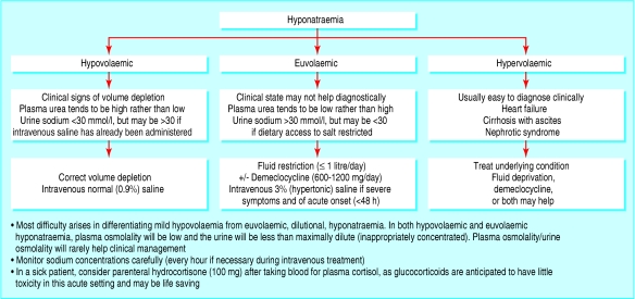
As the duration of hyponatraemia may be difficult to judge, the presence of symptoms and their severity should guide the treatment strategy (figure). Acute hyponatraemia developing within 48 hours carries a risk of cerebral oedema, so prompt treatment is indicated with apparently small risk of central pontine myelinolysis. This is presumed to occur if the blood-brain barrier becomes permeable with rapid correction of hyponatraemia and allows complement mediated oligodendrocyte toxicity (despite its name, central pontine myelinolysis can occur widely in the brain). Alcoholics with malnutrition, premenopausal or elderly women on thiazide diuretics, and patients with hypokalaemia or burns are at increased risk of central pontine myelinolysis.11,12 Neurological injury is typically delayed for two to six days after elevation of the sodium concentration, but the symptoms, which include dysarthria, dysphagia, spastic paraparesis, lethargy, seizures, coma, and even death, are generally irreversible, so prevention is key.
Figure 1.
Flowchart for assessing and managing patients with hyponatraemia
Box 2: Examination and investigations in patient with hyponatraemia
Evaluation of volume status
Skin turgor
Pulse rate
Postural blood pressure
Jugular venous pressure
Consider central venous pressure monitoring
Examination of fluid balance charts
General examination for underlying illness
Congestive cardiac failure
Cirrhosis
Nephrotic syndrome
Addison's disease
Hypopituitarism
Hypothyroidism
Investigations
Urinary sodium
Plasma glucose and lipids*
Renal function
Thyroid function
Peak cortisol during short synacthen test†
Plasma and urine osmolality‡
If indicated: chest x ray, and computed tomography and magnetic resonance imaging of head and thorax
*Pseudohyponatraemia due to artefactual reduction in plasma sodium in the presence of marked elevation of plasma lipids or proteins should no longer be seen with the measurement of sodium by ion specific electrodes; hyperglycaemia causes true hyponatraemia, irrespective of laboratory method.
†May be unhelpful in pituitary apoplexy, in which patients may still “pass” the test.
‡For SIADH: plasma osmolality < 270 mosm/kg with inappropriate urinary concentration (> 100 mosm/kg), in a euvolaemic patient after exclusion of hypothyroidism and glucocorticoid deficiency).
Animal data and correlative retrospective findings in humans suggest that slow correction of chronic hyponatraemia minimises central pontine myelinolysis. Unfortunately, no consensus exists about the optimal rate of correction of hyponatraemia. Although many people advocate a target rate not exceeding 8 mmol/l on any day of treatment, others suggest 12 mmol/l/day or even more if the patient has symptoms—for example, raising the sodium concentration by 1-2 mmol/l per hour until symptoms have resolved, with close monitoring of plasma sodium. The evidence base for using hypertonic saline (3% sodium chloride) in acute symptomatic hyponatraemia is slight, and we recommend that this should be used only after specialist advice has been sought and with frequent (one to two hourly) measurement of plasma sodium. Some authors recommend that a loop diuretic such as furosemide should be given with the hypertonic saline infusion to enhance free water clearance, but caution is needed as this may cause too rapid a rise in sodium.13
Additional educational resources
Useful websites for professionals
British Society for Endocrinology (www.endocrinology.org)
Endocrine Society (USA) (www.endo-society.org)
Useful websites for patients
Pituitary Foundation (www.pituitary.org.uk)—national UK charity that provides information and support to people with pituitary disorders
Pituitary Network Association (www.pituitary.com)—international non-profit organisation for patients with pituitary tumours and disorders
New developments for management of hyponatraemia
Fluid restriction (≤ 1 litre/day) is the initial approach to treating chronic asymptomatic hyponatraemia < 130 mmol/l. Depressingly, no long term trials of the efficacy in practice of this apparently simple approach have been done, but in short term trials it seems to have little effect.14,15 Demeclocycline, which inhibits arginine vasopressin action in the kidney collecting duct, is the current “drug of choice” for treating chronic asymptomatic hyponatraemia due to SIADH if fluid restriction alone does not restore sodium concentrations. Lithium exerts similar renal effects but is less desirable because of inconsistent effects and more side effects (renal impairment, central nervous system effects, thyroid disorders). Urea has been proposed as an alternative option but is poorly tolerated.
The development of orally active antagonists selective for the antidiuretic (renal V2 receptor) action of arginine vasopressin therefore has exciting therapeutic prospects in the management of hyponatraemia. Such “aquaretics” (for example, tolvaptan, lixivaptan) induce a water diuresis without affecting urinary electrolyte or solute excretion. Emerging, if short term, clinical trials have shown the expected effects of aquaresis and correction of hyponatraemia in cirrhosis, heart failure, and SIADH,16-18 and the drugs seem to be well tolerated; thirst is the only major side effect reported. Moreover, restriction of fluid intake may not be necessary with these agents.17 Although V1a (vasoconstrictor) receptor antagonism would not directly affect hyponatraemia, combined V1a/V2 receptor antagonists (such as conivaptan) are in phase III trials after showing promising effects in patients with heart failure in association with hyponatraemia, in which their additional antivasoconstrictor effects seem to helpfully reduce total peripheral resistance and increase cardiac output.17 Before their possible place in the pantheon of therapeutics can be determined, however, arginine vasopressin receptor antagonists need to show efficacy and lack of toxicity in long term trials with medically important morbidity and mortality end points.
Hypernatraemia
Hypernatraemia is much less common than hyponatraemia.3 It reflects a net water loss or a hypertonic sodium gain, with inevitable hyperosmolality. Severe symptoms are usually evident only with acute and large increases in plasma sodium concentrations to above 158-160 mmol/l. Importantly, the sensation of intense thirst that protects against severe hypernatraemia in health may be absent or reduced in patients with altered mental status or with hypothalamic lesions affecting their sense of thirst (adipsia) and in infants and elderly people. Non-specific symptoms such as anorexia, muscle weakness, restlessness, nausea, and vomiting tend to occur early. More serious signs follow, with altered mental status, lethargy, irritability, stupor, or coma. Acute brain shrinkage can induce vascular rupture, with cerebral bleeding and subarachnoid haemorrhage.
History, examination, and investigation
Often the cause is evident from the history (box 3). Measurement of urine osmolality in relation to the plasma osmolality and the urine sodium concentration help if the cause is unclear. Patients with diabetes insipidus present with polyuria and polydipsia (and not hypernatraemia unless thirst sensation is impaired). Central diabetes insipidus and nephrogenic diabetes insipidus may be differentiated by the response to water deprivation (failure to concentrate urine) followed by the V2 agonist desmopressin, causing concentration of urine in patients with central diabetes insipidus.
Management
In patients with hypernatraemia that has developed over a period of hours, rapid correction of plasma sodium (falling by 1 mmol/l per hour) improves the prognosis without the risk of convulsions and cerebral oedema.19 Management of a shocked patient needs specialist input and close monitoring, preferably in a high dependency unit. Intravenous normal saline should be used to correct the extracellular fluid depletion, with calculation of the free water deficit to determine how much 5% dextrose to give.3 In patients with hypernatraemia of longer or unknown duration, reducing the sodium concentration more slowly is prudent. Patients should be given intravenous 5% dextrose for acute hypernatraemia or half-normal saline (0.45% sodium chloride) for chronic hypernatraemia if unable to tolerate oral water. Central diabetes insipidus is treated with desmopressin, either as intranasal spray or tablets, with careful monitoring to avoid the complications of water intoxication (delaying one dose each week to allow polyuria and thirst to “breakthrough” in patients susceptible to hyponatraemia with desmopressin may be prudent). Treatment of nephrogenic diabetes insipidus includes removal of precipitating drugs (if possible) and sometimes initiation of thiazide diuretics, non-steroidal anti-inflammatory drugs, or both.
Final thoughts
Despite the frequent occurrence and the poor outcomes of serious disorders of sodium balance, few hard data are available to guide the clinician. This area needs clinical trials, notably of existing approaches (water restriction, demeclocyline, rates of dehydration and rehydration), to complement industrially funded randomised controlled trials of novel aquaretics.
Box 3: Classification of hypernatraemia
Hypovolaemia
Dermal losses—for example, burns, sweating
Gastrointestinal losses—for example, vomiting, diarrhoea, fistulas
Diuretics
Postobstruction
Acute and chronic renal disease
Hyperosmolar non-ketotic coma*
Hypervolaemia
Iatrogenic (hypertonic saline, tube feedings, antibiotics containing sodium, or hypertonic dialysis)
Hyperaldosteronism†
Euvolaemia
Diabetes insipidus (central, nephrogenic, or gestational)
Hypodipsia
Fever
Hyperventilation
Mechanical ventilation
*Sodium often raised, even after correction for glucose
†Typically mildly elevated sodium ∼147 mmol/l, so rarely a clinical problem
Contributors: All authors contributed to the literature search and review. RMR wrote the first draft. PLP designed the flowchart and revised the manuscript. JRS edited the final version.
Competing interests: None declared.
References
- 1.Arieff AI. Hyponatraemia, convulsions, respiratory arrest, and permanent brain damage after elective surgery in healthy women. N Engl J Med 1986;314: 1529-35. [DOI] [PubMed] [Google Scholar]
- 2.Anderson RJ. Hospital acquired hyponatraemia. Kidney Int 1986;29: 1237-47. [DOI] [PubMed] [Google Scholar]
- 3.Kumar S, Berl T. Sodium. Lancet 1998;352: 220-2. [DOI] [PubMed] [Google Scholar]
- 4.Klein L, O'Connor CM, Leimberger JD, Gattis-Stough W, Pina IL, Felker GM, et al. Lower serum sodium is associated with increased short-term mortality in hospitalized patients with worsening heart failure: results from the outcomes of a prospective trial of intravenous milrinone for exacerbations of chronic heart failure (OPTIME-CHF) study. Circulation 2005;111: 2454-60. [DOI] [PubMed] [Google Scholar]
- 5.Adrogué, HJ, Madias NE. Hyponatraemia. N Engl J Med 2000;342: 1581-9. [DOI] [PubMed] [Google Scholar]
- 6.Smith DM, McKenna K, Thompson CJ. Hyponatraemia. Clin Endocrinol 2000;52: 667-78. [DOI] [PubMed] [Google Scholar]
- 7.Ishikawa S, Schrier RW. Pathophysiological roles of arginine vasopressin and aquaporin-2 in impaired water excretion. Clin Endocrinol 2003;58: 1-17. [DOI] [PubMed] [Google Scholar]
- 8.Reynolds RM, Seckl JR. Hyponatraemia for the clinical endocrinologist. Clin Endocrinol 2005;63: 366-74. [DOI] [PubMed] [Google Scholar]
- 9.Ellis SJ. Severe hyponatraemia: complications and treatment. QJM 1995;88: 905-9. [PubMed] [Google Scholar]
- 10.Chung H-M, Kluge R, Schrier RW, Anderson RJ. Clinical assessment of extracellular fluid volume in hyponatraemia. Am J Med 1987;83: 905-8. [DOI] [PubMed] [Google Scholar]
- 11.Arieff AI. Hyponatraemia, convulsions, respiratory arrest, and permanent brain damage after elective surgery in healthy women. N Engl J Med 1986;314: 1529-35. [DOI] [PubMed] [Google Scholar]
- 12.Ayus JC, Wheeler JM, Arieff AI. Postoperative hyponatraemic encephalopathy in menstruant women. Ann Int Med 1992;117: 891-7. [DOI] [PubMed] [Google Scholar]
- 13.Hantman D, Rossier B, Zohlman R, Schrier R. Rapid correction of hyponatraemia in the syndrome of inappropriate antidiuretic hormone: an alternative treatment to hypertonic saline. Ann Int Med 1973;78: 870-5. [DOI] [PubMed] [Google Scholar]
- 14.Ayus JC, Arieff AI. Chronic hyponatremic encephalopathy in postmenopausal women—association of therapies with morbidity and mortality. JAMA 1999;281: 2299-304. [DOI] [PubMed] [Google Scholar]
- 15.Gerbes AL, Gulberg V, Gines P, Decaux G, Gross P, Gandjini H, et al. Therapy of hyponatremia in cirrhosis with a vasopressin receptor antagonist: a randomized double-blind multicenter trial. Gastroenterology 2003;124: 933-9. [DOI] [PubMed] [Google Scholar]
- 16.Ferguson JW, Therapondos G, Newby DE, Hayes PC. Therapeutic role of vasopressin receptor antagonism in patients with liver cirrhosis. Clin Sci 2003;105: 1-8. [DOI] [PubMed] [Google Scholar]
- 17.Goldsmith SR, Gheorghiade M. Vasopressin antagonism in heart failure. J Am Coll Cardiol 2005;46: 1785-91. [DOI] [PubMed] [Google Scholar]
- 18.Wong F, Blei AT, Blendis LM, Thuluvath PJ. A vasopressin receptor antagonist (VPA-985) improves serum sodium concentration in patients with hyponatraemia: a multicentre, randomised, placebo-controlled trial. Hepatology 2003;37: 182-91. [DOI] [PubMed] [Google Scholar]
- 19.Weiss-Guillet EM, Takala J, Jakob SM. Diagnosis and management of electrolyte emergencies. Best Pract Res Clin Endocrinol Metab 2003;17: 623-51. [DOI] [PubMed] [Google Scholar]
- 20.Saeed BO, Beaumont D, Handley GH, Weaver JH. Severe hyponatraemia: investigation and management in a district general hospital. J Clin Pathol 2002;55: 893-6. [DOI] [PMC free article] [PubMed] [Google Scholar]


