

Abstract
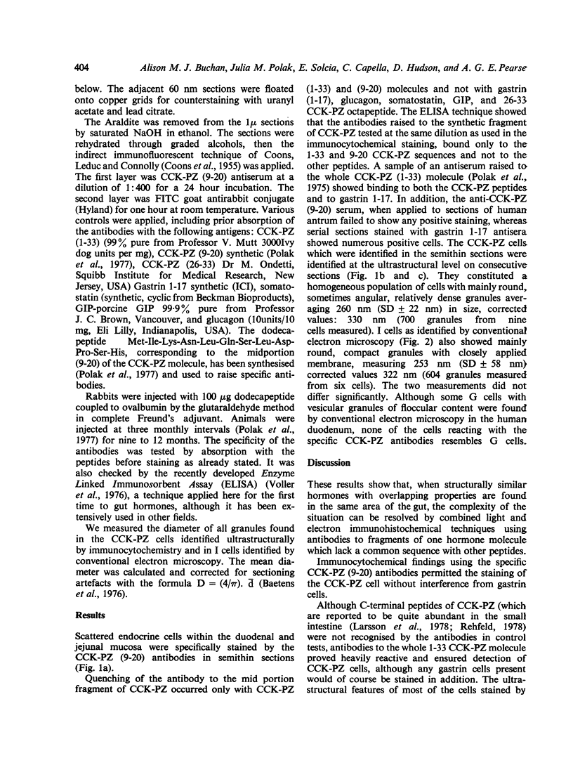
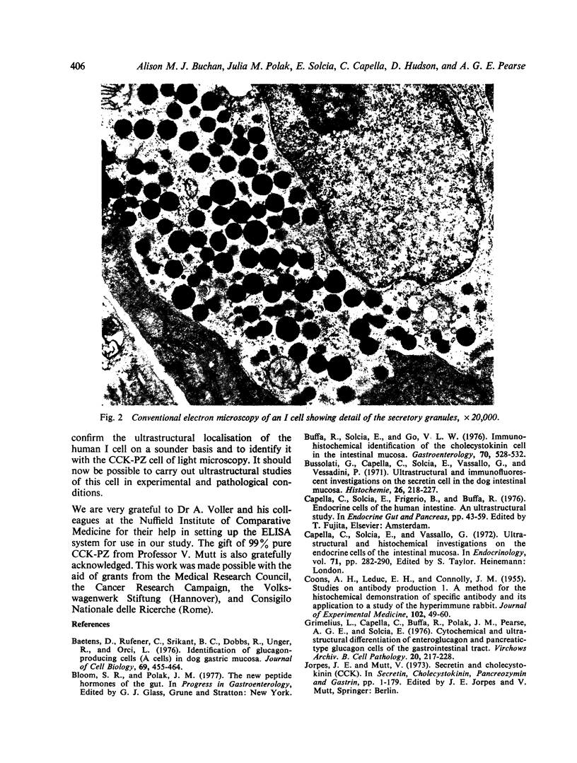
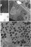
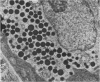
Evidence was obtained by the use of alternate semithin-thin serial secretions for light and electron microsocpy that the I cell is the source of CCK PZ. The antibodies used were raised to a synthetic fragment of the mid part (9-20) of the (1-33) CCK-PZ molecule, and were thus free from any contamination with cross-reacting subpopulations of antibodies that might bind to gastrin.
Full text
PDF

Images in this article
Selected References
These references are in PubMed. This may not be the complete list of references from this article.
- Baetens D., Rufener C., Srikant B. C., Dobbs R., Unger R., Orci L. Identification of glucagon-producing cells (A cells) in dog gastric mucosa. J Cell Biol. 1976 May;69(2):455–464. doi: 10.1083/jcb.69.2.455. [DOI] [PMC free article] [PubMed] [Google Scholar]
- Buffa R., Solcia E., Go V. L. Immunohistochemical identification of the cholecystokinin cell in the intestinal mucosa. Gastroenterology. 1976 Apr;70(4):528–532. [PubMed] [Google Scholar]
- Bussolati G., Capella C., Solcia E., Vassallo G., Vezzadini P. Ultrastructural and immunofluorescent investigations on the secretin cell in the dog intestinal mucosa. Histochemie. 1971;26(3):218–227. doi: 10.1007/BF00305655. [DOI] [PubMed] [Google Scholar]
- COONS A. H., LEDUC E. H., CONNOLLY J. M. Studies on antibody production. I. A method for the histochemical demonstration of specific antibody and its application to a study of the hyperimmune rabbit. J Exp Med. 1955 Jul 1;102(1):49–60. doi: 10.1084/jem.102.1.49. [DOI] [PMC free article] [PubMed] [Google Scholar]
- Grimelius L., Capella C., Buffa R., Polak J. M., Pearse A. G., Solcia E. Cytochemical and ultrastructural differentiation of enteroglucagon and pancreatic-type glucagon cells of the gastrointestinal tract. Virchows Arch B Cell Pathol. 1976 Apr 29;20(3):217–228. doi: 10.1007/BF02890341. [DOI] [PubMed] [Google Scholar]
- Polak J. M., Bloom S. R., Rayford P. L., Pearse A. G., Buchan A. M., Thompson J. C. Identification of cholecystokinin-secreting cells. Lancet. 1975 Nov 22;2(7943):1016–1018. doi: 10.1016/s0140-6736(75)90297-4. [DOI] [PubMed] [Google Scholar]
- Polak J. M., Pearse A. G., Szelke M., Bloom S. R., Hudson D., Facer P., Buchan A. M., Bryant M. G., Christophodes N., MacIntyre I. Specific immunostaining of CCK cells by use of synthetic fragment antisera. Experientia. 1977 Jun 15;33(6):762–763. doi: 10.1007/BF01944178. [DOI] [PubMed] [Google Scholar]
- Solcia E., Capella C., Vassallo G., Buffa R. Endocrine cells of the gastric mucosa. Int Rev Cytol. 1975;42:223–286. doi: 10.1016/s0074-7696(08)60982-1. [DOI] [PubMed] [Google Scholar]
- Voller A., Bidwell D. E., Bartlett A. Enzyme immunoassays in diagnostic medicine. Theory and practice. Bull World Health Organ. 1976;53(1):55–65. [PMC free article] [PubMed] [Google Scholar]