

Abstract
The cell population of the upper jejunal mucosa has been studied in cases of tropical sprue from the Far East and Middle East, and in similar cases arising in western Europe (`post-infective malabsorption'), and compared with cases of untreated coeliac disease and patients without small bowel disease.
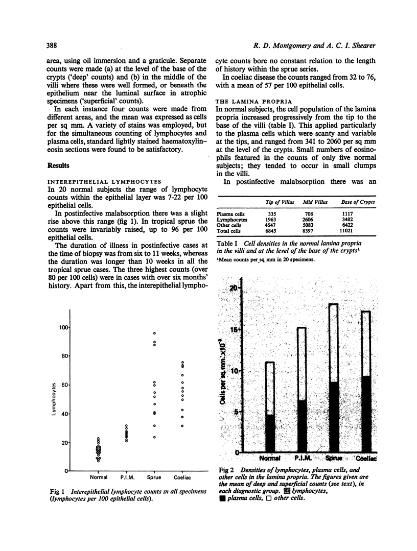
Infiltration of the epithelial layer of the upper jejunal mucosa by lymphocytes was found in tropical sprue to the same extent as in coeliac disease, and, to a lesser extent, in `postinfective malabsorption'.
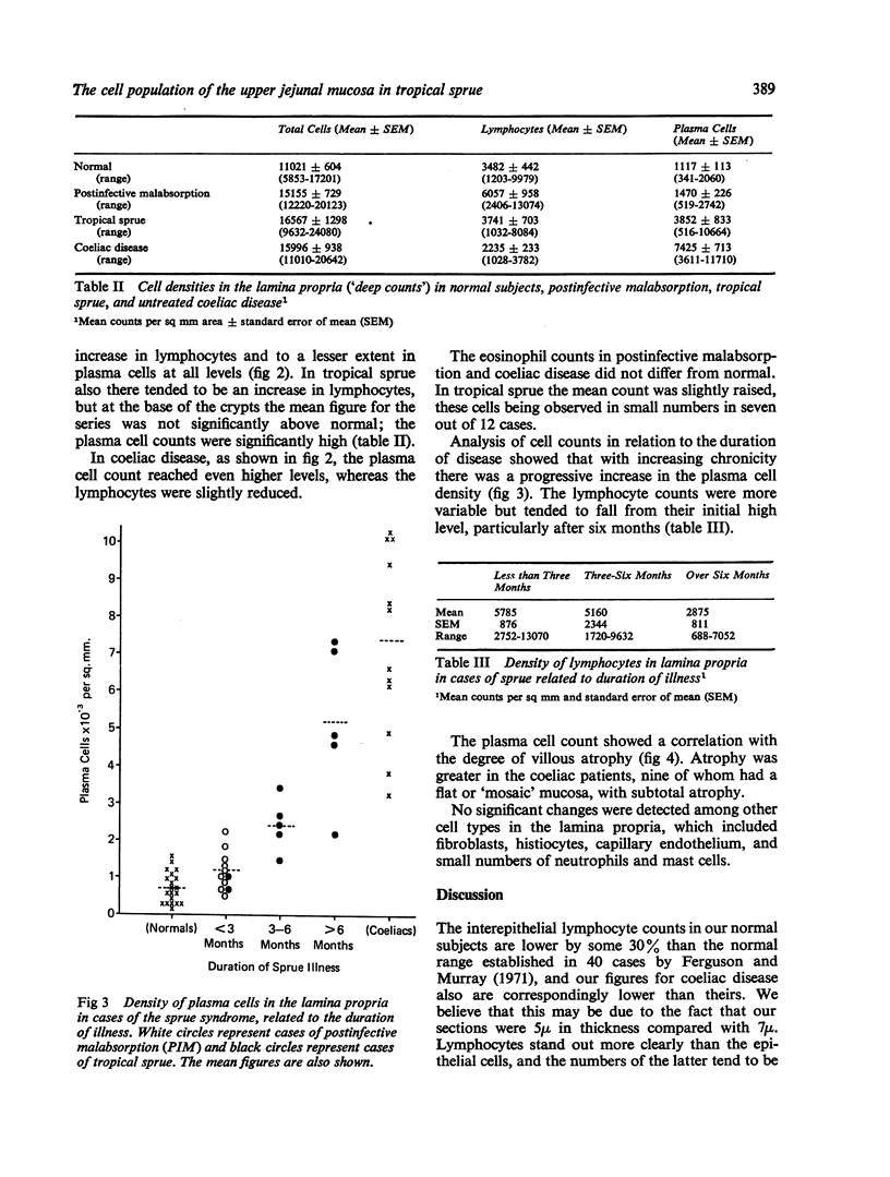
In the lamina propria, in all forms of acute sprue there was an increased density of lymphocytes. With increasing duration and with increasing mucosal atrophy, the lymphocytes were progressively replaced by plasma cells, and the cellular infiltration in chronic sprue was indistinguishable from that of coeliac disease.
The findings suggest that a humoral antibody response is a feature of sprue, and becomes more prominent as the condition becomes chronic.
Full text
PDF

Selected References
These references are in PubMed. This may not be the complete list of references from this article.
- CHACKO C. J., JOB C. K., JOHNSON S., BAKER S. J. Histopathological changes in the upper jejunum in tropical malabsorption syndrome studied by transoral biopsy. Indian J Pathol Bacteriol. 1961 Oct;4:203–213. [PubMed] [Google Scholar]
- Douglas A. P., Crabbé P. A., Hobbs J. R. Immunochemical studies on the serum, intestinal secretions and intestinal mucosa in patients with adult celiac disease and other forms of the celiac syndrome. Gastroenterology. 1970 Sep;59(3):414–425. [PubMed] [Google Scholar]
- England N. W., O'Brien W. Appearances of the jejunal mucosa in acute tropical sprue in Singapore. Gut. 1966 Apr;7(2):128–139. doi: 10.1136/gut.7.2.128. [DOI] [PMC free article] [PubMed] [Google Scholar]
- Ferguson A., Murray D. Quantitation of intraepithelial lymphocytes in human jejunum. Gut. 1971 Dec;12(12):988–994. doi: 10.1136/gut.12.12.988. [DOI] [PMC free article] [PubMed] [Google Scholar]
- Holmes G. K., Asquith P., Stokes P. L., Cooke W. T. Cellular infiltrate of jejunal biopsies in adult coeliac disease (ACD) in relation to gluten withdrawal. Gut. 1973 May;14(5):429–429. [PubMed] [Google Scholar]
- Montgomery R. D., Beale D. J., Sammons H. G., Schneider R. Postinfective malabsorption: a sprue syndrome. Br Med J. 1973 May 5;2(5861):265–268. doi: 10.1136/bmj.2.5861.265. [DOI] [PMC free article] [PubMed] [Google Scholar]
- Rubin C. E., Dobbins W. O., 3rd Peroral biopsy of the small intestine. A review of its diagnostic usefulness. Gastroenterology. 1965 Dec;49(6):676–697. [PubMed] [Google Scholar]
- THURLBECK W. M., BENSON J. A., Jr, DUDLEY H. R., Jr The histopathologic changes of sprue and their significance. Am J Clin Pathol. 1960 Aug;34:108–117. doi: 10.1093/ajcp/34.2.108. [DOI] [PubMed] [Google Scholar]