

Abstract
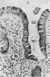
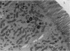
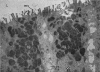
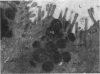
The significance of mucous change in the human duodenum in a series of patients with peptic ulcer disease has been appraised. No specific correlation was demonstrated with the acid output of the stomach if the extent of the change is considered, but it was shown to be more common in the higher acid states. Electron microscopic studies confirmed the specific structure of the mucous cells of the duodenum and suggest that they arise either by transformation of Brunner's gland cells or as a distinctive population in the crypts.
It is suggested that the mucous change is a protective mechanism involved in some way as yet unknown with the healing of ulcers.
Full text
PDF


Images in this article


Selected References
These references are in PubMed. This may not be the complete list of references from this article.
- BERTALANFFY F. D., NAGY K. P. Mitotic activity and renewal rate of the epithelial cells of human duodenum. Acta Anat (Basel) 1961;45:362–370. doi: 10.1159/000141762. [DOI] [PubMed] [Google Scholar]
- DONIACH I., SHINER M. Duodenal and jejunal biopsies. II. Histology. Gastroenterology. 1957 Jul;33(1):71–86. [PubMed] [Google Scholar]
- De Duve C., Wattiaux R. Functions of lysosomes. Annu Rev Physiol. 1966;28:435–492. doi: 10.1146/annurev.ph.28.030166.002251. [DOI] [PubMed] [Google Scholar]
- ECHLIN P. INTRA-CYTOPLASMIC MEMBRANOUS INCLUSIONS IN THE BLUE-GREEN ALGA, ANACYSTIS NIDULANS. Arch Mikrobiol. 1964 Oct 2;49:267–274. doi: 10.1007/BF00409749. [DOI] [PubMed] [Google Scholar]
- FARQUHAR M. G., PALADE G. E. Junctional complexes in various epithelia. J Cell Biol. 1963 May;17:375–412. doi: 10.1083/jcb.17.2.375. [DOI] [PMC free article] [PubMed] [Google Scholar]
- FELL H. B. The effect of excess vitamin A on cultures of embryonic chicken skin explanted at different stages of differentiation. Proc R Soc Lond B Biol Sci. 1956 Mar 26;146(923):242–256. doi: 10.1098/rspb.1957.0008. [DOI] [PubMed] [Google Scholar]
- Freeman J. A. Goblet cell fine structure. Anat Rec. 1966 Jan;154(1):121–147. doi: 10.1002/ar.1091540111. [DOI] [PubMed] [Google Scholar]
- GLAUERT A. M., GLAUERT R. H., ROGERS G. E. A new embedding medium for electron microscopy. Nature. 1956 Oct 13;178(4537):803–803. doi: 10.1038/178803a0. [DOI] [PubMed] [Google Scholar]
- Ito S. The enteric surface coat on cat intestinal microvilli. J Cell Biol. 1965 Dec;27(3):475–491. doi: 10.1083/jcb.27.3.475. [DOI] [PMC free article] [PubMed] [Google Scholar]
- JAMES A. H. GASTRIC EPITHELIUM IN THE DUODENUM. Gut. 1964 Aug;5:285–294. doi: 10.1136/gut.5.4.285. [DOI] [PMC free article] [PubMed] [Google Scholar]
- Johnston D., Jepson K. Use of pentagastrin in a test of gastric acid secretion. Lancet. 1967 Sep 16;2(7516):585–588. doi: 10.1016/s0140-6736(67)90739-8. [DOI] [PubMed] [Google Scholar]
- LATTA H., HARTMANN J. F. Use of a glass edge in thin sectioning for electron microscopy. Proc Soc Exp Biol Med. 1950 Jun;74(2):436–439. doi: 10.3181/00379727-74-17931. [DOI] [PubMed] [Google Scholar]
- LAWRIE J. H., SMITH G. M., FORREST A. P. THE HISTAMINE-INFUSION TEST. Lancet. 1964 Aug 8;2(7354):270–273. doi: 10.1016/s0140-6736(64)93043-0. [DOI] [PubMed] [Google Scholar]
- LILLIBRIDGE C. B. THE FINE STRUCTURE OF NORMAL HUMAN GASTRIC MUCOSA. Gastroenterology. 1964 Sep;47:269–290. [PubMed] [Google Scholar]
- MACDONALD W. C., TRIER J. S., EVERETT N. B. CELL PROLIFERATION AND MIGRATION IN THE STOMACH, DUODENUM, AND RECTUM OF MAN: RADIOAUTOGRAPHIC STUDIES. Gastroenterology. 1964 Apr;46:405–417. [PubMed] [Google Scholar]
- PALADE G. E. A study of fixation for electron microscopy. J Exp Med. 1952 Mar;95(3):285–298. doi: 10.1084/jem.95.3.285. [DOI] [PMC free article] [PubMed] [Google Scholar]
- REYNOLDS E. S. The use of lead citrate at high pH as an electron-opaque stain in electron microscopy. J Cell Biol. 1963 Apr;17:208–212. doi: 10.1083/jcb.17.1.208. [DOI] [PMC free article] [PubMed] [Google Scholar]
- RHODES J. EXPERMENTAL PRODUCTION OF GASTRIC EPITHELIUM IN THE DUODENUM. Gut. 1964 Oct;5:454–458. doi: 10.1136/gut.5.5.454. [DOI] [PMC free article] [PubMed] [Google Scholar]
- Rhodes J., Evans K. T., Lawrie J. H., Forrest A. P. Coarse mucosal folds in the duodenum. Q J Med. 1968 Jan;37(145):151–169. [PubMed] [Google Scholar]
- SABATINI D. D., BENSCH K., BARRNETT R. J. Cytochemistry and electron microscopy. The preservation of cellular ultrastructure and enzymatic activity by aldehyde fixation. J Cell Biol. 1963 Apr;17:19–58. doi: 10.1083/jcb.17.1.19. [DOI] [PMC free article] [PubMed] [Google Scholar]
- TRIER J. S. STUDIES ON SMALL INTESTINAL CRYPT EPITHELIUM. I. THE FINE STRUCTURE OF THE CRYPT EPITHELIUM OF THE PROXIMAL SMALL INTESTINE OF FASTING HUMANS. J Cell Biol. 1963 Sep;18:599–620. doi: 10.1083/jcb.18.3.599. [DOI] [PMC free article] [PubMed] [Google Scholar]
- TRIER J. S. STUDIES ON SMALL INTESTINAL CRYPT EPITHELIUM. II. EVIDENCE FOR THE MECHANISMS OF SECRETORY ACTIVITY BY UNDIFFERENTIATED CRYPT CELLS OF THE HUMAN SMALL INTESTINE. Gastroenterology. 1964 Nov;47:480–495. [PubMed] [Google Scholar]
- TRUMP B. F., SMUCKLER E. A., BENDITT E. P. A method for staining epoxy sections for light microscopy. J Ultrastruct Res. 1961 Aug;5:343–348. doi: 10.1016/s0022-5320(61)80011-7. [DOI] [PubMed] [Google Scholar]
- Trier J. S., Rubin C. E. Electron microscopy of the small intestine: a review. Gastroenterology. 1965 Nov;49(5):574–603. [PubMed] [Google Scholar]