

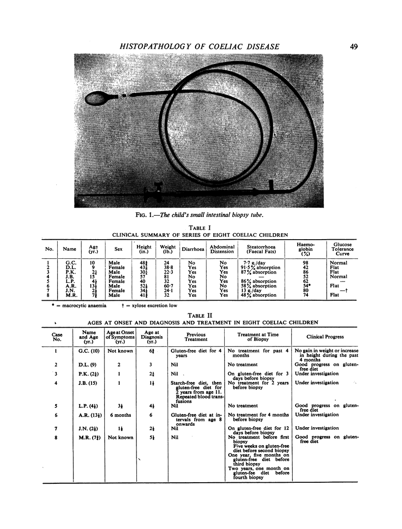
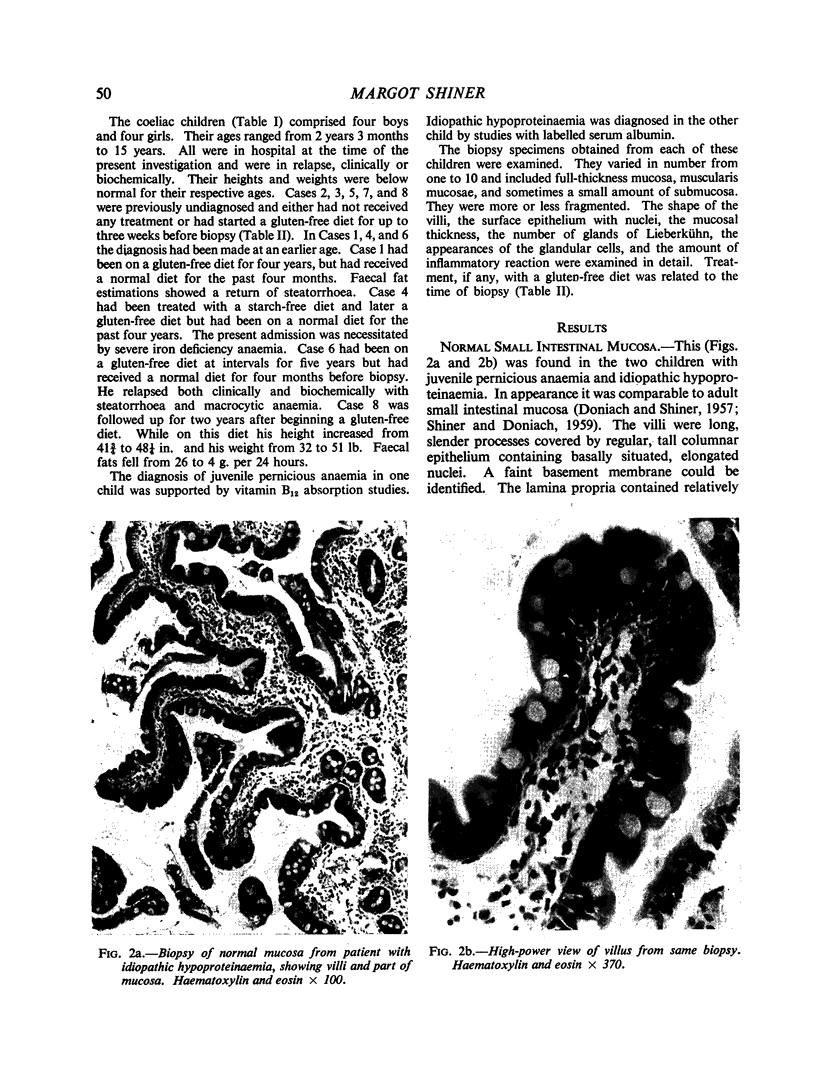
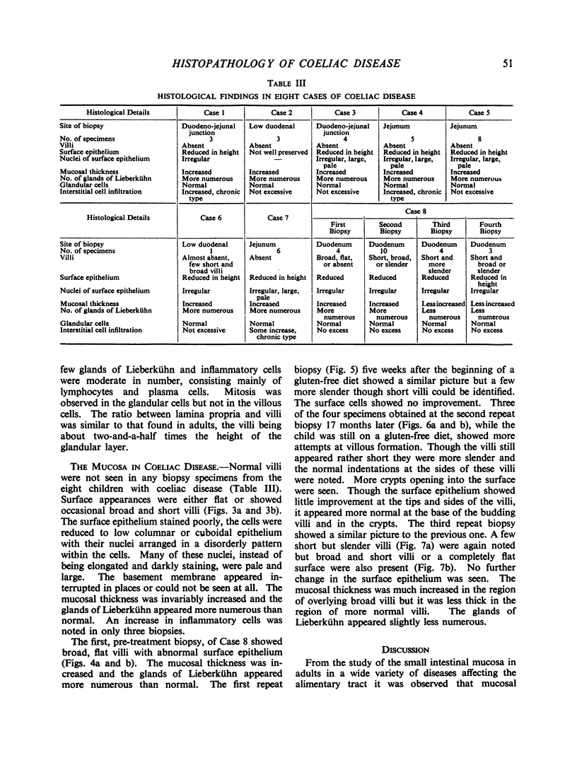
Abstract
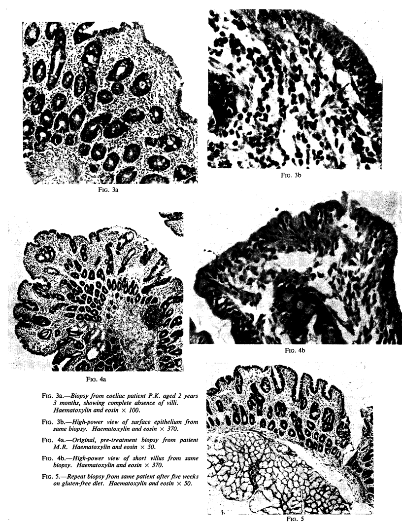
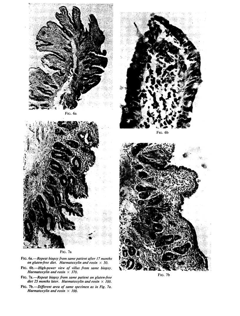
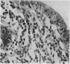
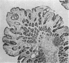
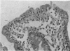
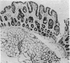
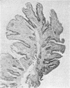
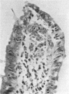
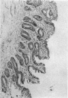
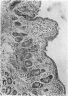
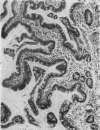
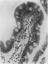
Villous atrophy, changes in the surface epithelium, mucosal thickening, and glandular hypertrophy were a feature of all the mucosal biopsies from the small intestine obtained from eight coeliac children. No histological differences were observed between the children previously treated with intermittent gluten-free diets and the untreated children. Serial biopsy studies were carried out on one coeliac child before and after treatment with a gluten-free diet over a period of two years. On the whole they confirmed the irreversible nature of the observed histopathological changes but minor improvements could not be excluded. Mucosal abnormalities in coeliac disease are the same as in adult idiopathic steatorrhoea, where they are observed in patients with or without a response to a gluten-free diet. It is concluded that the elimination of gluten from the diet has little if any influence on the histopathological abnormalities observed in coeliac disease.
Full text
PDF
Images in this article
Selected References
These references are in PubMed. This may not be the complete list of references from this article.
- DONIACH I., SHINER M. Duodenal and jejunal biopsies. II. Histology. Gastroenterology. 1957 Jul;33(1):71–86. [PubMed] [Google Scholar]
- SAKULA J., SHINER M. Coeliac disease with atrophy of the small-intestine mucosa. Lancet. 1957 Nov 2;273(7001):876–877. doi: 10.1016/s0140-6736(57)90010-7. [DOI] [PubMed] [Google Scholar]