Short abstract
A new genome-wide analysis of human kinases using RNA interference shows an unexpected depth and complexity to the interactions between signal transduction and vesicular transport.
Abstract
The mechanisms of signal transduction and vesicular transport have traditionally been studied in isolation, but recent studies make it clear that the two processes are inextricably linked. A new genome-wide analysis of human kinases using RNA interference shows an unexpected depth and complexity to the interactions between these processes.
Since the discovery in the nematode Caenorhabditis elegans that the introduction of a double-stranded RNA (dsRNA) trigger can lead to the selective inhibition of gene expression in a sequence-specific manner [1], this phenomenon - now widely known as RNA interference (RNAi) - has spawned many new applications. Among these is the invention of a whole area of genomics research dedicated to studying RNAi-induced phenotypes. Such phenotypes generally mimic reduction-of-function or loss-of-function mutations that have formed the backbone of traditional genetics research for more than a century. Most large-scale screens using this method have been performed in model organisms such as C. elegans [2-4] and Drosophila melanogaster [5,6]. As a result of recent insights into the mechanism of RNAi and the resulting identification of small interfering RNAs (siRNAs), such large-scale approaches are now also feasible in cultured mammalian cells [7,8]. The quantity of data derived from large-scale or whole-genome RNAi-based screens, while sometimes overwhelming, has begun to provide insights into complex biological phenomena that had previously proved intractable [4,9]. Endocytosis is one such complex cellular process that has begun to give up its secrets to the RNAi cognoscenti. In this article we focus on a recent report by Marino Zerial and colleagues [10] who utilize cutting-edge techniques to perform a genome-wide RNAi screen exploring the role of each human kinase in the regulation of endocytosis.
Endocytosis uses membrane-bound vesicles to internalize macromolecules and fluid from the plasma membrane and extracellular space and is a crucial process for all eukaryotic cells. Endocytosis mediates a plethora of biological processes including nutrient uptake, regulation of growth factor receptors, synaptic vesicle recycling by the nervous system, and antigen processing by the immune system. Recent RNAi-based studies provide new insights into the regulation of the best studied pathway, clathrin-dependent endocytosis [11], as well as the less well understood endocytosis pathway mediated by lipid rafts [12].
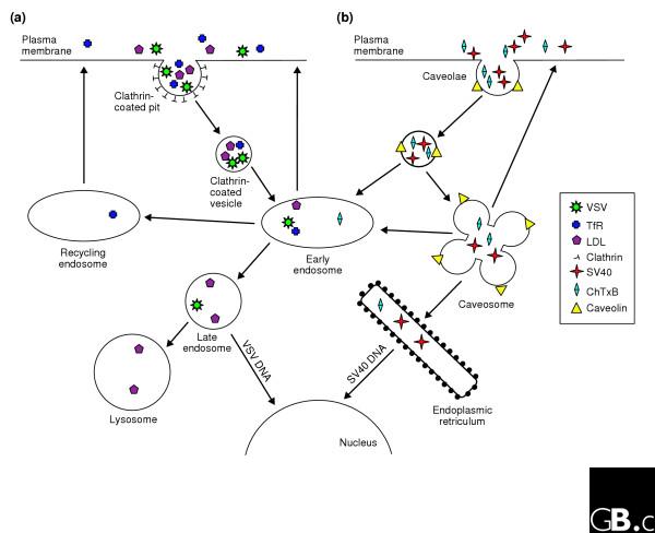
The basic steps in the clathrin-dependent endocytic pathway as currently understood are shown in Figure 1a. After recruitment of cargo molecules into a clathrin-coated pit, the pit is pinched off into a vesicle. The clathrin coat is actively removed, allowing fusion of the vesicle with an early endosome. Early endosomes are sorting stations within the cell, delivering some cargo molecules to late endosomes and eventually lysosomes, while other cargo molecules are instead recycled, either directly or indirectly, to the cell surface.
Figure 1.

Schematic representation of two endocytosis pathways. (a) Cargo trafficking mediated by clathrin-dependent endocytosis. This pathway is typically initiated by the recruitment of cargo into clathrin-coated pits at the plasma membrane. After pinching off from the plasma membrane, clathrin-coated vesicles are transported to the early endosome. Here cargo is sorted for delivery to the degradative pathway, that is, the late endosome and lysosome, or is recycled to the plasma membrane directly, or via the recycling endosome. (b) Caveolae-dependent endocytosis. This pathway starts at the plasma membrane. After leaving the plasma membrane caveolar vesicles can either briefly fuse with early endosomes or fuse with caveosomes. From caveosomes, cargo can either traffic to the endoplasmic reticulum or early endosomes, or back to the plasma membrane. Vesicular stomatitis virus (VSV) and transferrin receptor (TfR) enter cells via the clathrin-mediated pathway, whereas simian virus 40 (SV40) and cholera toxin B subunit (ChTxB) use the caveolae and raft-mediated pathway. LDL, low density lipoprotein.
Over the past decade the importance of alternative clathrin-independent routes of endocytosis for certain cell-surface cargo has become increasingly clear (Figure 1b). One uptake route for some, but not all, clathrin-independent cargo is through caveolae, specialized invaginations in the plasma membrane [12]. Caveolae are considered to represent a specialized form of a cholesterol- and sphingolipid-rich lipid raft domain that is chemically distinguished by the presence of caveolins, cholesterol-binding proteins essential to the structure and function of these invaginations [13]. At least some raft-dependent cargo is thought to enter cells through caveolae, which inside the cell form endosome-like structures known as caveosomes [14].
Pelkmans et al. [10] now describe an RNAi-based screen they developed to explore the role of all human kinases (also called the kinome) in the regulation of clathrin-dependent and caveolae-dependent endocytic pathways. The authors developed a clever method to assay quickly for defective endocytosis taking advantage of two well studied viruses that infect cells via endocytic uptake. Vesicular stomatitis virus (VSV) enters cells via the clathrin-mediated pathway (Figure 1a) [15], whereas simian virus 40 (SV40) uses caveolae/raft-mediated endocytosis for host-cell infection (Figure 1b) [16,17]. By systematically knocking down the level of each human kinase individually, 590 in total, and monitoring the changes in viral infection rates in HeLa cells (which reflect regulation of the two endocytic routes), the authors were able to gain new insights into endocytic regulation [10]. The screen was designed to detect both decreases and increases in viral infection rates. Lower rates of viral infection suggest a reduction in the uptake or trafficking of viral particles, whereas an increased infection rate suggests enhanced uptake or trafficking of virus.
Surprisingly, more than a third of all kinases affected either VSV and/or SV40 infection, indicating that the kinome contains large numbers of endocytosis regulators. Most kinases previously implicated in endocytosis were detected in this work [10] as regulators of viral infection, indicating the high sensitivity of the assay used for the screen. Nearly a quarter of all of the kinases found to be regulators of endocytosis in the screen are currently poorly characterized or uncharacterized, and thus represent fertile ground for new research. In particular, the next challenge will be to identify the specific substrates of these kinases, potentially revealing new components in the endocytic process, and new signaling pathways that impinge on endocytic trafficking. Interestingly, kinases whose loss resulted in less efficient VSV infection through the clathrin pathway were much more numerous that those whose loss led to more efficient infection. Conversely, the number of kinases whose loss resulted in increased SV40 infection was relatively high. A significant group of kinases were identified whose loss affected both viruses, many in a reciprocal manner.
These observations lead to several interesting hypotheses. First, the fact that few perturbations of the clathrin pathway result in higher output indicates that in HeLa cells the clathrin pathway is constitutively highly active. On the other hand, many perturbations in kinase function result in higher throughput in the caveolar-raft pathway, suggesting that this pathway is under significant phosphorylation-dependent negative regulation. The identification of a group of kinases with opposite effects on the two pathways may indicate that the pathways are physiologically linked. Some of these opposite effects appear to be mediated through the regulation of actin (see [10] and references therein). Actin dynamics have been proposed to be required for clathrin-dependent endocytosis, whereas actin microfilaments may inhibit caveolar uptake. Such interpretations are highly tentative, however, as viral infection is a complex process that does not simply reflect the ability of the virus to enter the endosomal pathway, but also its ability to break free and reach the cytoplasm, and express viral genes. Furthermore, more rapid delivery of viral particles to the destination compartment, such as the late endosome for VSV, does not necessarily guarantee greater infection rates.
To address these questions and validate the role of specific kinases in regulating endocytosis, one additional kinome-wide phenotypic screen was performed [10]. This analyzed the localization of fluorescently labeled transferrin, a classic assay of clathrin-mediated uptake and transit through the recycling pathway. Pelkmans et al. [10] further analyzed 50 of the positive kinases showing an altered phenotype in the viral screens by documenting the morphology and distribution of early and late endosomes, of caveolin labeled with green fluorescent protein (GFP), and of several cargo molecules, providing clues to which particular trafficking step had been perturbed. Hierarchical clustering of kinases on the basis of these phenotypic observations identified functional groups that are likely to work in concert to regulate particular aspects of endocytosis. In theory, these phenotypes can be used to help identify the direct or indirect targets of these kinases - that is, the molecules that directly regulate membrane trafficking events through a similar detailed comparison of phenotypes after knockdown of well studied trafficking factors. It should be noted that a small number of kinases showed defects in transferrin uptake after RNAi but were not picked up as regulators of viral infection, suggesting that the actual number of kinases regulating endocytic trafficking could be even higher than revealed by the primary screen.
Pelkmans et al. [10] also clustered the kinases into 11 groups on the basis of their known functions in various signaling pathways. Although one might suspect that the large number of metabolic kinases found in the screen indicated indirect effects of the metabolic state of the cell on trafficking, the fact that most of the metabolic kinases showed specificity for only one endocytic pathway or the other suggests otherwise. In addition, several signaling pathways also showed selective effects on only one of the endocytic uptake routes. Wnt signaling pathway kinases, cell-cycle-regulating kinases, mTOR pathway kinases, and signaling pathways originating from G-protein-coupled receptors were all shown to be regulators of clathrin-mediated endocytosis or trafficking. Interestingly, kinases participating in integrin signaling were shown to specifically control only non-clathrin-mediated endocytosis. A recent article by Wu et al. [18], however, indicates that focal adhesion kinase (FAK), one of the kinases downstream of integrins, regulates endocytosis of matrix metalloproteinases through a clathrin-dependent mechanism. This apparent conflict is likely to be due to the different cargo molecules assayed, suggesting that a more comprehensive screen of cargo molecule types will be required to clarify the relationships between regulatory molecules and their targets. It is also important to remember that signaling pathways in the cell are not isolated but are highly interactive, implying that many of the effects described are likely to be quite indirect.
Genomic approaches are increasingly being used to address many aspects of membrane trafficking. Sieburth et al. [9] recently published a genome-wide screen in C. elegans, identifying proteins necessary for the structure and function of the neuromuscular synapse. Clathrin-mediated endocytosis in particular is thought to be a critical component of the synaptic-vesicle cycle, and this new screen identified some known clathrin-associated molecules as well as many completely new synaptic components [9]. A significant number of kinases were also identified. Other groups have also recently applied a systems biology or genomics approach to understanding the functional relationships among eukaryotic membrane-trafficking components. Gurkan et al. [19] analyzed expression profiles of Rab GTPases and their effectors in a wide variety of mammalian tissues and cells. This important study provides a wealth of new information relevant to understanding mammalian Rab proteins, of which there are more than 50, and their effectors, most of which are currently very poorly characterized.
A key remaining question for all such genomic screens will be the directness of the effects shown. In particular, identification of the most proximal endocytotic pathway substrates of the kinases identified by Pelkmans et al. [10] will be essential. It remains to be determined whether kinases from the same signaling pathway all regulate the same step of trafficking. Early evidence provided by the secondary screens [10] suggests that multiple aspects of trafficking are targeted simultaneously, probably leading to more precise outcomes. The large number of kinases controlling endocytic trafficking also suggests that a large number of phosphatases will contribute to this process. Finally, it will be of great interest to see how these regulatory mechanisms contribute to higher-order processes such as cell polarity and tissue formation and organization. The information derived from each of these genomics approaches has provided a plethora of valuable leads that are likely to drive the vesicle-trafficking field for years to come.
References
- Fire A, Xu S, Montgomery MK, Kostas SA, Driver SE, Mello CC. Potent and specific genetic interference by double-stranded RNA in Caenorhabditis elegans. Nature. 1998;391:806–811. doi: 10.1038/35888. [DOI] [PubMed] [Google Scholar]
- Kamath RS, Fraser AG, Dong Y, Poulin G, Durbin R, Gotta M, Kanapin A, Le Bot N, Moreno S, Sohrmann M, et al. Systematic functional analysis of the Caenorhabditis elegans genome using RNAi. Nature. 2003;421:231–237. doi: 10.1038/nature01278. [DOI] [PubMed] [Google Scholar]
- Fraser AG, Kamath RS, Zipperlen P, Martinez-Campos M, Sohrmann M, Ahringer J. Functional genomic analysis of C. elegans chromosome I by systematic RNA interference. Nature. 2000;408:325–330. doi: 10.1038/35042517. [DOI] [PubMed] [Google Scholar]
- Ashrafi K, Chang FY, Watts JL, Fraser AG, Kamath RS, Ahringer J, Ruvkun G. Genome-wide RNAi analysis of Caenorhabditis elegans fat regulatory genes. Nature. 2003;421:268–272. doi: 10.1038/nature01279. [DOI] [PubMed] [Google Scholar]
- Boutros M, Kiger AA, Armknecht S, Kerr K, Hild M, Koch B, Haas SA, Heidelberg Fly Array Consortium. Paro R, Perrimon N. Genome-wide RNAi analysis of growth and viability in Drosophila cells. Science. 2004;303:832–835. doi: 10.1126/science.1091266. [DOI] [PubMed] [Google Scholar]
- Ivanov AI, Rovescalli AC, Pozzi P, Yoo S, Mozer B, Li HP, Yu SH, Higashida H, Guo V, Spencer M, et al. Genes required for Drosophila nervous system development identified by RNA interference. Proc Natl Acad Sci USA. 2004;101:16216–16221. doi: 10.1073/pnas.0407188101. [DOI] [PMC free article] [PubMed] [Google Scholar]
- Elbashir SM, Harborth J, Lendeckel W, Yalcin A, Weber K, Tuschl T. Duplexes of 21-nucleotide RNAs mediate RNA interference in cultured mammalian cells. Nature. 2001;411:494–498. doi: 10.1038/35078107. [DOI] [PubMed] [Google Scholar]
- Paddison PJ, Silva JM, Conklin DS, Schlabach M, Li M, Aruleba S, Balija V, O'Shaughnessy A, Gnoj L, Scobie K, et al. A resource for large-scale RNA-interference-based screens in mammals. Nature. 2004;428:427–431. doi: 10.1038/nature02370. [DOI] [PubMed] [Google Scholar]
- Sieburth D, Ch'ng Q, Dybbs M, Tavazoie M, Kennedy S, Wang D, Dupuy D, Rual JF, Hill DE, Vidal M, et al. Systematic analysis of genes required for synapse structure and function. Nature. 2005;436:510–517. doi: 10.1038/nature03809. [DOI] [PubMed] [Google Scholar]
- Pelkmans L, Fava E, Grabner H, Hannus M, Habermann B, Krausz E, Zerial M. Genome-wide analysis of human kinases in clathrin- and caveolae/raft-mediated endocytosis. Nature. 2005;436:78–86. doi: 10.1038/nature03571. [DOI] [PubMed] [Google Scholar]
- Mukherjee S, Ghosh RN, Maxfield FR. Endocytosis. Physiol Rev. 1997;77:759–803. doi: 10.1152/physrev.1997.77.3.759. [DOI] [PubMed] [Google Scholar]
- Nichols B. Caveosomes and endocytosis of lipid rafts. J Cell Sci. 2003;116:4707–4714. doi: 10.1242/jcs.00840. [DOI] [PubMed] [Google Scholar]
- Rothberg KG, Heuser JE, Donzell WC, Ying YS, Glenney JR, Anderson RG. Caveolin, a protein component of caveolae membrane coats. Cell. 1992;68:673–682. doi: 10.1016/0092-8674(92)90143-Z. [DOI] [PubMed] [Google Scholar]
- Harder T, Simons K. Caveolae, DIGs, and the dynamics of sphingolipid-cholesterol microdomains. Curr Opin Cell Biol. 1997;9:534–542. doi: 10.1016/S0955-0674(97)80030-0. [DOI] [PubMed] [Google Scholar]
- Sieczkarski SB, Whittaker GR. Differential requirements of Rab5 and Rab7 for endocytosis of influenza and other enveloped viruses. Traffic. 2003;4:333–343. doi: 10.1034/j.1600-0854.2003.00090.x. [DOI] [PubMed] [Google Scholar]
- Anderson HA, Chen Y, Norkin LC. Bound simian virus 40 translocates to caveolin-enriched membrane domains, and its entry is inhibited by drugs that selectively disrupt caveolae. Mol Biol Cell. 1996;7:1825–1834. doi: 10.1091/mbc.7.11.1825. [DOI] [PMC free article] [PubMed] [Google Scholar]
- Damm EM, Pelkmans L, Kartenbeck J, Mezzacasa A, Kurzchalia T, Helenius A. Clathrin- and caveolin-1-independent endocytosis: entry of simian virus 40 into cells devoid of caveolae. J Cell Biol. 2005;168:477–488. doi: 10.1083/jcb.200407113. [DOI] [PMC free article] [PubMed] [Google Scholar]
- Wu X, Gan B, Yoo Y, Guan JL. FAK-Mediated Src phosphorylation of endophilin A2 inhibits endocytosis of MT1-MMP and promotes ECM degradation. Dev Cell. 2005;9:185–196. doi: 10.1016/j.devcel.2005.06.006. [DOI] [PubMed] [Google Scholar]
- Gurkan C, Lapp H, Alory C, Su AI, Hogenesch JB, Balch WE. Large-scale profiling of Rab GTPase trafficking networks: the membrome. Mol Biol Cell. 2005;16:3847–3864. doi: 10.1091/mbc.E05-01-0062. [DOI] [PMC free article] [PubMed] [Google Scholar]


