Abstract
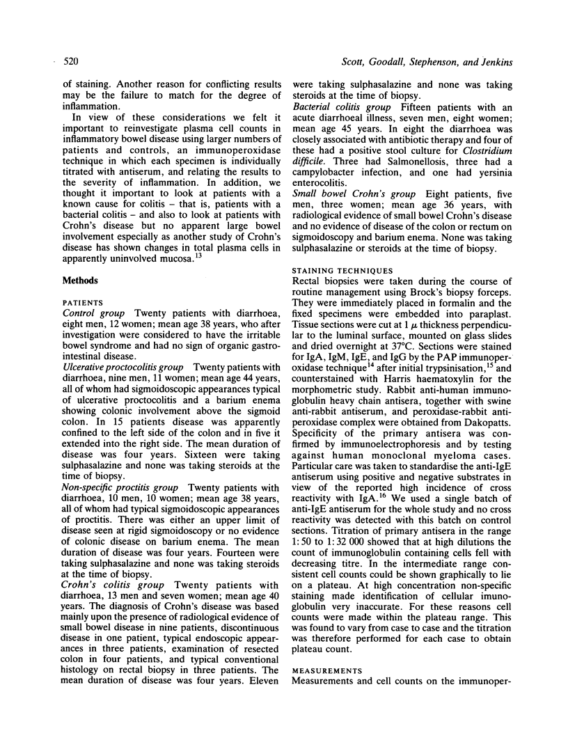
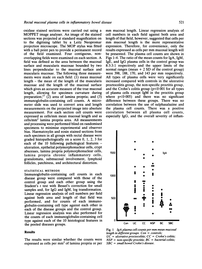
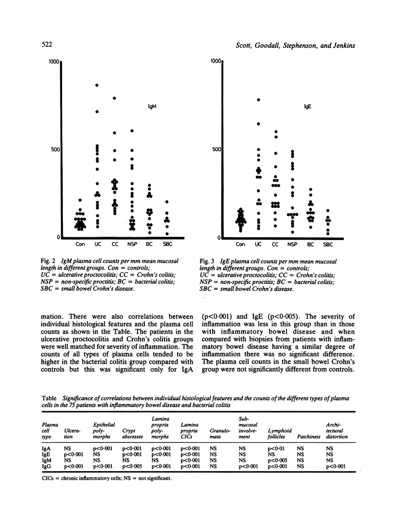
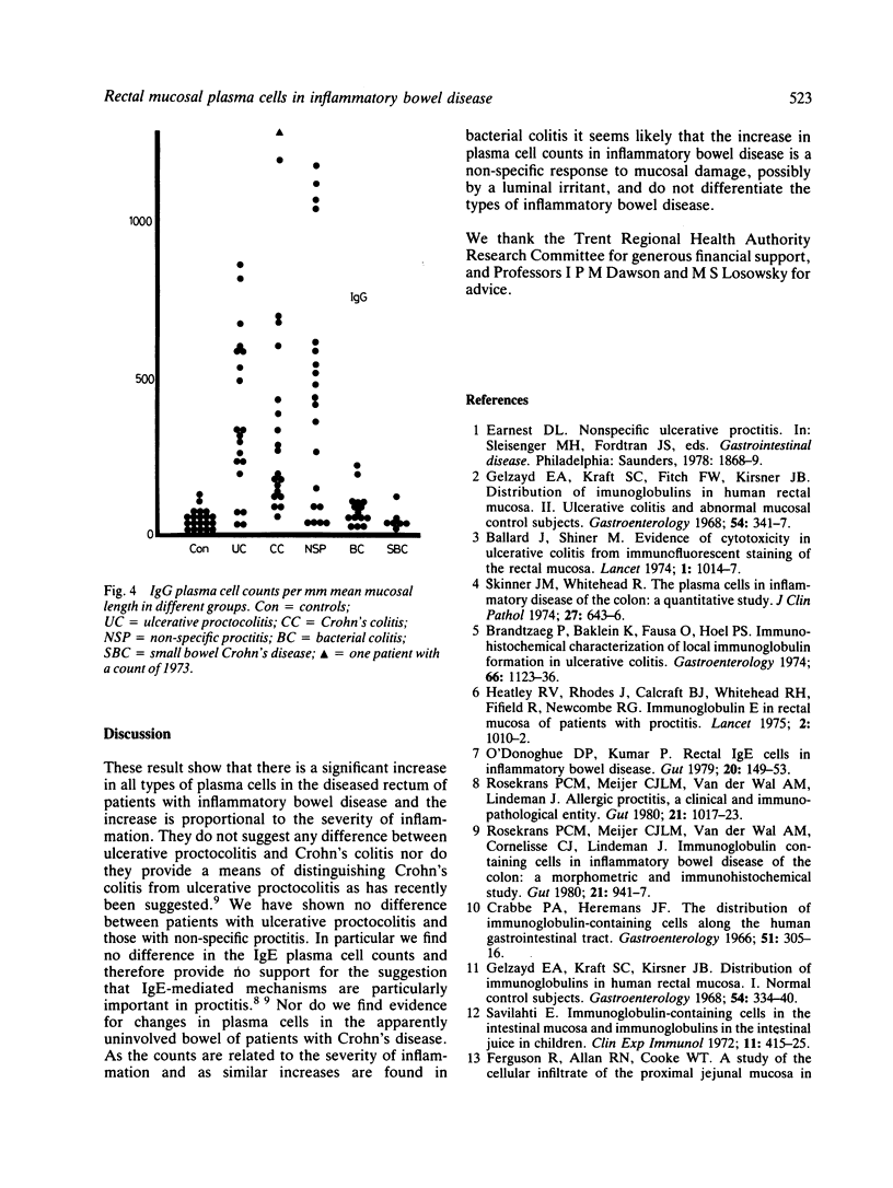
To achieve optimum staining and reproducible counts of plasma cells in paraffin embedded tissue with the immunoperoxidase technique we have found it essential to obtain a plateau count by titration of antisera for each specimen. This modification was used to study IgA, IgM, IgE, and IgG plasma cells in rectal biopsies from 20 controls, 20 patients with ulcerative proctocolitis, 20 with Crohn's colitis, 20 with non-specific proctitis, 15 with bacterial colitis, and seven with Crohn's disease but no apparent large bowel involvement. Counts were correlated with the characteristic histological features of inflammatory bowel disease. In controls the ratio of the mean counts for IgA, IgM, IgE, and IgG plasma cells was 8:3:3:1. All types of plasma cells were very significantly increased in the patients with ulcerative proctocolitis, Crohn's colitis, and non-specific proctitis and counts correlated with the severity of inflammation. There was no significant difference between the counts in these three groups. All counts tended to be higher in bacterial colitis than in controls, the difference being significant for IgA and IgE. When matched for severity of inflammation there was no significant difference between the counts in bacterial colitis and inflammatory bowel disease. The counts in patients with Crohn's disease but no large bowel involvement were not significantly different from controls. These results suggest that changes in plasma cell counts in inflammatory bowel disease are a non-specific response to mucosal damage, possible by a luminal irritant, and do not differentiate the type of inflammatory bowel disease.
Full text
PDF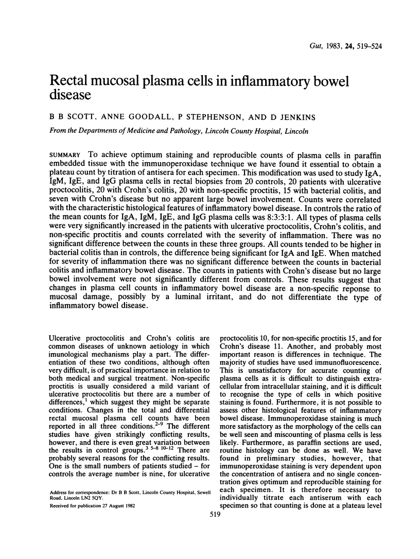
Selected References
These references are in PubMed. This may not be the complete list of references from this article.
- Ballard J., Shiner M. Evidence of cytotoxicity in ulcerative colitis from immunofluorescent staining of the rectal mucosa. Lancet. 1974 May 25;1(7865):1014–1017. doi: 10.1016/s0140-6736(74)90416-4. [DOI] [PubMed] [Google Scholar]
- Brandtzaeg P., Baklien K., Fausa O., Hoel P. S. Immunohistochemical characterization of local immunoglobulin formation in ulcerative colitis. Gastroenterology. 1974 Jun;66(6):1123–1136. [PubMed] [Google Scholar]
- Crabbé P. A., Heremans J. F. The distribution of immunoglobulin-containing cells along the human gastrointestinal tract. Gastroenterology. 1966 Sep;51(3):305–316. [PubMed] [Google Scholar]
- Curran R. C., Gregory J. The unmasking of antigens in paraffin sections of tissue by trypsin. Experientia. 1977 Oct 15;33(10):1400–1401. doi: 10.1007/BF01920206. [DOI] [PubMed] [Google Scholar]
- Ferguson R., Allan R. N., Cooke W. T. A study of the cellular infiltrate of the proximal jejunal mucosa in ulcerative colitis and Crohn's disease. Gut. 1975 Mar;16(3):205–208. doi: 10.1136/gut.16.3.205. [DOI] [PMC free article] [PubMed] [Google Scholar]
- Gelzayd E. A., Kraft S. C., Fitch F. W., Kirsner J. B. Distribution of immunoglobulins in human rectal mucosa. II. Ulcerative colitis and abnormal mucosal control subjects. Gastroenterology. 1968 Mar;54(3):341–347. [PubMed] [Google Scholar]
- Gelzayd E. A., Kraft S. C., Kirsner J. B. Distribution of immunoglobulins in human rectal mucosa. I. Normal control subjects. Gastroenterology. 1968 Mar;54(3):334–340. [PubMed] [Google Scholar]
- Heatley R. V., Calcraft B. J., Fifield R., Rhodes J., Whitehead R. H., Newcombe R. G. Immunoglobulin E in rectal mucosa of patients with proctitis. Lancet. 1975 Nov 22;2(7943):1010–1012. doi: 10.1016/s0140-6736(75)90294-9. [DOI] [PubMed] [Google Scholar]
- McIntyre R., Piris J. Quantification of human gastric G cell density in endoscopic biopsy specimens: effect of shape of specimen. J Clin Pathol. 1980 Jun;33(6):513–516. doi: 10.1136/jcp.33.6.513. [DOI] [PMC free article] [PubMed] [Google Scholar]
- O'Donoghue D. P., Kumar P. Rectal IgE cells in inflammatory bowel disease. Gut. 1979 Feb;20(2):149–153. doi: 10.1136/gut.20.2.149. [DOI] [PMC free article] [PubMed] [Google Scholar]
- Rosekrans P. C., Meijer C. J., van der Wal A. M., Cornelisse C. J., Lindeman J. Immunoglobulin containing cells in inflammatory bowel disease of the colon: a morphometric and immunohistochemical study. Gut. 1980 Nov;21(11):941–947. doi: 10.1136/gut.21.11.941. [DOI] [PMC free article] [PubMed] [Google Scholar]
- Rosekrans P. C., Meijer C. J., van der Wal A. M., Lindeman J. Allergic proctitis, a clinical and immunopathological entity. Gut. 1980 Dec;21(12):1017–1023. doi: 10.1136/gut.21.12.1017. [DOI] [PMC free article] [PubMed] [Google Scholar]
- Savilahti E. Immunoglobulin-containing cells in the intestinal mucosa and immunoglobulins in the intestinal juice in children. Clin Exp Immunol. 1972 Jul;11(3):415–425. [PMC free article] [PubMed] [Google Scholar]
- Skinner J. M., Whitehead R. The plasma cells in inflammatory disease of the colon: a quantitative study. J Clin Pathol. 1974 Aug;27(8):643–646. doi: 10.1136/jcp.27.8.643. [DOI] [PMC free article] [PubMed] [Google Scholar]
- Taylor C. R. Immunoperoxidase techniques: practical and theoretical aspects. Arch Pathol Lab Med. 1978 Mar;102(3):113–121. [PubMed] [Google Scholar]


