FIGURE 5.
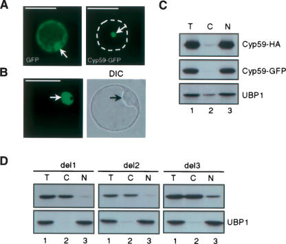
Cellular localization of AtCyp59. (A) Localization of AtCyp59-GFP fusion protein and GFP alone in transiently transformed tobacco protoplasts. Dashed line delineates the shape of protoplasts. Arrows point to nuclei. (B) Localization of AtCyp59-GFP fusion protein in transiently transformed Arabidopsis protoplasts. Single confocal image with corresponding differential interference contrast (DIC) image of whole cell is shown. Arrows point to nuclei. Protoplasts were analyzed 24 h after transformation by using Zeiss Axiovert epifluorescence microscope (A) or Leica TCS confocal microscope (B). Bars, 50 μm (A,B). (C) Cellular localization of AtCyp59 studied by cellular fractionation of Arabidopsis protoplasts transiently expressing AtCyp59-GFP or AtCyp59-HA. Western blots were analyzed with monoclonal antibodies against GFP or HA tags. The distribution of endogenous nuclear UBP1 protein was used to control quality of the fractionation procedure. (Lane 1) Total protein extracts (T), (lane 2) cytoplasmic fraction (C), (lane 3) nuclear fraction (N). (D) Localization of AtCyp59-HA deletions in transiently transformed Arabidopsis protoplasts as determined by cellular fractionation. Details are as in C.

