Abstract
Enhanced signaling in myocytes by the G protein Gq has been implicated in cardiac hypertrophy and the transition to heart failure. α1-Adrenergic receptors (α1-ARs) are members of the 7-transmembrane-spanning domain (7-TM) receptor family and signal via interaction with Gq in the heart. The specific effects of a loss of α1-AR signaling in the heart are explored by O’Connell et al. in this issue of the JCI (see the related article beginning on page 1005). Paradoxically, gene ablation of the α1A and α1B subtypes in mice results in a maladaptive form of reactive cardiac hypertrophy from pressure overload, with a predisposition to heart failure. Thus signaling to the α1-AR (compared with signaling from other receptors such as angiotensin receptors, which also couple to Gq) appears to be specifically required for a normal hypertrophic response. This represents another example of how receptors that share common G proteins have diversified, developing unique signaling programs. These findings may have particular clinical relevance because of the widespread use of α1-AR antagonists in the treatment of hypertension and symptomatic prostate enlargement.
It is now recognized that there are 2 distinct subgroups of α-adrenergic receptors, designated as α1-adrenergic receptors (α1-ARs) and α2-ARs, all of which are members of the superfamily of 7-transmembrane-spanning domain (7-TM) receptors (also termed G protein–coupled receptors). There are 3 human α1-AR subtypes, denoted α1A, α1B, and α1D. Since α1-ARs expressed on vascular smooth muscle act to constrict and thus increase peripheral vascular resistance, there has been substantial development and widespread use of α1-AR antagonists for the treatment of hypertension. What has not been well acknowledged is the fact that α1-ARs are also expressed on cardiomyocytes, and thus treatment of hypertension with α1-AR antagonists may also have effects on the heart that are distinct from afterload reduction. All α1-AR subtypes couple to the heterotrimeric G protein Gq. Upon agonist activation, the Gα subunit activates the effector phospholipase C, which produces at least 2 intracellular second messengers, inositol-1,4,5-triphosphate and diacylglycerol. The former increases intracellular calcium, while the latter activates several PKC isoenzymes that modify heart failure (1). Since catecholamines are elevated in heart failure, cardiac α1-AR/Gq signaling is activated to various extents in the syndrome.
The Gαq pathway has been studied extensively as to its role in cardiac hypertrophy and heart failure (2). Cardiac overexpression of Gαq in transgenic mice (3) results in hypertrophy, decreased ventricular function, loss of β-adrenergic receptor inotropic responsiveness, and induction of a classic hypertrophy gene expression profile. In these mice, pressure overload by surgical transverse aortic constriction (TAC), pregnancy, or higher transgenic overexpression of Gαq resulted in cardiomyocyte apoptosis and decompensated heart failure (3, 4). Other studies showed that transgenic overexpression of a Gαq dominant-negative minigene resulted in the lack of a hypertrophy response to TAC (5). Furthermore, cardiac overexpression of a constitutively activated α1B-AR resulted in cardiac hypertrophy (6), while a more severe cardiomyopathy developed as a result of overexpression of the Gq-coupled angiotensin II type 1 (AT1) receptor (7). These studies, then, began to point toward hyperactive Gαq signaling as a key mechanism causing hypertrophy, depressed ventricular function, and failure. A readily drawn conclusion from such studies might be that in the human heart, factors that increase Gαq signaling predispose to cardiac hypertrophy and, potentially, the transition from hypertrophy to decompensated heart failure. In addition, approaches that decrease this signaling might be protective against the development of heart failure or be beneficial in treatment.
Ablation of α1-ARs and cardiac hypertrophy
In the report by O’Connell et al. in this issue of the JCI (8), the hypertrophic response to TAC was assessed in mice in which the genes encoding α1A-AR and α1B-AR had been ablated (α1A/B KO mice). Mice without these Gαq-coupled receptors demonstrated rapid decompensation and heart failure after TAC. In those that survived, echocardiographic studies showed lower ejection fractions than in WT mice. Although both sets of mice exhibited hypertrophy, the α1A/B KO mice had increased apoptosis and interstitial fibrosis. Furthermore, they had an atypical hypertrophy-associated gene profile, with minimal changes in expression of β-myosin heavy chain, α-skeletal actin, and atrial natriuretic factor transcripts. These data suggest that α1-AR/Gq signaling is necessary for adaptation to pressure overload. This issue is of substantial clinical importance because of the extensive use of α1-AR antagonists for the treatment of hypertension and symptomatic prostate enlargement. In a large cohort of hypertensive patients, those treated with the α1-AR antagonist doxazosin had a relative risk of 2.04 (95% confidence interval = 1.79–2.32, P < 0.001) of developing heart failure compared with those receiving a diuretic (9). Other studies with smaller cohorts have also observed this relationship but indicate an attenuation of this risk after adjustment for systolic blood pressure (10). Of note, this latter study found that systolic blood pressure was lower in the diuretic group compared with the α1-AR antagonist group, particularly in women, in whom the risk of heart failure was greatest. This may indicate that normotensive men treated with α1-AR antagonists for symptomatic prostatic enlargement are not at significantly increased risk for developing heart failure, but this hypothesis has not been tested in adequately powered trials. Of particular concern would be the scenario in which an individual taking α1-AR antagonists develops a stressed myocardium (e.g., new-onset hypertension, myocardial infarction). The inability to develop the appropriate hypertrophic response could predispose such individuals to cardiac failure.
A matter of timing
The data obtained from this study of α1A/B KO mice (8) provide additional support for the potential deleterious effects of α1-AR blockade. How, though, are these results reconciled with the generally perceived notion that enhanced (as opposed to suppressed) receptor/Gαq signaling evokes hypertrophy and/or failure? There are several components to this issue. First, examination of earlier mouse studies (3) indicates that the effects observed in the Gαq mouse appear to be mostly due to Gαq overexpression being present from birth (because of the properties of the α-myosin light chain promoter that was used in the transgenic construct). When an inducible promoter was used, and Gαq overexpression was induced after the heart had reached normal size, no pathologic effect was noted (11). In other studies, mice lacking Gαq (and the similarly functioning Gα11 protein) died in utero with underdeveloped, hypoplastic hearts (12). So, it appears that for cardiac development (one form of growth), a certain critical level of Gαq signaling is necessary to achieve normal morphology. Gαq/Gα11 KO mice that survive the perinatal period are unable to mount a hypertrophic response to TAC as adults (13).
Thus, similarly to the requirements for normal growth during development, finely tuned Gαq signaling appears to be necessary for reactive growth. Taking these studies into account, then, the O’Connell report (8) does not fully define the role of α1-AR signaling in the hypertrophic or failure response to pressure overload, because the α1A/B KO mice have abnormal hearts as adults. Prior to TAC, these hearts are smaller with decreased myocyte surface area and volume (14). So whether the lack of α1-AR signaling in the normal, adult heart has an effect on hypertrophy or the transition to failure cannot be fully addressed in mice that have a lack of α1A/B-AR from conception. An inducible, cell type–specific knockout would be required to explore this issue in the most rigorous manner. Secondly, it is important to realize that cardiac hypertrophy per se is a form of growth whereby the heart responds to states, such as the increased afterload from hypertension, in order to maintain performance. Thus, under these circumstances hypertrophy can be considered as a compensatory and initially advantageous response of the heart. The inability to achieve the normal biochemical and anatomic hypertrophy response, such as when Gαq signaling is suppressed, may thus predispose to maladaptive hypertrophy and a more rapid transition to pump failure.
7-TM receptor diversification
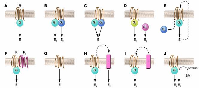
It is not entirely clear why α1-AR/Gαq signaling is specifically necessary for a normal response to pressure overload. There are numerous other cardiac neurohumoral receptors that also couple to Gαq and are activated in heart failure. These include the AT1 receptor, which is an effective pharmacologic target for angiotensin-converting enzyme inhibitors (15) and angiotensin receptor antagonists (16) in the treatment of heart failure. What is apparent, though, is that 7-TM signaling is not a simple, linear, 1-way “switch.” Indeed, in the most straightforward model, the hundreds of 7-TM receptors couple to effectors by only a handful of G proteins. So a major question is how receptor subtypes within cells have evolved to carry out specialized functions. Such “signaling diversification” has been shown to be due to a variety of mechanisms (Figure 1). At issue now for the hypertrophy-related 7-TM receptors is to understand how their signaling has differentiated such that specific influences on cardiac function arise. Indeed, the α1B-AR appears to be in a signaling complex that includes EGFR and PDGFR, which when activated modulate α1B-AR function (17). The Gq-coupled AT1 receptor may be in a different complex with the EGFRs, where each receptor can counterregulate the other (18). Thus these receptor complexes must be delineated and their effects as a unit considered, rather than pathophysiologic effects being ascribed to a single member of the unit. The challenge for the future, then, is to think in terms of signaling complexes, rather than individual components, in order to ascertain critical intracellular events in myocytes that modulate the hypertrophic response and transition to failure.
Figure 1. Mechanisms of 7-TM receptor diversification.
The classic paradigm is shown in A, where a receptor (denoted “R,” “R1,” or “R2”) couples to a G protein (denoted “G”), which activates an effector (denoted “E”). In B, 1 receptor couples to 2 G proteins, thereby activating 2 effectors. In C, receptor coupling to 2 G proteins modulates a single effector. Scenario D involves coupling to a single G protein, but the α and βγ subunits carry out distinct signaling to 2 different effectors. In E, feedback modulation of the receptor (such as phosphorylation) by 1 effector (or an effector-activated event such as protein kinase activation) causes the receptor to couple to the second G protein. In F, the functional coupling of R1 to the G protein is altered by its heterodimerization with another receptor, R2. In contrast to the above, 7-TM receptors can couple directly to effectors without the G protein intermediary (G). In H and I, the 7-TM receptor is affected by, or acts upon, other types of receptors (denoted “r”), such as receptor tyrosine kinases, which alter signaling via the respective cognate pathways. In J, a signaling moiety (SM) is redistributed to a microdomain by the scaffolding action of the arrestins. These proteins act both to partially desensitize the receptor and to evoke a second effector signal by this scaffolding. One or more of these mechanisms may be responsible for the specialized signaling of the α1-AR required for normal hypertrophy, as elucidated by O’Connell et al. (8).
Footnotes
Nonstandard abbreviations used: AR, adrenergic receptor; AT1, angiotensin II type 1; TAC, transverse aortic constriction; 7-TM, 7-transmembrane-spanning domain.
Conflict of interest: The author has declared that no conflict of interest exists.
See the related article beginning on page 1005.
References
- 1.Dorn G.W., Force T. Protein kinase cascades in the regulation of cardiac hypertrophy. J. Clin. Invest. 2005;115:527–537. doi: 10.1172/JCI200524178. [DOI] [PMC free article] [PubMed] [Google Scholar]
- 2.Dorn G.W. Physiologic growth and pathologic genes in cardiac development and cardiomyopathy. Trends Cardiovasc. Med. 2005;15:185–189. doi: 10.1016/j.tcm.2005.05.009. [DOI] [PubMed] [Google Scholar]
- 3.D’Angelo D.D., et al. Transgenic Gαq overexpression induces cardiac contractile failure in mice. Proc. Natl. Acad. Sci. U. S. A. 1997;94:8121–8126. doi: 10.1073/pnas.94.15.8121. [DOI] [PMC free article] [PubMed] [Google Scholar]
- 4.Sakata Y., Hoit B.D., Liggett S.B., Walsh R.A., Dorn G.W., II. Circulation. 97:1488–1495. doi: 10.1161/01.cir.97.15.1488. [DOI] [PubMed] [Google Scholar]
- 5.Akhter S.A., et al. Targeting the receptor-Gq interface to inhibit in vivo pressure overload myocardial hypertrophy. Science. 1998;280:574–577. doi: 10.1126/science.280.5363.574. [DOI] [PubMed] [Google Scholar]
- 6.Milano C.A., et al. Myocardial expression of a constitutively active alpha 1B-adrenergic receptor in transgenic mice induces cardiac hypertrophy. Proc. Natl. Acad. Sci. U. S. A. 1994;91:10109–10113. doi: 10.1073/pnas.91.21.10109. [DOI] [PMC free article] [PubMed] [Google Scholar]
- 7.Hein L., et al. Overexpression of angiotensin AT1 receptor transgene in the mouse myocardium produces a lethal phenotype associated with myocyte hyperplasia and heart block. Proc. Natl. Acad. Sci. U. S. A. 1997;94:6391–6396. doi: 10.1073/pnas.94.12.6391. [DOI] [PMC free article] [PubMed] [Google Scholar]
- 8.O’Connell T.D., et al. α1 -Adrenergic receptors prevent a maladaptive cardiac response to pressure overload. . J. Clin. Invest. 2006;116:1005–1015. doi: 10.1172/JCI22811. [DOI] [PMC free article] [PubMed] [Google Scholar]
- 9.ALLHAT Collaborative Research Group. . Major cardiovascular events in hypertensive patients randomized to doxazosin vs chlorthalidone. JAMA. 2000;283:1967–1975. [PubMed] [Google Scholar]
- 10.Bryson C.L., et al. Risk of congestive heart failure in an elderly population treated with peripheral alpha-1 antagonists. J. Am. Geriatr. Soc. 2004;52:1648–1654. doi: 10.1111/j.1532-5415.2004.52456.x. [DOI] [PubMed] [Google Scholar]
- 11.Syed F., et al. Physiological growth synergizes with pathological genes in experimental cardiomyopathy. Circ. Res. 2004;95:1200–1206. doi: 10.1161/01.RES.0000150366.08972.7f. [DOI] [PubMed] [Google Scholar]
- 12.Offermanns S., et al. Embryonic cardiomyocyte hypoplasia and craniofacial defects in G alpha q/G alpha 11-mutant mice. EMBO J. 1998;17:4304–4312. doi: 10.1093/emboj/17.15.4304. [DOI] [PMC free article] [PubMed] [Google Scholar]
- 13.Wettschureck N., et al. Absence of pressure overload induced myocardial hypertrophy after conditional inactivation of Galphaq/Galpha11 in cardiomyocytes. Nat. Med. 2001;7:1236–1240. doi: 10.1038/nm1101-1236. [DOI] [PubMed] [Google Scholar]
- 14.O’Connell T.D., et al. The α(1A/C)- and α(1B)-adrenergic receptors are required for physiological cardiac hypertrophy in the double-knockout mouse. J. Clin. Invest. 2003;111:1783–1791. doi: 10.1172/JCI200316100. [DOI] [PMC free article] [PubMed] [Google Scholar]
- 15.Garg R., Yusuf S. Overview of randomized trials of angiotensin-converting enzyme inhibitors on mortality and morbidity in patients with heart failure. Collaborative Group on ACE Inhibitor Trials. JAMA. 1995;273:1450–1456. [PubMed] [Google Scholar]
- 16.Sharma D., Buyse M., Pitt B., Rucinska E.J. Meta-analysis of observed mortality data from all-controlled, double-blind, multiple-dose studies of losartan in heart failure. Losartan Heart Failure Mortality Meta-analysis Study Group. Am. J. Cardiol. 2000;85:187–192. doi: 10.1016/s0002-9149(99)00646-3. [DOI] [PubMed] [Google Scholar]
- 17.del Carmen Medina L., Vazquez-Prado J., Garcia-Sainz J.A. Cross-talk between receptors with intrinsic tyrosine kinase activity and α1b-adrenoceptors. Biochem. J. 2000;350:413–419. doi: 10.1042/0264-6021:3500413. [DOI] [PMC free article] [PubMed] [Google Scholar]
- 18.Olivares-Reyes J.A., et al. Agonist-induced interactions between angiotensin AT1 and epidermal growth factor receptors. Mol. Pharmacol. 2005;68:356–36. doi: 10.1124/mol.104.010637. [DOI] [PubMed] [Google Scholar]


