Abstract
MicroRNAs (miRNAs), which are non-coding RNAs 18–25 nt in length, regulate a variety of biological processes, including vertebrate development. To identify new species of miRNA and to simultaneously obtain a comprehensive quantitative profile of small RNA expression in mouse embryos, we used the massively parallel signature sequencing technology that potentially identifies virtually all of the small RNAs in a sample. This approach allowed us to detect a total of 390 miRNAs, including 195 known miRNAs covering ∼80% of previously registered mouse miRNAs as well as 195 new miRNAs, which are so far unknown in mouse. Some of these miRNAs showed temporal expression profiles during prenatal development (E9.5, E10.5 and E11.5). Several miRNAs were positioned in polycistron clusters, including one particular large transcription unit consisting of 16 known and 23 new miRNAs. Our results indicate existence of a significant number of new miRNAs expressed at specific stages of mammalian embryonic development and which were not detected by earlier methods.
INTRODUCTION
A small RNA termed micRNA (mRNA-interfering complementary RNA) that interferes with a specific mRNA was first described in Escherichia coli in 1984 (1). Since then, small RNA repressors for gene expression have been widely reported from bacteria to humans (2–4). RNA repressors bind to target complementary mRNAs leading to direct inhibition of mRNA translation and/or destabilization of the target mRNAs. In plants and animals, a large number of small RNAs of 18–25 bases in length, termed microRNAs (miRNAs) and siRNAs have been found to play important roles in silencing specific target genes. miRNAs are the transcripts which are cleaved from a 70 base hairpin precursor by Dicer (5,6). The total estimated number of reasonably conserved miRNAs in vertebrates varies from 250 (7) to ∼600 (8). Recently, number of new miRNAs, which are not conserved beyond primates, have been identified, and humans are reported to contain at least 800 miRNAs (9).
Diverse roles ranging from developmental patterning or cell differentiation to genome rearrangement and DNA excision are proposed for this novel class of small RNA molecules (2,10). In contrast to plants, in Caenorhabditis elegans and Drosophila, mammalian miRNAs appear to target a significantly broader diversity of cellular functions and the majority of documented cases of exact targets are involved in regulation of transcription factors (11). Nevertheless, the notion that a significant fraction of miRNAs function in early and late development is supported by (i) differential expression profiles, (ii) similarities in expression patterns of miRNAs during development in both mammals and lower organisms, (iii) cell culture studies and (iv) analysis of dicer mouse mutants (11). The elucidation of the spatial and temporal patterns of their expression is critical for understanding the precise role of the mammalian miRNAs in development.
The methods for quantification of the expression level of specific miRNAs include northern blotting, RNase protection assay, RT–PCR and microarray (8). Among semi-quantitative assays, in situ hybridization and miRNA reporter transgenic expression analysis have been carried out (12,13). All of these approaches depend on prior knowledge of the miRNA sequences. In contrast, the massively parallel signature sequencing technology (MPSS) allows one to quantitatively identify millions of small RNAs in a single reaction without prior knowledge of their sequences (14). If such an analysis is conducted on embryos or tissues at specific stages of development, one can obtain expression patterns at the transcriptome level. This method also allows one to identify distinct species of miRNAs that exist only at specific stages of development.
MATERIALS AND METHODS
Samples preparation and cDNA library construction
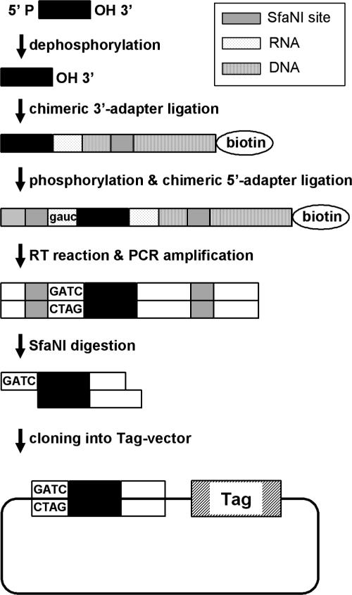
The method for cDNA library construction for the MPSS analysis was modified and is shown in Figure 1. Total RNA was isolated from BALB/c whole embryos (E9.5, E10.5 and E11.5) using Trizol Reagent (Invitrogen). Of the purified 21–27 nt RNA fraction 20 ng from each sample was treated with bacterial alkaline phosphatase (Takara Bio Inc.), followed by ligation with phosphorylated RNA–DNA chimeric 3′-adaptor (5′-cagcagGAATGCTCAATGATGCTGACGGCTCCCTATAGTGAGTCGTATTA-3′, RNA is shown in lowercase). The 3′ end of the adaptor was biotinylated to prevent self-ligation. Ligation was carried out using T4 RNA ligase (Takara Bio Inc.) at 15°C for 15 h. RNA fraction with attached adapters was purified using PAGE gel, phosphorylated by T4 polynucleotide kinase (Takara Bio Inc.) and followed by second round of ligation with the DNA–RNA chimeric 5′-adaptor containing the GAUC site (5′-CCATGTTCGCATCGGCaggauc-3′, RNA is shown in lowercase). The ligated product was converted to cDNA by reverse transcriptase (M-MLV RTase, Takara Bio Inc.) with the following primer, 5′-TAATACGACTCACTATAGGG-3′. The cDNA was amplified by 12 cycles of PCR using Pyrobest DNA Polymerase (Takara Bio Inc.) and PCR primers (5′-CCATGTTCGCATCGGCAGGATC-3′, 5′-AGCCGTCAGCATCATTGAGCAT-3′) in the presence of 5-methylated-dCTP, dATP, dGTP, dTTP mixture. PCR products were purified, digested by SfaNI (NEB) and cloned into the Tag vector pMBS I (Solexa), linearized with BamHI and BbsI.
Figure 1.
Construction of a small RNA-derived cDNA library. Small RNAs were dephosphorylated, followed by ligation with a phosphorylated RNA–DNA chimeric 3′-adaptor (3′ end of the adaptor was biotinylated to prevent self-ligation.) RNA fraction with the attached adapter was purified using PAGE gel, phosphorylated and followed by a second round of T4 RNA ligation with a DNA–RNA chimeric 5′-adaptor containing a GAUC site. The ligated product was converted to cDNA by reverse transcriptase. The cDNA was amplified by PCR with primers having a SfaNI site. PCR products were purified, digested by SfaNI and cloned into the BamHI–BbsI site of Tag vector pMBS I, which contains distinct 32mer oligonucleotide tags along with the cloning site.
MPSS analysis
MPSS analysis was carried out at Takara Bio Inc. (Otsu, Japan). Reagents and procedures used for MPSS analysis have been described (14). We eliminated the use of a cell sorter for the micro-beads selection step as follows: PCR was performed with the cDNA library using the mixture of biotin-labeled and FAM-labeled-forward primers and non-labeled reverse primers. Samples were then treated with T4 DNA ligase, and incubated with streptavidin-conjugated magnetic beads (Dynal) to capture DNA bound micro-beads. The DNA bound micro-beads were released from magnetic beads by digestion with MboI (Takara Bio Inc.). Samples were divided into two portions and two double-stranded adaptors (two-stepper adaptor and four-stepper adaptor) were separately ligated to the beads. About 1 million of each adapter-ligated micro-beads were loaded into separate flow cells and set on a sequencing instrument GenIII (Solexa). MPSS sequencing determined four bases by hybridization of labeled linker-probes (decoder probes) after ligation of double-stranded adapter mixture (encoded adaptor). These bases were removed by digestion with BbvI and the process was repeated to determine the next set of four bases. By extending each sequencing reaction, we were able to obtain 22 base sequences, which did not include the GATC site of the 5′ primer.
Identifying miRNA candidates
More than 500 000 of 22 base sequences or ‘signatures’ were obtained for each library after removing ambiguities arising from low-quality sequences. Distinct signatures were summed to obtain an abundance value for each signature sequence, normalized in ‘transcripts per million (TPM)’ for each library. A significance filter was applied to obtain more reliable signatures, such that a given signature is found in any library at ≥8 TPM. MiRNA candidates were then selected by analyzing each signature. A homology search was carried out for these candidates using the mouse genome database (May 2004 Assembly) (http://genome.ucsc.edu). From this analysis, repeat sequences were subtracted by RepeatMasker, using the NCBI BLAST program, and the signatures that were 100% homologous with the database sequence were extracted (Matched Signatures). A total of 8026 types of Matched Signatures were analyzed for homology using the following databases, the European ribosomal RNA database (http://www.psb.ugent.be/rRNA/), the Genomic tRNA database (http://lowelab.ucsc.edu/GtRNAdb/) and Non-coding RNA database (http://ncrna.bioinformatics.com.au/ and http://www.bioinfo.org.cn/NONCODE/index.htm). These analyses helped to remove rRNA, tRNA and sn/snoRNA from the sequences obtained. After these filtering, we obtained 3374 types of signatures (total 852 686 TPM from all three libraries) as miRNA pre-candidates. At this stage, the abundance value for each signature sequence was normalized to ‘transcripts per quarter million (TPQ)’. To identify whether these mature miRNA pre-candidates are mature miRNA candidates, we created potential miRNA precursors by sliding a 110 nt window containing mature miRNA pre-candidate sequence throughout the mouse genome (mouse genome database May 2004 Assembly) and folding the window with the program RNAfold14 of Vienna RNA package. Three criteria were applied to each potential miRNA precursor for prediction of miRNA; (i) hairpin precursor structure that contained 22 nt mature miRNA sequence in one arm and folded with the lowest free energy (predicted using RNAfold14); (ii) at least 16 nt matches between 22 nt of the mature miRNA and the other arm of the hairpin structure and (iii) absence of large internal loops or bulges. MiRNAs selected according to the above mentioned criteria were grouped. Finally, 1480 types of signatures (total 734 075 TPM) were selected by these three criteria mentioned above. These were grouped into 390 sequence signatures owing to the sequence redundancy. These 390 signatures were subjected to homology search. The other 1894 types of signatures (total 118 611 TPM), which did not meet the three criteria were grouped into 1439 sequence signatures.
Northern blotting and RT–PCR
Total RNA (30 µg) prepared from BALB/c whole embryos was separated on denaturing PAGE gels and transferred to Nytran SuPerCharge membranes (Schleicher & Schuell) followed by ultraviolet cross-linking and baking at 80°C for 1 h. Oligonucleotides complementary to the mature miRNA were labeled with [γ-32P]ATP using Megalabel kit (Takara Bio Inc.). The membranes were pre-hybridized in DIG Easy Hyb solution (Roche) at 25°C for 1 h, hybridized with labeled probe in DIG Easy Hyb at room temperature overnight and washed with 2× SSC solution containing 0.1% SDS prior to exposure to X-ray film. Oligonucleotide sequences are listed in Table S5 (Supplementary Data). DNase I treated total RNAs were used for the first-strand cDNA synthesis with MMLV-RTase (Takara Bio Inc.), PCR were carried out with Ex Taq Hot Start Ver (Takara Bio Inc.).
Determination of novel miRNA conservation
Novel mature miRNAs, which have no homology in mouse miRBase, were searched for homology in miRBase of other organisms, and 23 miRNAs were conserved in human, rat, chicken or zebrafish miRBase. Homology search was carried out for novel mature miRNAs using human genome database (data assembled in May 2004, UCSC) and rat genome database (assembled in June 2003, UCSC). From this analysis, repeat sequences were subtracted by RepeatMasker using the NCBI BLAST. A total of 16 miRNAs, 18 miRNAs and 139 miRNAs were homologous (>18/22 nt hit) with only human, only rat and both human and rat, respectively. To identify whether these 174 sequences of 22 nt are part of the miRNA precursor in each genome, potential miRNA precursors were made and analyzed using the three criteria mentioned in ‘Identifying miRNA candidates’. Finally, we obtained 119 mature miRNAs (128 precursors) conserved in human and rat genomes. The 23 miRNAs mentioned above are included in these 119 miRNAs. Thus, 23 miRNAs are conserved in miRBase of other organisms and 96 miRNAs (105 precursors) are conserved in human and rat but are not described in the miRBase of human and rat.
RESULTS AND DISCUSSION
miRNA expression profiling with MPSS
The MPSS technology is based on cloning cDNA library on micro-beads and the acquisition of 17 base sequences (signatures) from these cDNAs (14). cDNA fragments 20–21 bases in length are cloned into a vector for sequencing according to the original cloning method for analysis of mRNA expression levels. In this study, in order to carry out MPSS analysis of the expression level of small RNA species from whole mouse embryos at E9.5, E10.5 and E11.5, we modified the cloning method to make small RNA-derived micro-beads libraries. Our modified sequencing method enables us to sequence up to 22 bases. We obtained a total of 2 590 717 TPM signatures or 20 014 unique signatures after significance filtering from the three libraries (Table 1). After removing rRNA, tRNA and sn/snoRNA, we obtained 3374 distinct signatures as miRNA pre-candidates. A total of 390 distinct signatures were identified as mature miRNAs and 517 were identified as their precursors as some signatures had multiple precursors (Table S2, Supplementary Data). Remaining 1439 types of signatures did not meet even one of the three criteria (Table S5, Supplementary Data). The top 20 signatures ranked by abundance are known miRNAs (Table S1, Supplementary Data) and out of these 19 hairpin structures are same as those for registered ones, confirming that our method and adopted criteria for searching miRNAs are correct. To identify the location of the precursors on the mouse genome, we used two types of mRNA databases, one is a pre-existing curated RefSeq (accession prefix NM) database and the other is GenBank mRNAs database. They were intergenic (81.5%), exonic (1.5%) or intronic (17.0%) in case of RefSeq, and intergenic (43.3%), exonic (13.5%) or intronic (43.1%) in case of GenBank mRNAs. We found 195 mature miRNAs previously registered in the mouse miRBase release 7.0, covering 80% of all known mouse miRNAs (Table S2, Supplementary Data). Other 195 miRNAs (264 precursors) have not been previously reported. Among these, 86 miRNAs (103 precursors) are found on the opposite arms of known mouse miRNA precursors, 23 miRNAs (23 precursors) are conserved in other organisms' mature miRNAs registered in the miRBase and 96 miRNAs (105 precursors) are conserved in human and/or rat whole genome, but have not been registered in the miRBase. The remaining 61 miRNAs (111 precursors) are novel and display no homology to any miRNA in the miRBase (Release 7.0). A total of 71 miRNAs (78 precursors) are overlapping, such as miRNA007, which was found on the opposite arm of a known mouse miRNA precursor (mmu-mir-26b) and is also conserved in human. Among the new 195 miRNAs (264 precursors), 76 miRNAs (136 precursors) may be mouse-specific, which were not homologous to miRNAs found in human and rat genomes. A total of 86 miRNAs (147 precursors) may be rodent-specific, which were found only in mouse and rat genomes, but not in human genome. Remaining 109 miRNAs (117 precursors), found in human genome were conserved in mammals.
Table 1.
Composition of small RNA populations cloned from mouse embryo
| Class | distinct signature | TPMa | |||||
|---|---|---|---|---|---|---|---|
| E9.5 | E10.5 | E11.5 | |||||
| Genome | 8026 | 557 735 | 65.8% | 609 573 | 70.4% | 589 808 | 67.2% |
| RNAb | 4652 | 336 011 | 39.7% | 238 675 | 27.6% | 329 744 | 37.6% |
| rRNA | 4393 | 329 338 | 38.9% | 234 620 | 27.1% | 326 604 | 37.2% |
| tRNA | 122 | 2257 | 0.3% | 1784 | 0.2% | 1927 | 0.2% |
| snoRNA | 94 | 1302 | 0.2% | 934 | 0.1% | 368 | 0.0% |
| snRNA | 44 | 3142 | 0.4% | 1337 | 0.2% | 896 | 0.1% |
| MiRNA pre-candidates | 3374 | 221 724 | 26.2% | 370 898 | 42.8% | 260 064 | 29.6% |
| Satisfying the criteria | 1480 | 173 576 | 20.5% | 335 419 | 38.7% | 225 080 | 25.6% |
| Grouped by sequence | 390 | ||||||
| Not satisfying the criteria | 1894 | 48 148 | 5.7% | 35 479 | 4.1% | 34 984 | 4.0% |
| Grouped by sequence | 1439 | ||||||
| No hit in genome | 11 988 | 289 620 | 34.2% | 256 062 | 29.6% | 287 919 | 32.8% |
| Total | 20 014 | 847 355 | 100.0% | 865 635 | 100.0% | 877 727 | 100.0% |
aSummed TPM after significance filtering to have more reliable signatures, which determines if a given signature is found in any library at more than 7 TPM.
bSummed number of signature which are homologous with rRNA, tRNA, snoRNA and snRNA by BLAST NCBI (more than 90%). One RNA has homology with both rRNA and tRNA.
Another class of small RNAs, siRNAs, which are produced from long hairpin-less precursor dsRNA by Dicer (15,16) exist in significant number in plants, worm, fly and fission yeast. In these organisms, siRNAs can silence repetitive element expression, control genomic recombination, or promote transcriptional silencing through histone and/or DNA modification (17,18). Although RNAi is widely used as a tool for gene knockdown and regulation of gene expression in mammalian systems (19,20), there is no direct evidence for similar in vivo functions of endogenous siRNA in animals. Our raw data analysis would have shown presence of siRNAs in our mouse samples, by detecting double-stranded small RNAs. However, only two small RNAs having double-stranded structures were detected in our analysis; one from micro satellite-like sequences (CT repeat) and the other from tandem repeat sequences on the mouse genome (not listed in the RepeatMasker). Therefore, it appears that the mouse embryo transcriptome obtained using MPSS method described here, does not contain siRNAs.
Hierarchical clustering of miRNA expression levels
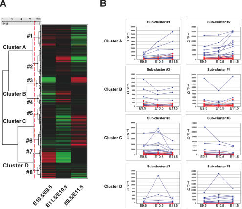
A hierarchical clustering analysis of all miRNAs was carried out by converting the ratio for the TPQ of E9.5 to E10.5, E10.5 to E11.5 and E11.5 to E9.5 to log2, which was then clustered using SpotFire (Figure 2). If TPQ was 0, it was converted to ‘1’. The following four clusters (A–D) and eight sub-clusters (#1–8) were identified; Cluster A, up-regulated at E11.5 (sub-cluster #1 and #2); Cluster B, down-regulated at E10.5 (#3 and #4); Cluster C, down-regulated at E11.5 (#5 and #6) and Cluster D, up-regulated at E10.5 (cluster #7 and #8). miRNAs in sub-cluster #1 increased during the three embryonic stages (E11.5 > E10.5 > E9.5), these include some previously reported species of miRNA to be enriched in the development stage (mmu-mir-21, 22, 124a and 125b) (21) and others that are ubiquitously expressed or activated after birth (22). Expression of sub-cluster #7 members increased from E9.5 to E10.5, and decreased at E11.5 to the level of E9.5. The proportion of newly discovered miRNAs among members of this sub-cluster is highest (78% is novel) and these 28 new miRNAs from sub-cluster #7 are predominantly E10.5-stage specific (Table S2, Supplementary Data).
Figure 2.
Hierarchical clustering analysis of the expression of 390 miRNAs obtained from whole embryos of E9.5, E10.5 and E11.5. (A) Heatmap and dendrogram (a tree graph). The ratio for the TPQ of E9.5 to E10.5, E10.5 to E11.5 and E11.5 to E9.5 (log2) were clustered (unweigted average, correlation similarity). There are four Clusters (A, B, C and D) with two Sub-clusters each, if the cut-off line is set as the red dot-line in the dendrogram. (B) Expression pattern of each sub-cluster. TPQ of all the member in each Sub-cluster are blotted (blue squares; known miRNAs and red circles; novel miRNAs). miRNAs in the Cluster A mainly increase during the three embryonic stages (E9.5 < E10.5 < E11.5), and a ratio of Sub-cluster #1 is E10.5/E9.5>E11.5/E10.5 and that of #2 is E11.5/E10.5 > E10.5/E9.5. miRNAs in the Cluster B are down-regulated at E10.5 (E9.5 > E10.5 < E11.5), and ratio from E10.5 to E11.5 of Sub-cluster #4 is higher than that of Sub-cluster #3. MiRNAs in the Cluster C are down-regulated at E11.5 (E9.5, E10.5>E11.5), Sub-cluster #5 is E9.5 < E10.5 and Sub-cluster #6 is E9.5 > E10.5. miRNAs in the Cluster D are up-regulated at E10.5, and miRNAs of Sub-cluster #7 is decreased at E11.5 to the level of E9.5 but Sub-cluster #8 is E9.5 < E11.5. Many miRNAs (72%) in Sub-cluster #7 are not detectable at both E9.5 and E11.5.
Tissue specific expression of several miRNAs at certain stages of mammalian embryonic development has been reported (12,18,23–26). Interestingly, several stem–cell-specific miRNAs such as mmu-mir-290, 291-3p, 292-3p, 293, 294 and 295, were found, while mmu-mir-296, potentially specific to ES cells (18,25) was not detected. Mmu-mir-21 and 22 which are known as differentiation-state-related miRNAs (18), were detected and their highest expression was observed at E11.5. Mmu-mir-9 and 125b, which were shown to be brain-enriched with the highest level of expression in the forebrain of prenatal E21 followed by down-regulation after birth (21), were highly expressed at E10.5 and E11.5 respectively (Table S3, Supplementary Data). Mmu-miR-124a, previously reported to increase during the embryonic period (21), was also observed to increase in our samples (E11.5 > E10.5 > E9.5, 10-fold).
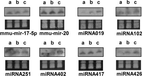
Using total RNAs isolated from BALB/c mouse whole embryos (E9.5, E10.5 and E11.5), northern blotting analysis was carried out for two known miRNAs, mmu-miR-17-5p and mmu-mir-20, and six new miRNAs (Figure 3). Overall, the result of northern blot analysis correlated with TPQ among the samples of each probe (Table 2).
Figure 3.
Northern blot analysis of different miRNAs in whole embryos. Northern blot analysis with P32-labeled antisense oligonucleotides complementary to the mature miRNA used as a probe. Prior to transfer, using ethidium bromide staining, it was verified that equal amount of total RNA is loaded in each lane, as shown in the lower panel for each hybridization figure. a, E9.5; b, E10.5; c, E11.5.
Table 2.
TPM results from MPSS data for each miRNA sample analyzed by northern blotting
| miRNA name | TPQ | ||
|---|---|---|---|
| E9.5 | E10.5 | E11.5 | |
| mmu-miR-17-5p | 51 487 | 47 339 | 34 526 |
| mmu-miR-20 | 1108 | 15 555 | 1103 |
| miRNA019 | 2861 | 4813 | 8834 |
| miRNA102 | 2831 | 3338 | 1084 |
| miRNA251 | 2181 | 2151 | 2264 |
| miRNA402 | 2394 | 2600 | 3819 |
| miRNA417 | 5995 | 3443 | 1126 |
| miRNA426 | 2979 | 6210 | 15 400 |
Clustering of miRNA genes in the genome
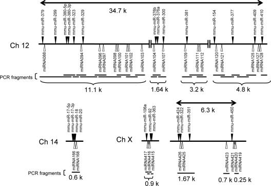
Comparative genomic analyses of miRNAs from different species suggest a variety of mechanisms regulating their expression (2). Some miRNA genes are clustered in the genome, implying that they are transcribed as a single transcriptional unit (27,28). We found several clusters of miRNA genes; e.g. there is a large cluster of miRNAs within a 35 kb sequence on chromosome 12 (104 457 239–104 491 958; UCSC genome database, Mouse May 2004 assembly). It contains 16 registered and 23 new miRNAs, all positioned in the same orientation and not separated by a transcription unit. To identify whether these miRNAs are processed from a single transcript, we selected four clusters and performed RT–PCR analysis, using RNA samples from E10.5 embryo, since all miRNAs to be examined were expressed at this particular stage (Figure 4 and Table S2, Supplementary Data). Data indicate that cluster I (from mmu-mir-17 to miRNA168) on chromosome 14 and II (from mmu-mir-106a to 363) on the X chromosome are transcribed as a single transcriptional unit. For cluster III (from mmu-mir-424 to 450) on the X chromosome, we found that at least mmu-mir-424–351 are co-transcribed as a single unit. For cluster IV (from mmu-mir-379 to 410) on chromosome 12, four transcripts containing mmu-mir-379-miRNA-l03, miRNA104-mmu-mir-300, miRNA-l09–112 and mmu-mir-154–409 were detected, though it is not clear whether all miRNAs in this cluster are transcribed from a single transcriptional unit. Interestingly, each polycistronic cluster contains miRNA members from different expression sub-clusters (Table S4, Supplementary Data), suggesting that the expression level of each miRNA is controlled by additional mechanisms. The major differences in expression levels of microRNAs found in the same cluster were previously reported by others (26,29). For example, MIR-23B is expressed in humans at much lower levels in all tissues, compared with MIR-27B, MIR-189 and MIR-24, all residing in the same cluster on chromosome 9 (29). It has been suggested that the existence of a variety of post-trancriptional regulation modes may be responsible for these differences. Additional studies will be needed to clear this issue.
Figure 4.
Genomic miRNA clusters and RT–PCR expression analysis. Relative locations of known (filled arrows) and novel (open arrows) miRNA cistrons in clusters on chromosomes 12, 14 and X are shown. To investigate whether miRNAs are expressed as a single transcriptional unit, RT–PCR analysis was conducted using RNA samples from E10.5 embryos with primers generating overlapping fragments. Amplified DNA fragments are shown as horizontal bars.
The miRNA profile obtained in this study provides insight into the embryonic stage-specific miRNA transcriptome, and will facilitate the identification of the primary target for each miRNA and thereby the pathways regulated by embryonic specific mRNAs. Recent reports suggest that certain miRNAs regulate a significant number of target mRNAs (2,30). Therefore, MPSS profiling of miRNAs will be useful for the investigation of the regulatory network of miRNA–mRNA interactions at the global genomic level. Recently, Green and her associates used MPSS to identify a large number of small RNAs (both siRNAs and miRNAs) in Arabidopsis thaliana, suggesting that there are extensive and complex regulatory roles of small RNAs in this plant (31).
SUPPLEMENTARY DATA
Supplementary data are available at NAR Online.
Supplementary Material
Acknowledgments
The authors thank Toshiaki Inoue for help with whole mouse embryos preparation, Tomonori Oura for help with sequence and structure data analysis, discussion and comments, Monica Roth, Sangita Phadtare and Raymond Habas for discussion and comments. The authors also thank members of the Dragon Genomics Center of Takara Bio Inc. for oligonucleotide synthesis and DNA sequencing of Tag vector libraries. Funding to pay the Open Access publication charges for this article was provided by Takara Bio Inc.
Conflict of interest statement. None declared.
REFERENCES
- 1.Coleman J., Green P.J., Inouye M. The use of RNAs complementary to specific mRNAs to regulate the expression of individual bacterial genes. Cell. 1984;37:429–436. doi: 10.1016/0092-8674(84)90373-8. [DOI] [PubMed] [Google Scholar]
- 2.Bartel D.P. MicroRNAs: genomics, biogenesis, mechanism, and function. Cell. 2004;116:281–297. doi: 10.1016/s0092-8674(04)00045-5. [DOI] [PubMed] [Google Scholar]
- 3.Ambros V. The functions of animal microRNAs. Nature. 2004;431:350–355. doi: 10.1038/nature02871. [DOI] [PubMed] [Google Scholar]
- 4.Meister G., Tuschl T. Mechanisms of gene silencing by double-stranded RNA. Nature. 2004;431:343–349. doi: 10.1038/nature02873. [DOI] [PubMed] [Google Scholar]
- 5.Hutvagner G., McLachlan J., Pasquinelli A.E., Balint E., Tuschl T., Zamore P.D. A cellular function for the RNA-interference enzyme Dicer in the maturation of the let-7 small temporal RNA. Science. 2001;293:834–838. doi: 10.1126/science.1062961. [DOI] [PubMed] [Google Scholar]
- 6.Ketting R.F., Fischer S.E., Bernstein E., Sijen T., Hannon G.J., Plasterk R.H. Dicer functions in RNA interference and in synthesis of small RNA involved in developmental timing in C. elegans. Genes Dev. 2001;15:2654–2659. doi: 10.1101/gad.927801. [DOI] [PMC free article] [PubMed] [Google Scholar]
- 7.Lim L.P., Glasner M.E., Yekta S., Burge C.B., Bartel D.P. Vertebrate microRNA genes. Science. 2003;299:1540. doi: 10.1126/science.1080372. [DOI] [PubMed] [Google Scholar]
- 8.Aravin A., Tuschl T. Identification and characterization of small RNAs involved in RNA silencing. FEBS Lett. 2005;579:5830–5840. doi: 10.1016/j.febslet.2005.08.009. [DOI] [PubMed] [Google Scholar]
- 9.Bentwich I., Avniel A., Karov Y., Aharonov R., Gilad S., Barad O., Barzilai A., Einat P., Einav U., Meiri E., et al. Identification of hundreds of conserved and nonconserved human microRNAs. Nature Genet. 2005;37:766–770. doi: 10.1038/ng1590. [DOI] [PubMed] [Google Scholar]
- 10.Baulcombe D. DNA events. An RNA microcosm. Science. 2002;297:2002–2003. doi: 10.1126/science.1077906. [DOI] [PubMed] [Google Scholar]
- 11.Wienholds E., Plasterk R.H. MicroRNA function in animal development. FEBS Lett. 2005;579:5911–5922. doi: 10.1016/j.febslet.2005.07.070. [DOI] [PubMed] [Google Scholar]
- 12.Mansfield J.H., Harfe B.D., Nissen R., Obenauer J., Srineel J., Chaudhuri A., Farzan-Kashani R., Zuker M., Pasquinelli A.E., Ruvkun G., et al. MicroRNA-responsive ‘sensor’ transgenes uncover Hox-like and other developmentally regulated patterns of vertebrate microRNA expression. Nature Genet. 2004;36:1079–1083. doi: 10.1038/ng1421. [DOI] [PubMed] [Google Scholar]
- 13.Wienholds E., Kloosterman W.P., Miska E., Alvarez-Saavedra E., Berezikov E., de Bruijn E., Horvitz H.R., Kauppinen S., Plasterk R.H. MicroRNA expression in zebrafish embryonic development. Science. 2005;309:310–311. doi: 10.1126/science.1114519. [DOI] [PubMed] [Google Scholar]
- 14.Brenner S., Johnson M., Bridgham J., Golda G., Lloyd D.H., Johnson D., Luo S., McCurdy S., Foy M., Ewan M., et al. Gene expression analysis by massively parallel signature sequencing (MPSS) on microbead arrays. Nat. Biotechnol. 2000;18:630–634. doi: 10.1038/76469. [DOI] [PubMed] [Google Scholar]
- 15.Storz G., Altuvia S., Wassarman K.M. An abundance of RNA regulators. Annu. Rev. Biochem. 2005;74:199–217. doi: 10.1146/annurev.biochem.74.082803.133136. [DOI] [PubMed] [Google Scholar]
- 16.Filipowicz W., Jaskiewicz L., Kolb F.A., Pillai R.S. Post-transcriptional gene silencing by siRNAs and miRNAs. Curr. Opin. Struct. Biol. 2005;15:331–341. doi: 10.1016/j.sbi.2005.05.006. [DOI] [PubMed] [Google Scholar]
- 17.Seitz H., Youngson N., Lin S.P., Dalbert S., Paulsen M., Bachellerie J.P., Ferguson-Smith A.C., Cavaille J. Imprinted microRNA genes transcribed antisense to a reciprocally imprinted retrotransposon-like gene. Nature Genet. 2003;34:261–262. doi: 10.1038/ng1171. [DOI] [PubMed] [Google Scholar]
- 18.Houbaviy H.B., Murray M.F., Sharp P.A. Embryonic stem cell-specific MicroRNAs. Dev. Cell. 2003;5:351–358. doi: 10.1016/s1534-5807(03)00227-2. [DOI] [PubMed] [Google Scholar]
- 19.Prawitt D., Brixel L., Spangenberg C., Eshkind L., Heck R., Oesch F., Zabel B., Bockamp E. RNAi knock-down mice: an emerging technology for post-genomic functional genetics. Cytogenet. Genome Res. 2004;105:412–421. doi: 10.1159/000078214. [DOI] [PubMed] [Google Scholar]
- 20.Bayne E.H., Allshire R.C. RNA-directed transcriptional gene silencing in mammals. Trends Genet. 2005;21:370–373. doi: 10.1016/j.tig.2005.05.007. [DOI] [PubMed] [Google Scholar]
- 21.Krichevsky A.M., King K.S., Donahue C.P., Khrapko K., Kosik K.S. A microRNA array reveals extensive regulation of microRNAs during brain development. RNA. 2003;9:1274–1281. doi: 10.1261/rna.5980303. [DOI] [PMC free article] [PubMed] [Google Scholar]
- 22.Alvarez-Garcia I., Miska E.A. MicroRNA functions in animal development and human disease. Development. 2005;132:4653–4662. doi: 10.1242/dev.02073. [DOI] [PubMed] [Google Scholar]
- 23.Mattick J.S., Makunin I.V. Small regulatory RNAs in mammals. Hum. Mol. Genet. 2005;14:R121–R132. doi: 10.1093/hmg/ddi101. [DOI] [PubMed] [Google Scholar]
- 24.Dostie J., Mourelatos Z., Yang M., Sharma A., Dreyfuss G. Numerous microRNPs in neuronal cells containing novel microRNAs. RNA. 2003;9:180–186. doi: 10.1261/rna.2141503. [DOI] [PMC free article] [PubMed] [Google Scholar]
- 25.Suh M.R., Lee Y., Kim J.Y., Kim S.K., Moon S.H., Lee J.Y., Cha K.Y., Chung H.M., Yoon H.S., Moon S.Y., et al. Human embryonic stem cells express a unique set of microRNAs. Dev. Biol. 2004;270:488–498. doi: 10.1016/j.ydbio.2004.02.019. [DOI] [PubMed] [Google Scholar]
- 26.Sempere L.F., Freemantle S., Pitha-Rowe I., Moss E., Dmitrovsky E., Ambros V. Expression profiling of mammalian microRNAs uncovers a subset of brain-expressed microRNAs with possible roles in murine and human neuronal differentiation. Genome Biol. 2004;5:R13. doi: 10.1186/gb-2004-5-3-r13. [DOI] [PMC free article] [PubMed] [Google Scholar]
- 27.Lagos-Quintana M., Rauhut R., Lendeckel W., Tuschl T. Identification of novel genes coding for small expressed RNAs. Science. 2001;294:853–858. doi: 10.1126/science.1064921. [DOI] [PubMed] [Google Scholar]
- 28.Lau N.C., Lim L.P., Weinstein E.G., Bartel D.P. An abundant class of tiny RNAs with probable regulatory roles in Caenorhabditis elegans. Science. 2001;294:858–862. doi: 10.1126/science.1065062. [DOI] [PubMed] [Google Scholar]
- 29.Barad O., Meiri E., Avniel A., Aharonov R., Barzilai A., Bentwich I., Einav U., Gilad S., Hurban P., Karov Y., et al. MicroRNA expression detected by oligonucleotide microarrays: system establishment and expression profiling in human tissues. Genome Res. 2004;14:2486–2494. doi: 10.1101/gr.2845604. [DOI] [PMC free article] [PubMed] [Google Scholar]
- 30.Lim L.P., Lau N.C., Garrett-Engele P., Grimson A., Schelter J.M., Castle J., Bartel D.P., Linsley P.S., Johnson J.M. Microarray analysis shows that some microRNAs downregulate large numbers of target mRNAs. Nature. 2005;433:769–773. doi: 10.1038/nature03315. [DOI] [PubMed] [Google Scholar]
- 31.Lu C., Tej S.S., Luo S., Haudenschild C.D., Meyers B.C., Green P.J. Elucidation of the small RNA component of the transcriptome. Science. 2005;309:1567–1569. doi: 10.1126/science.1114112. [DOI] [PubMed] [Google Scholar]
Associated Data
This section collects any data citations, data availability statements, or supplementary materials included in this article.


