

Abstract
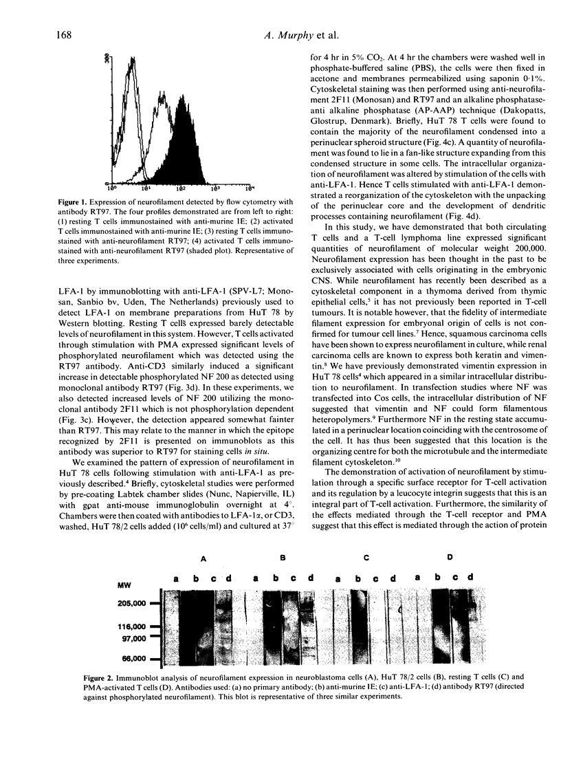
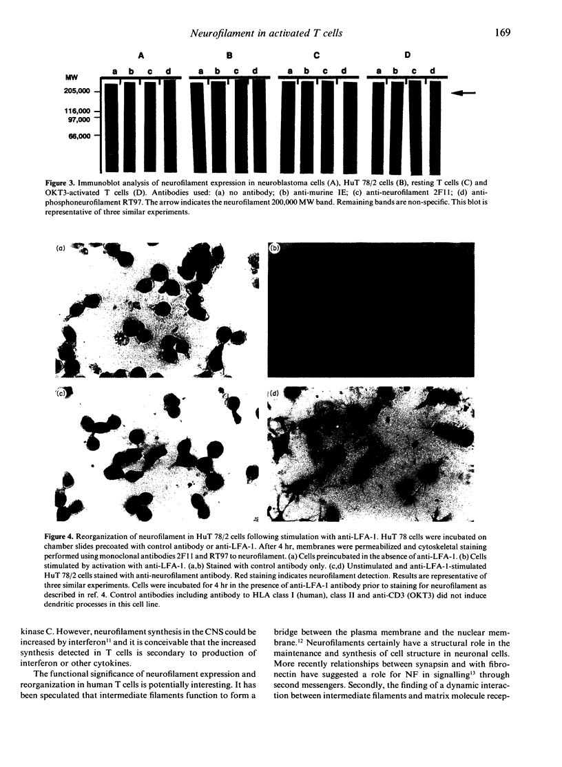
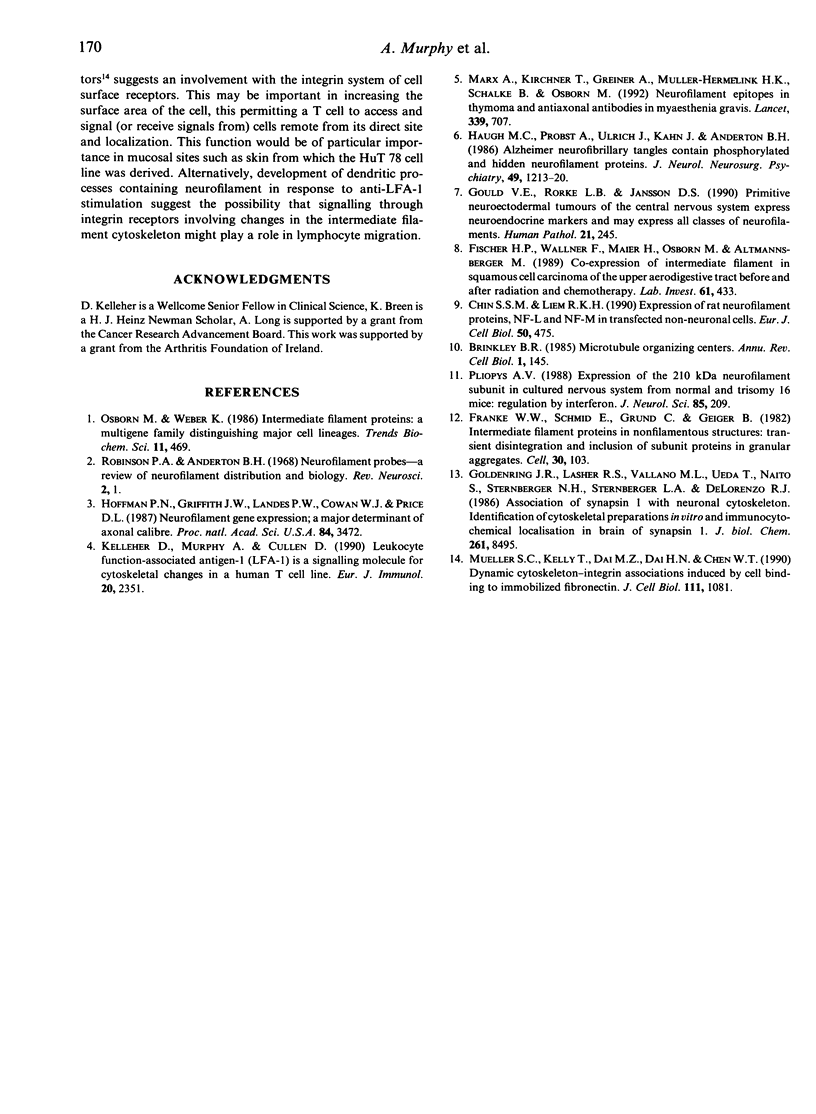
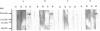
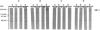
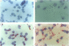
The expression of intermediate filaments in normal cells is mainly determined by their embryonal developmental origin. Flow cytometry using monoclonal antibody RT97 demonstrated that neurofilament was detectable in the human HuT 78 T-cell line and on resting T lymphocytes. Expression was greatly increased on lymphocytes activated for 3 days with phorbol ester. Western blotting confirmed the presence of the 200,000 MW form of neurofilament in T lymphocytes. Stimulation of peripheral blood T cells with phorbol myristate acetate (PMA) or with anti-CD3 monoclonal antibodies resulted in a marked increase in detection of phosphorylated neurofilament on Western blotting. Stimulation of HuT 78 cells with anti-LFA-1 resulted in redistribution of neurofilament from a perinuclear spheroid core into dendritic processes. These data indicate that T cells activated through the T-cell receptor associated complex express an intermediate filament usually associated with neurally derived cells. The finding that neurofilament expression and organization are regulated by T-cell surface molecules suggests a role for this intermediate filament in T-cell function.
Full text
PDF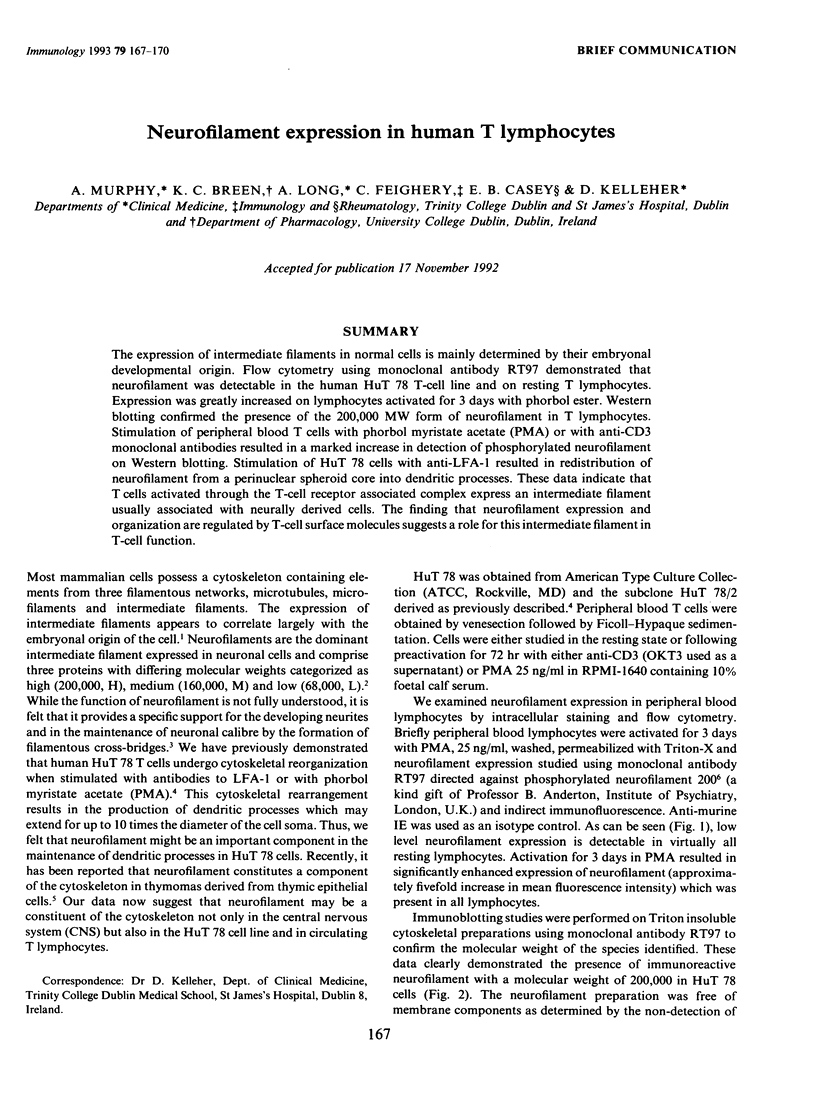
Images in this article
Selected References
These references are in PubMed. This may not be the complete list of references from this article.
- Brinkley B. R. Microtubule organizing centers. Annu Rev Cell Biol. 1985;1:145–172. doi: 10.1146/annurev.cb.01.110185.001045. [DOI] [PubMed] [Google Scholar]
- Chin S. S., Liem R. K. Expression of rat neurofilament proteins NF-L and NF-M in transfected non-neuronal cells. Eur J Cell Biol. 1989 Dec;50(2):475–490. [PubMed] [Google Scholar]
- Fischer H. P., Wallner F., Maier H., Weber K., Osborn M., Altmannsberger M. Coexpression of intermediate filaments in squamous cell carcinomas of upper aerodigestive tract before and after radiation and chemotherapy. Lab Invest. 1989 Oct;61(4):433–439. [PubMed] [Google Scholar]
- Franke W. W., Schmid E., Grund C., Geiger B. Intermediate filament proteins in nonfilamentous structures: transient disintegration and inclusion of subunit proteins in granular aggregates. Cell. 1982 Aug;30(1):103–113. doi: 10.1016/0092-8674(82)90016-2. [DOI] [PubMed] [Google Scholar]
- Furuhashi K., Hatano S. Control of actin filament length by phosphorylation of fragmin-actin complex. J Cell Biol. 1990 Sep;111(3):1081–1087. doi: 10.1083/jcb.111.3.1081. [DOI] [PMC free article] [PubMed] [Google Scholar]
- Goldenring J. R., Lasher R. S., Vallano M. L., Ueda T., Naito S., Sternberger N. H., Sternberger L. A., DeLorenzo R. J. Association of synapsin I with neuronal cytoskeleton. Identification in cytoskeletal preparations in vitro and immunocytochemical localization in brain of synapsin I. J Biol Chem. 1986 Jun 25;261(18):8495–8504. [PubMed] [Google Scholar]
- Gould V. E., Rorke L. B., Jansson D. S., Molenaar W. M., Trojanowski J. Q., Lee V. M., Packer R. J., Franke W. W. Primitive neuroectodermal tumors of the central nervous system express neuroendocrine markers and may express all classes of intermediate filaments. Hum Pathol. 1990 Mar;21(3):245–252. doi: 10.1016/0046-8177(90)90223-r. [DOI] [PubMed] [Google Scholar]
- Haugh M. C., Probst A., Ulrich J., Kahn J., Anderton B. H. Alzheimer neurofibrillary tangles contain phosphorylated and hidden neurofilament epitopes. J Neurol Neurosurg Psychiatry. 1986 Nov;49(11):1213–1220. doi: 10.1136/jnnp.49.11.1213. [DOI] [PMC free article] [PubMed] [Google Scholar]
- Hoffman P. N., Cleveland D. W., Griffin J. W., Landes P. W., Cowan N. J., Price D. L. Neurofilament gene expression: a major determinant of axonal caliber. Proc Natl Acad Sci U S A. 1987 May;84(10):3472–3476. doi: 10.1073/pnas.84.10.3472. [DOI] [PMC free article] [PubMed] [Google Scholar]
- Kelleher D., Murphy A., Cullen D. Leukocyte function-associated antigen 1 (LFA-1) is a signaling molecule for cytoskeletal changes in a human T cell line. Eur J Immunol. 1990 Oct;20(10):2351–2354. doi: 10.1002/eji.1830201028. [DOI] [PubMed] [Google Scholar]
- Marx A., Kirchner T., Greiner A., Müller-Hermelink H. K., Schalke B., Osborn M. Neurofilament epitopes in thymoma and antiaxonal autoantibodies in myasthenia gravis. Lancet. 1992 Mar 21;339(8795):707–708. doi: 10.1016/0140-6736(92)90601-x. [DOI] [PubMed] [Google Scholar]