Abstract
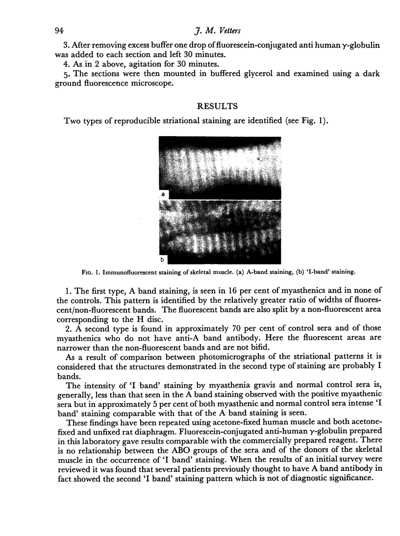
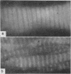
Using indirect immunofluorescent tests upon the sera of patients with myasthenia gravis and of normal subjects, two types of staining pattern are found. The first, A band staining, is seen only with myasthenic sera. The second, or `I band' pattern has not previously been described and is seen in an equal proportion of normal controls and of myasthenic patients. `I band' staining is a possible source of confusion in immunofluorescent tests for muscle antibody.
Full text
PDF


Images in this article
Selected References
These references are in PubMed. This may not be the complete list of references from this article.
- BEUTNER E. H., WITEBSKY E., RICKEN D., ADLER R. H. Studies on autoantibodies in myasthenia gravis. JAMA. 1962 Oct 6;182:46–58. [PubMed] [Google Scholar]
- DJANIAN A. Y., BEUTNER E. H., WITEBSKY E. TANNED-CELL HEMAGGLUTINATION TEST FOR DETECTION OF ANTIBODIES IN SERA OF PATIENTS WITH MYASTHENIA GRAVIS. J Lab Clin Med. 1964 Jan;63:60–70. [PubMed] [Google Scholar]
- NASTUK W. L., PLESCIA O. J., OSSERMAN K. E. Changes in serum complement activity in patients with myasthenia gravis. Proc Soc Exp Biol Med. 1960 Oct;105:177–184. doi: 10.3181/00379727-105-26050. [DOI] [PubMed] [Google Scholar]


