Abstract
We provide the first evidence for a link between polyamines and biofilm levels in Yersinia pestis, the causative agent of plague. Polyamine-deficient mutants of Y. pestis were generated with a single deletion in speA or speC and a double deletion mutant. The genes speA and speC code for the biosynthetic enzymes arginine decarboxylase and ornithine decarboxylase, respectively. The level of the polyamine putrescine compared to the parental speA+ speC+ strain (KIM6+) was depleted progressively, with the highest levels found in the Y. pestis ΔspeC mutant (55% reduction), followed by the ΔspeA mutant (95% reduction) and the ΔspeA ΔspeC mutant (>99% reduction). Spermidine, on the other hand, remained constant in the single mutants but was undetected in the double mutant. The growth rates of mutants with single deletions were not altered, while the ΔspeA ΔspeC mutant grew at 65% of the exponential growth rate of the speA+ speC+ strain. Biofilm levels were assayed by three independent measures: Congo red binding, crystal violet staining, and confocal laser scanning microscopy. The level of biofilm correlated to the level of putrescine as measured by high-performance liquid chromatography-mass spectrometry and as observed in a chemical complementation curve. Complementation of the ΔspeA ΔspeC mutant with speA showed nearly full recovery of biofilm to levels observed in the speA+ speC+ strain. Chemical complementation of the double mutant and recovery of the biofilm defect were only observed with the polyamine putrescine.
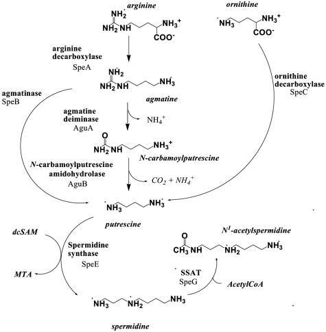
Polyamine biosynthesis in bacteria has two alternative pathways (Fig. 1): one pathway starts with the conversion of ornithine to putrescine by ornithine decarboxylase (ODC) (EC 4.1.1.17), and a second pathway involves the conversion of arginine to agmatine by arginine decarboxylase (ADC) (EC 4.1.1.19) (62). The bacterial ODC is regulated at the level of transcription by cyclic AMP (11, 41, 69), posttranslationally by nucleotides (45), and by a bacterial antizyme, AtoC, which binds to ODC, rendering it inactive (36). While Yersinia pestis ADC was enzymatically characterized previously by Balbo et al. (3), ADC function in bacteria is poorly understood. ADC is found in bacteria and plants but has no homologues in humans (8).
FIG. 1.
Putative polyamine pathway of Y. pestis. The proposed pathway is based on BLAST searches using functionally characterized protein sequences in Y. pestis, E. coli, or P. aeruginosa as queries. The enzyme designations are given next to each arrow. With the exception of agmatinase (SpeB), all of the other proteins indicated have been identified in Y. pestis KIM10+ and CO92. AcetylCoA, acetyl coenzyme A.
The polyamines putrescine and spermidine are involved in modulating the synthesis of DNA, RNA, and protein (25, 62, 70). Specific functions associated with the polyamine putrescine have been identified. For example, putrescine is a constituent of the outer membrane in some gram-negative bacteria (33, 67) and is involved in the activation of the expression of oxyR and katG in Escherichia coli (65) and in the repression of the speAB operon, thereby regulating its own production (41). Additionally, in E. coli, the levels of cellular putrescine are modulated by growth rate (66), and polyamines have recently been shown to protect E. coli from toxic effects of oxygen (6). In Pseudomonas aeruginosa, exogenous putrescine can increase transcription of several loci including the spuABCDEFGH operon (37), which is involved in polyamine uptake and utilization. Furthermore, putrescine has been shown to act as an extracellular signal required for swarming in Proteus mirabilis (59). The latter suggests a role for polyamines in bacterial differentiation and in signaling events that coordinate multicellular actions.
In this report, we investigate the importance of polyamines to biofilm formation in Yersinia pestis, the etiological agent of bubonic and pneumonic plague (51). Plague is a zoonotic disease, having a rodent-flea life cycle with humans as an accidental host. Transmission of Y. pestis from some fleas to mammals involves colonization and blockage of the proventricular valve, which separates the midgut from the esophagus (24, 27). This blockage depends upon a set of genes in Y. pestis, designated the hemin storage (hms) locus, that are involved in the production of an extracellular matrix or biofilm and causes the flea to regurgitate a bacterium-laced blood meal (9, 24, 34). The hmsHFRS and hmsT genes were first identified as essential for temperature-dependent hemin and Congo red (CR) binding by Y. pestis. Biofilm formation, as measured by CR binding, occurs at the lower temperatures of 26 to 34°C associated with fleas but not at 37°C or mammalian temperature (28, 49, 52). The term biofilm refers to surface-attached bacteria that have formed a protective matrix through intercellular communication (22). The initial steps in biofilm formation are regulated by signals that are specific to the bacterial species. For example, in urinary tract infections, fatty acids have been implicated in P. mirabilis biofilm formation (35). In contrast, later stages involving biofilm maturation, such as depth and architecture, appear to be regulated by bacterial signals referred to as quorum sensing (16, 17).
We have utilized the genomic sequence of Y. pestis (12) to identify putative polyamine biosynthetic enzymes (Fig. 1). We characterized the enzymatic activity and phenotype of Y. pestis mutants with deletions in two key polyamine biosynthetic genes, speA and speC. We show for the first time a link between levels of polyamines and the capacity to form a plague biofilm, suggesting a new function for polyamines.
MATERIALS AND METHODS
Bacterial strains and cultivation.
All bacterial strains used in this study are listed in Table 1. E. coli cells were grown in Luria broth (LB). Where appropriate, ampicillin (Ap) (100 μg/ml), kanamycin (Km) (40 μg/ml), or chloramphenicol (Cm) (15 μg/ml) was added to cultures. Y. pestis cells were streaked onto CR (60) plates from fresh cultures made from buffered glycerol stocks stored at −80°C and incubated at 26 to 30°C for 48 h. For most experiments, individual colonies picked from LB, CR, or PMH2 agar plates were inoculated into a fresh chemically defined medium, PMH2 (19), that had not been treated with Chelex 100 resin to remove iron or into heart infusion broth (HIB). Cell growth was monitored with a Cary 50 UV or a Spectronic Genesys5 spectrophotometer at 620 nm.
TABLE 1.
Bacterial strains, plasmids and primers useda
| Strain, plasmid, or primer | Relevant characteristics or sequence | Source and/or reference or use |
|---|---|---|
| Strains | ||
| Y. pestis | ||
| KIM6(pKD46)+ | Pgm+ Lcr− Pla+ SpeA+ SpeC+ Apr pKD46 | 14; this study |
| KIM6-2110(pKD46)+ | Pgm+ Lcr− Pla+ SpeA+ (ΔspeC::kan2110) Apr pKD46 | This study |
| KIM6-2111(pKD46)+ | Pgm+ Lcr− Pla+ SpeC+ (ΔspeA::cam2111) Apr pKD46 | This study |
| KIM6-2112(pKD46)+ | Pgm+ Lcr− Pla+ (ΔspeA::cam2111) (ΔspeC::kan2110) pKD46 | This study |
| E. coli | ||
| BL21(DE3) | F−ompT hsdSB(rB− mB−) gal dcm (DE3) | Novagen |
| Top10F′ | F′ [lacIq Tn10 (Tetr)] mcrA Δ(mrr-hsdRMS-mcrBC) φ80lacZΔM15 ΔlacX74 recA1 araD139 Δ(ara-leu)7697 galU galK rpsL (Strr) endA1 nupG | Invitrogen |
| Plasmids | ||
| pET-28a-c | 5.4 kb, Kmr, six-histidine-tagged fusion protein plasmid | Novagen |
| pET-28b-speA | 7.4 kb, Kmr, His-ADC expression vector, 2-kb speA PCR product ligated into EcoRI-XhoI sites of pET-28b | 3 |
| pET-28b-speC | 7.6 kb, Kmr, His-ODC expression vector, 2.2-kb speC PCR product ligated into BamHI-XhoI sites of pET-28b | This study |
| pBAD/Hisab | 4.1 kb, Apr, six-histidine-tagged fusion protein plasmid | Invitrogen |
| pBAD/HisbADC | 6.1 kb, Apr, His-ADC expression vector, 2-kb speA PCR product ligated into SacI-HindIII sites of pBAD/Hisb | This study |
| pKD3 | 2.8 kb, Cmr, template plasmid | 10 |
| pKD4 | 3.3 kb, Kmr, template plasmid | 10 |
| pKD46 | 6.3 kb, Apr, Red recombinase expression plasmid | 10 |
| Primers | ||
| ODC_KOf | 5′-ATGACACTATTAAAAATAGCAACAAGCGCCCGCCTGGCTTCTT ATGTAGAGTGTAGGCTGGAGCTGCTTC-3′ | For insertion of Km or Cm cassette into speC |
| ODC_KOr | 5′-TTGCGTAATCACATAACCAAAGATACGCCGCCAAGACACAT CACCTTTTTCATATGAATATCCTCCTTAGT-3′ | For insertion of Km or Cm cassette into speC |
| ADC-KO2f | 5′-TTCTTTACGCTCCATGCAGGAGGTAGCCATGAATGATCGAAAT GCCAGCAGTGTAGGCTGGAGCTGCTTC-3′ | For insertion of Km or Cm cassette into speA |
| ADC-KO2r | 5′-CAGGAACTGCGCCTGCAATTCGGTATCCAGATCGGTTTCTTTT ACCTGACCATATGAATATCCTCCTTAGT-3′ | For insertion of Km or Cm cassette into speA |
| YpADCecofb | TCCGAATTCGATGTCTGATGATAACTTGATTAGCCGTCCG | Cloning into expression vector for speA |
| YpADCXhor | TAACTCGAGTTCGTCTTCAAGATATGTGTAGCCG | Cloning into expression vector for speA |
| ODCBamf | 5′-GGATCCGATGACACTATTAAAAATAGCAACAAGCGCCCGCC-3′ | Cloning into expression vector for speC |
| ODCXhor | 5′-CTCGAGTTGCGTAATCACATAACCAAAGATACGCCGCC-3′ | Cloning into expression vector for speC |
| YPadcSacfb | 5′-TCCGAGCTCGATGTCTGATGATAACTTGATTAGCCGTCCG-3′ | Clone speA for insertion into the pBAD/Hisb expression vector for complementation |
| YPadcHindr | 5′-TAAAAGCTTTTCGTCTTCAAGATATGTGTAGCCG-3′ | Clone speA for insertion into the pBAD/Hisb expression vector for complementation |
| YPADC+50f | 5′-CGTCAGTATCGCAATAGAGGCG-3′ | For sequencing from 50 bp outside speA |
| YPADC+50r | 5′-CAAACCTGAGTTTGCCC-3′ | For sequencing from 50 bp outside speA |
| YPODC+50r | 5′-GGCAGCCTCATACCTGACCG-3′ | For sequencing from 50 bp outside speC |
| YPODC+50f | 5′-GGCCTCGGCTGATGGAGCGGAG-3′ | For sequencing from 50 bp outside speC |
All Y. pestis strains are avirulent due to the lack of the low-calcium response plasmid pCD1. Strains with a plus sign possess an intact 102-kb pgm locus containing the genes for hemin storage (hms) and yersiniabactin (Ybt) iron transport system. Abbreviations: Apr, Cmr, and Kmr, resistance to ampicillin, chloramphenicol, and kanamycin, respectively.
Plasmids and recombinant DNA techniques.
Plasmids were purified from cultures grown overnight by alkaline lysis (4). Standard cloning and recombinant DNA methods (55) were used to construct the plasmids listed in Table 1. Plasmids were introduced into chemically competent E. coli Top10F′ cells (Invitrogen), a standard cloning strain. Y. pestis cells were transformed by electroporation as previously described (13). Plasmid DNA was sequenced at the Davis Sequencing Center when appropriate. Synthetic oligonucleotide primers were purchased from Integrated DNA Technologies. Sequences of primers used in this study are listed in Table 1.
Identification of polyamine biosynthetic genes in Y. pestis and comparison to E. coli and P. aeruginosa.
We used BLAST (1) and the query sequence ODC (gi:13432139; NCBI) from E. coli and AguA, AguB, and agmatinase sequences from P. aeruginosa (43) to identify homologous Y. pestis sequences. The BLOSUM62 matrix was used for scoring; 0.01 or 0.001 was used as E-value thresholds for inclusion of sequences in the profile calculation. The sequences were then aligned using VectorNTI 9.0 (InforMax).
Construction of polyamine biosynthetic deletion mutants.
Y. pestis mutants were constructed using the Red recombinase system described previously by Datsenko and Wanner (10). A PCR product containing speC sequences flanking a Cm resistance gene cassette was amplified using primers ODC KO1f and ODC KO1r (Table 1) with pKD3 as a template. Primers ADC KO2f and ADC KO2r were used to amplify the Cm resistance gene cassette from pKD3 to generate the speA mutant. Approximately 0.5 to 2 μg of purified PCR product was electroporated into KIM6(pKD46)+ cells (referred to as speA+ speC+ in the following text and figures). Electrocompetent cells were made from cultures grown at 30°C in LB to an optical density (OD) at 620 nm of ∼0.6 and then incubated for 1.5 h with 0.2% arabinose to induce the Red recombinase encoded on pKD46. The resulting mutants, KIM6-2110(pKD46)+ (ΔspeC::cam2110) and KIM6-2111(pKD46)+ (ΔspeA::cam2111), were selected for growth on LB agar plates containing the appropriate antibiotic. Gene replacements were confirmed by PCR using the primers indicated in Table 1. To generate the double deletion mutant, we started with KIM6-2111(pKD46)+ and followed the procedure for generation of the speC mutation in this strain. The resultant mutant, KIM6-2112(pKD46)+ (ΔspeA::cam2111 ΔspeC::kan2110), was selected for growth on LB agar supplemented with Cm and Km.
ADC and ODC cloning, expression purification, and radiometric assay.
Cloning of speA has been previously described (pET-28b-speA) (3) (Table 1). The expression vector for ODC (pET-28b-speC) (Table 1) was constructed by ligating the 2.2-kb speC PCR product into the BamHI-XhoI sites of pET-28b. The PCR product was amplified from KIM6+ genomic DNA. The nucleotide sequence of the PCR product was verified by DNA sequencing. Expression vectors were transformed into BL21(DE3) competent cells (Novagen). Expression of ADC and ODC was performed according to the procedure described previously by Balbo et al. (3). Purification of the recombinant ADC and ODC was achieved by absorption to nickel-nitrilotriacetic acid resin (QIAGEN) according to the manufacturer's instructions. The quality of the purified polypeptides was assessed by 12% sodium dodecyl sulfate-polyacrylamide gel electrophoresis and Coomassie blue staining (44).
For radiometric assays, the Y. pestis speA+ speC+ strain and its derivatives were grown for ∼8 h in PMH2 to an OD at 620 nm of 1 to 1.5, pelleted, and stored at −20°C. For enzyme assays, 100 mg of wet cell weight was resuspended in 1 ml of activity buffer (50 mM Tris-HCl, pH 8.35, 0.1 mM pyridoxal-5′-phosphate [PLP], 0.1 mM EDTA, 1 mM dithiothreitol, 1 mg/ml lysozyme [5 mM MgCl2 was added to arginine decarboxylase activity assays only]) and disrupted by vortexing with 0.1-mm Zirconia/Silica beads (Biospec) at 1-min intervals for 3 min. Total protein was determined using the clarified lysate as described previously by Gallaguer (18). Reactions were performed with minor modifications as described previously by Bachrach and Ben-Joseph (2) and Coleman et al. (8). Briefly, reaction mixtures consisted of 300 μl of activity buffer, 100 μl of 200 mM substrate (containing 8 nCi of carboxy-14C-labeled l-arginine or l-ornithine), and 100 μl of 100 mg/ml cell lysate to start the reaction. The reaction mixtures were incubated at 37°C with shaking in 10-ml sidearm Erlenmeyer flasks with stoppers and polypropylene center wells (Kontes). Two hundred microliters of 5 M H2SO4 was used to stop the reaction; the released radiolabeled CO2 was trapped in 200 μl of fresh 1 M NaOH placed into the center well. The well was cut, placed into 10 ml of scintillation fluid (Fisher Scintisafe gel), and vortexed with 800 μl of H2O to solubilize samples. The activities of purified ADC and ODC were also measured using the same radiometric assay conditions.
CR binding assays.
We modified an assay described previously by Hartzell et al. to assess CR binding that measures the extent of exopolysaccharides (EPSs) produced (9, 23, 32). Y. pestis cells were grown overnight (∼16 h) in HIB at 26°C. Cells were pelleted and resuspended in HIB-CR medium (1% [wt/vol] HIB containing 0.2% galactose and 30 μg CR/ml) such that all cultures had an equivalent wet weight of cells per microliter of medium. A ∼200-μg aliquot of each culture was incubated for 3 h on a rocking platform at room temperature. The amount of CR bound by the cells was determined by measuring the OD of cell supernatants at 500 nm with a Spectronic Genesys5 spectrophotometer and subtracting this value from the reading obtained with a medium-only control.
CV staining.
Cells attached to glass test tubes were detected with crystal violet staining essentially as described previously by O'Toole et al. (46). Briefly, cells grown overnight on PMH2 slants were diluted into fresh PMH2 to an OD at 620 nm of 0.1 and grown for 16 to 18 h with shaking at room temperature. The cultures were incubated with 0.1% crystal violet (CV) for 15 to 20 min before draining the liquid and washing the test tubes three times with water. CV retained by attached bacterial cells was solubilized with a mixture of 80% ethanol and 20% acetone. The amount of dye bound, representing the mass of attached bacterial cells, was monitored by measuring the absorbance at 570 nm on a Cary 50 UV spectrophotometer.
Confocal laser scanning microscopy.
As previously described (32), Y. pestis strains containing pGFPmut3.1 grown overnight on tryptose blood agar base (Difco Laboratories) slants were used to inoculate PMH2 to an OD at 620 nm of 0.1. Five milliliters of each culture was placed into a 50-ml conical tube containing a glass coverslip and incubated overnight at 26°C in a shaking water bath. The growth rates and final yields of Hms+ and Hms− cells are equivalent in PMH2. The coverslips were rinsed well with distilled water and examined with a Leica TCS laser scanning confocal microscope system. The samples were viewed with the 63X1.2 HCX PL APO objective on a Leica DM RXE microscope equipped with an argon laser emitting at 488 nm.
Quantification of polyamines using HPLC-MS.
The procedure used here was described previously by Morgan (42) and by Tabor et al. (64). Briefly, cells were grown from an OD of 0.1 to an OD of ∼1 at 30°C. Cells were pelleted, and the wet cell weight was recorded. Cell pellets were resuspended in H2O at 10 μl/mg of wet cells and lysed using 0.1-mm Zirconia/Silica beads (Biospec). Trichloroacetic acid (100%) and 2 mM cadaverine (internal standard) were each added to the lysate at 5 μl per 100 μl of lysate. The lysate was centrifuged at 20,000 × g. One hundred microliters of the clarified lysate was combined with 400 μl of H2O and then benzoylated by adding 2 ml of 2 M NaOH and 20 μl of a 50:50 benzoyl chloride-methanol solution. The mixture was vortexed for 1 min and then incubated for 1 h at room temperature with continuous shaking. Benzoylated polyamines were extracted by vortexing with 1 ml of chloroform. The chloroform layer was extracted, washed with 1 ml of H2O, reextracted, dried, and then dissolved in 100 μl mobile phase (60% methanol, 40% water). Known amounts of spermine, spermidine, and putrescine were prepared in duplicate to produce standard curves. We used a Spherisorb ODS-2 (Waters) column fitted with a 50- by 4.6-mm guard column with a flow rate of 0.4 ml/min, and UV absorption was measured in the range of 200 to 500 nm. Polyamines were eluted using a gradient from 60 to 100% methanol. Mass spectroscopy was used to verify the identity of each peak observed in high-performance liquid chromatography (HPLC) fractions. HPLC-atmospheric pressure chemical ionization-mass spectrometry (MS) analysis was performed on a Waters Alliance 2695 instrument coupled with a Waters 2996 photodiode array detector and a Waters/Micromass ZQ mass spectrometer.
Y. pestis ΔspeA ΔspeC mutant genetic complementation.
The double deletion mutant was complemented using full-length speA amplified from pET-28b-speA with the primer pairs indicated in Table 1, cloned into the pBAD/Hisb (Invitrogen) expression vector using SacI and HindIII restriction sites, and sequenced. Y. pestis KIM6-2112+ (ΔspeA ΔspeC) cells were cured of pKD46 by growth in the absence of Ap. KIM6-2112+ cells were then made electrocompetent and transformed with pBAD/Hisb-ADC plasmid for complementation studies.
Chemical complementation of the double ΔspeA ΔspeC mutant with exogenous polyamines.
We screened for chemical complementation of the ΔspeA ΔspeC mutant by adding select polyamines to the defined, polyamine-free PMH2 medium. Concentrations of 1 mM putrescine, spermidine, or agmatine were added to PMH2. Biofilm formation by the various Y. pestis strains was quantified using the CV staining assay. Since putrescine was the only polyamine that complemented the biofilm-deficient phenotype of the ΔspeA ΔspeC mutant, we then generated a dose-response curve where we measured CV as a function of exogenous putrescine from 0.1 μM to 10 mM.
RESULTS
Identification of the putative polyamine biosynthetic pathway in Y. pestis and comparison with E. coli and P. aeruginosa pathways.
Polyamine biosynthesis in bacteria is carried out by a set of PLP-dependent enzymes (Fig. 1) (21). In E. coli, there are two forms of ornithine and arginine decarboxylases, one constitutive and another inducible, all of which are involved in the synthesis of polyamines (31, 38). The inducible arginine decarboxylase gene adiA has been shown to be necessary for acid resistance of pathogenic E. coli (53). Both of the ODC genes along with adiA share a high degree of sequence similarity (21). Only the constitutive ADC has a sequence that is different from all other decarboxylases and belongs to a separate structural class (20, 40).
A BLAST search of the Y. pestis KIM10+ (12) and CO92 (48) genomes in TIGR Comprehensive Microbial Resource database (July 2005) using the sequence of E. coli constitutive ADC (gi:16130839; NCBI) as a query revealed a single speA gene (y3313 in KIM10+, with an E value of 4.9e−293; ypo0929 in CO92, with an E value of 4.5e−293). A BLAST search using the constitutive E. coli ODC (gi:13432139; NCBI) revealed two genes in both Y. pestis strains (CO92 and KIM10+), one annotated as ODC isozyme (y3347 in KIM10+, with an E value of 2.0e−287; ypo1201 in CO92, with an E value of 1.3e−222) and a second annotated as ODC-like or AdiA-like (y2987 in KIM10+, with an E value of 4.1e−90; ypo0960 in CO92, with an E value of 3.6e−91). We have cloned, expressed, purified, and performed the kinetic characterization of the constitutive ADC Y. pestis KIM10+, establishing its function as a PLP-dependent arginine decarboxylase (3). The functional characterization of Y. pestis ODC (y3347) is presented here.
A BLAST search using the agmatinase amino acid sequence from E. coli (gi:48474302; NCBI) did not show any significant hits (E values > 0.8), suggesting that there is no related Y. pestis sequence in either KIM10+ or CO92. An alternative pathway for the conversion of agmatine to putrescine, called the agu pathway (Fig. 1), is found in P. aeruginosa but not in E. coli. Using the amino acid sequences of the functionally characterized AguA (gi:9946136; NCBI) and AguB (gi:9946137; NCBI) proteins from P. aeruginosa (PAO1) (43), we identified homologous open reading frames in KIM10+ and CO92 (aguA, y3325 KIM10+, with an E value of 1.2e−106, and ypo0939 CO92, with an E value of 1.1e−106; aguB, y3324 KIM10+, with an E value of 7.1e−102, and ypo0938 CO92, with an E value of 6.6e−102). Thus, our in silico analysis suggests that the Y. pestis polyamine pathway is similar to that of P. aeruginosa with regard to the processing of agmatine to putrescine.
Growth characteristics of polyamine deletion mutants.
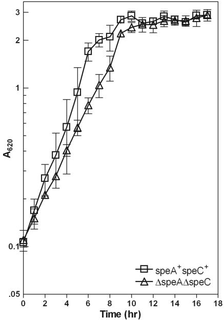
The ΔspeA and ΔspeC single deletion mutants did not affect the growth rate of Y. pestis (data not shown). Comparisons of the slopes of the exponential growth curves indicate that the double deletion mutant grew at ∼65% of the exponential growth rate of speA+ speC+ cells (Fig. 2) in the a defined medium PMH2, which contains no polyamines. All biofilm assays occurred in the 16- to 18-h time range at which the cell density measurements indicated no significant differences between the mutant and parental strains (Fig. 2). The steady growth of the double deletion mutant combined with high cell densities observed after 16 h of growth indicate that the growth effect is relatively minor. This observation is in sharp contrast to growth rates of polyamine-deficient mutants of P. aeruginosa and E. coli, which show either little growth (43) or a third of the growth (63) observed in the parental strain.
FIG. 2.
Growth curves of Y. pestis strains KIM6+ (speA+ speC+) and KIM6-2112+ (ΔspeA::cam2111 ΔspeC::kan2110) in PMH2 at 30°C.
Depletion of polyamines in ΔspeA, ΔspeC, and ΔspeA ΔspeC mutants.
Polyamines were quantified in clarified cellular extracts using HPLC-MS as described in Materials and Methods. The HPLC analysis of the speA+ speC+ parental strain (Table 2) indicates that Y. pestis, like other bacteria, has both putrescine and spermidine but not spermine (7). In the ΔspeA ΔspeC double mutant, no significant levels of putrescine or spermidine were observed (Table 2). Despite the significantly low levels of polyamines, Y. pestis was still able to grow (Fig. 2), suggesting that there may still be polyamines below our level of detection as has been observed in a polyamine-deficient E. coli K-12 strain (47). Putrescine levels were reduced ∼95% in the ΔspeA mutant (Table 2) and 55% in ΔspeC mutant relative to the parental speA+ speC+ strain. However, the levels of spermidine remained constant in both single deletion mutants (Table 2). Thus, based on direct measurement of polyamine levels, our results show that speA and speC represent two alternative pathways for polyamine biosynthesis and that the speA gene product is part of the primary biosynthetic pathway.
TABLE 2.
Polyamine concentrations in Y. pestis strains
| Strain | Concn [μmol/g wet cells (±SD)]a
|
|
|---|---|---|
| Putrescine | Spermidine | |
| speA+speC+ | 3.8 (±1.5) | 2.3 (±0.7) |
| ΔspeC | 1.7 (±0.9) | 1.6 (±0.4) |
| ΔspeA | 0.2 (±0.1) | 1.4 (±0.3) |
| ΔspeA ΔspeC | <0.03 | <0.1 |
| ΔspeA ΔspeC (pBAD/HisbADC) | 0.4 (±0.1) | 3.2 (±0.8) |
Values are the averages of three or more experiments.
The mutation of speA and speC abolishes the enzymatic activities of their gene products, ADC and ODC, respectively.
To provide an independent confirmation that the speA and speC gene products have the predicted decarboxylase functions, we performed in vitro enzyme assays using radioactive [14C]l-arginine and [14C]l-ornithine as substrates. Cellular extracts from the ΔspeA and ΔspeC mutants had the expected reductions in ADC and ODC activities, respectively (Table 3). In the double deletion mutant, only background levels of activity for both ODC and ADC were observed. Finally, the expected enzymatic activity of heterologously expressed and purified ADC (pET28b-speA) and ODC (pET28b-speC) was demonstrated (Table 3). Thus, Y. pestis speA and speC are enzymatically active, and disruption of these genes causes a loss of activity in the respective mutants.
TABLE 3.
ODC and ADC activity in cellular extracts of Y. pestis speA+ speC+ (KIM6+) and polyamine-deficient mutants
| Enzyme | nmol CO2/min/mg ± SDa
|
μmol CO2/min/mg ± SDa
|
|||||
|---|---|---|---|---|---|---|---|
| speA+speC+ | ΔspeA | ΔspeC | ΔspeA ΔspeC | ΔspeA ΔspeC (pBAD/HisbADC) | Purified ADC | Purified ODC | |
| ADC | 20.86 ± 4.22 | 2.01 ± 0.85 | 15.36 ± 0.62 | 0.94 ± 0.35 | 26.14 ± 0.77 | 29.10 ± 7.93 | NDb |
| ODC | 8.98 ± 1.02 | 19.11 ± 4.92 | 0.67 ± 0.13 | 0.48 ± 0.39 | NDb | NDb | 1.89 ± 0.48 |
Values are the averages of three or more experiments.
ND, not determined.
Polyamine metabolism plays a role in biofilm formation.
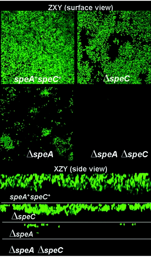
Biofilm formation was measured using three independent methods. Y. pestis cells expressing green fluorescent protein examined by CLSM showed a clear and progressive disruption in biofilm formation that corresponded with the reduction in putrescine levels (Fig. 3 and Table 2). In the ΔspeC mutant, we saw a small effect on attachment and thickness of the biofilm when qualitatively compared to the Y. pestis speA+ speC+ strain (Fig. 3). The effects of the ΔspeA mutation were more dramatic: there was a severe reduction in the attachment of the ΔspeA mutant to the glass coverslips. Finally, the ΔspeA ΔspeC double deletion mutant was incapable of forming any biofilm on the surface of the coverslips. The results indicate that polyamine levels modulate biofilm formation.
FIG. 3.
Confocal laser scanning microscopy images of Y. pestis cells expressing green fluorescent protein in order to visualize the extent of biofilm formation. The ZXY plane (surface view) shows the extent of attachment to the coverslip, while the XZY plane (side view) shows a cross section visualizing the thickness of the biofilm. There was a progressive decrease in biofilm formation: speA+ speC+ strain > ΔspeC mutant > ΔspeA mutant > ΔspeA ΔspeC mutant.
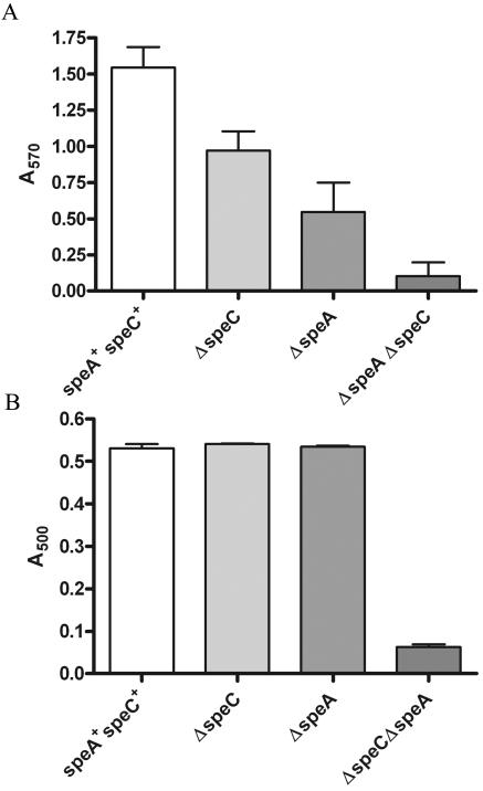
The CV staining assay (46) of cells grown at room temperature showed that the ΔspeC and ΔspeA single deletion mutants had a ∼33% and 66% reduction in attachment compared to the parental strain (Fig. 4A). The double deletion mutant showed a low level of attachment similar to that of hms mutants unable to form biofilm (32).
FIG. 4.
CV staining (A) and analysis of CR binding assays (B) as a measure of biofilm formation by Y. pestis strains. Y. pestis strains were grown overnight at 26°C for both assays. CV bound to attached bacterial cells was solubilized, and absorbance was measured at 570 nm. For CR binding, equivalent wet cell weights were incubated for 3 h in PMH2 containing 30 μg CR/ml. The amount of CR absorbed by the bacterial cells is expressed in terms of absorbance at 500 nm. CV staining and CR binding values are averages of three or more independent experiments, with error bars indicating standard deviations.
Another measure of biofilm production is related to EPS production, which is an essential component of the extracellular matrix that makes up biofilm. EPS can be analyzed separately from attachment using a dye, CR, that binds to carbohydrates in basic or neutral EPS (68). CR binding is a feature of the hms-dependent biofilm of Y. pestis that occurs during growth at temperatures of up to 34°C (26, 28, 60). The observed CR binding was severely reduced only in the ΔspeA ΔspeC double mutant (Fig. 4B), where no significant levels of putrescine or spermidine were formed (Table 2).
Complementation.
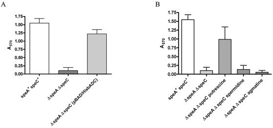
We tested for complementation of the defect in biofilm formation in two ways, genetically and chemically. Genetic complementation of the ΔspeA ΔspeC double mutant with full-length speA increased CV staining. The expression of speA from the pBAD/HisbADC vector restored biofilm levels as measured by CV staining (Fig. 5A). Chemical complementation of the ΔspeA ΔspeC double mutant with select exogenous polyamines showed restoration of biofilm with 1 mM putrescine. In contrast, agmatine and spermidine had little to no effect on biofilm formation (Fig. 5B) as measured by CV staining.
FIG. 5.
CV staining assay of genetically (A) and chemically (B) complemented Y. pestis ΔspeA ΔspeC mutant. (A) The ΔspeA ΔspeC mutant was complemented by the expression of full-length speA from pBAD/HisbspeA. The speA+ speC+ strain and the ΔspeA ΔspeC mutant are included for comparison. (B) PMH2 medium was supplemented with 1 mM concentrations of putrescine, spermidine, or agmatine. CV staining values are the averages of three or more independent experiments, with error bars indicating standard deviations. The speA+ speC+ strain is included for comparison.
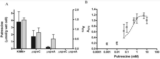
Our observation of the specific recovery of the double mutant with putrescine is consistent with the observed correlation between endogenous putrescine levels and biofilm formation in the three different mutants (Fig. 6A). Furthermore, the recovery of biofilm formation with exogenous putrescine was dose dependent (Fig. 6B). Thus, putrescine appears to be the polyamine of primary importance for biofilm formation in Y. pestis.
FIG. 6.
Correlation between putrescine and biofilm levels as measured by CV staining in each of the different polyamine deletion mutants. (A) The black bars show intracellular putrescine levels, while the gray bars show absorbance at 570 nm of CV from attached bacterial cells; error bars indicate standard deviations. (B) Dose-response curve of CV staining observed in the presence of putrescine in the ΔspeA ΔspeC double mutant. Below a threshold level of 0.1 mM putrescine, biofilm is clearly absent. CV staining and putrescine values are the averages of three or more independent experiments, with error bars indicating standard errors of the means. The dashed line indicates the general trend in biofilm reduction as a function of reduction in exogenous putrescine concentration.
DISCUSSION
Polyamine biosynthetic pathway in Y. pestis.
In bacteria, there are two known pathways for the conversion of agmatine to putrescine (Fig. 1). One pathway is found in E. coli, involving agmatinase (EC 3.5.3.11; the speB product), which catalyzes the direct conversion of agmatine to putrescine (57, 61). A second pathway occurs in P. aeruginosa, which has two enzymes responsible for converting agmatine to putrescine. Agmatine deiminase (EC 3.5.3.12; the aguA product) catalyzes the conversion of agmatine to N-carbamoylputrescine, which in turn is converted to putrescine by N-carbamoylputrescine amidohydrolase (EC 3.5.1.53; the aguB product) (43) (Fig. 1). Our genomic analysis of polyamine biosynthetic genes in Y. pestis suggests a P. aeruginosa-like polyamine biosynthetic pathway utilizing AguA-like and AguB-like enzymes for the conversion of agmatine to putrescine.
We observed no detectable levels of polyamines in the ΔspeA ΔspeC double mutant (Table 2). However, we cannot exclude a low level of decarboxylase activity attributable to lysine decarboxylases or another uncharacterized but weakly active arginine and/or ornithine decarboxylase (47). In contrast to E. coli and P. aeruginosa (43, 63), Y. pestis polyamine deficiency causes little to no effect on planktonic growth (Fig. 2). This suggests that the investigation of Y. pestis polyamine-deficient mutants may be useful in uncovering potentially specific roles for putrescine and spermidine that are usually masked in bacteria where a severe growth defect is the dominant effect. Our results indicate that both the speA and speC gene products participate in polyamine biosynthesis in Y. pestis. HLPC-MS analysis directly determined polyamine levels in cellular extracts (Table 2) while enzymatic activities of purified ADC and ODC were measured directly (Table 3). These assays show a loss of appropriate enzyme activity in the mutants and show that mutation of both speA and speC caused a depletion of polyamine pools below our detection limit. The deletion of speA alone had a much greater effect on putrescine levels than the speC deletion, suggesting that ADC is responsible for the bulk of putrescine biosynthesis under the conditions tested. However, ODC also functions in polyamine biosynthesis. In both single deletion mutants, we observed reduced levels of putrescine while spermidine levels were constant, within margins of error. The importance of ADC and ODC for polyamine production is apparent in the double deletion mutant, where spermidine was depleted below our detection limit.
It is also noteworthy that in the single deletion mutants, spermidine levels were not affected (Fig. 6), while putrescine levels progressively decreased. One possible explanation for the relative invariable levels of spermidine in the single deletion mutants may be the control of spermidine via polyamine degradation by spermidine acetyltransferase (SAT) (SpeG) (Fig. 1). Experiments performed using E. coli that focused on the regulation of polyamine levels have shown that polyamine degradation via SAT is involved in controlling levels of spermidine (15). Another mechanism of regulation of spermidine described for E. coli involves S-adenosylmethionine decarboxylase as proposed previously by Kashiwagi and Igarashi (30). The overexpression of full-length ADC in the double mutant raises the spermidine concentration above the level observed in the speA+ speC+ strain, likely triggering its down-regulation via SAT or S-adenosylmethionine decarboxylase along with the excretion of putrescine via PotE (30), lowering putrescine levels. Thus, the complementation of the double mutant with speA is sufficient for recovery of the biofilm phenotype: however, the toxic effects of high polyamine levels are likely triggering its down-regulation (30).
The effect of polyamines on biofilm formation in Y. pestis.
Our investigation of polyamine-deficient strains of Y. pestis has shown a link between the reduction in the levels of biofilm formation and the depletion of polyamines. The ΔspeC mutant showed a moderate reduction in putrescine levels with a decline in CV staining, while the ΔspeA mutant had significantly reduced levels of putrescine and CV staining (Fig. 6A). The CLSM images of the mutants and parental Y. pestis speA+ speC+ strains (Fig. 3) also reflect this correlation. This suggests that there is a certain putrescine concentration threshold that is required for biofilm formation. Complementation of the biofilm-deficient ΔspeA ΔspeC mutant with speA was sufficient for recovery of the biofilm defect (Fig. 5A). Chemical complementation occurred only with exogenous putrescine in a dose-dependent manner (Fig. 6B). CR binding, a measure of EPS production, was not affected in the single deletion mutants but was significantly reduced in the ΔspeA ΔspeC double deletion mutant, when cellular levels of both putrescine and spermidine were depleted. While this indicates that polyamines are important in EPS production, the results with CV staining and CLSM suggest that polyamines may affect several steps in biofilm formation in Y. pestis. Thus, our results demonstrate a link between biofilm formation and polyamine metabolism.
Biofilm formation in Y. pestis is controlled by the levels and activities of six hms gene products. Temperature regulation of biofilm formation is achieved by degradation of HmsH, HmsR, and HmsT at 37°C. HmsT contains a GGDEF domain and is required for the synthesis of cyclic di-GMP (c-di-GMP), a molecule that is presumably needed for the optimal activity of the EPS synthase, likely HmsR in Y. pestis (32). HmsP, on the other hand, has a phosphodiesterase activity and likely functions to degrade c-di-GMP (5, 32, 50, 54, 58). Thus, HmsT and HmsP likely regulate the levels of c-di-GMP to control biofilm formation. Here, we show that polyamine metabolism also affects biofilm formation. A comparison of the results obtained here with polyamine deletion mutants to those of hms deletions shows that the ΔspeC ΔspeA double deletion mutant has a reduction in biofilm formation similar to that of a number of hms mutants, including the hmsT mutant (32). The role of polyamines in biofilm formation remains to be elucidated. They may serve as signaling molecules affecting gene or protein expression, an intermediate in biofilm synthesis, or a structural component of biofilm.
This is the first report linking polyamine biosynthesis and biofilm formation. However, other studies have identified a link between polyamine transport systems and biofilm deficiencies. Genes encoding proteins with similarity to components of polyamine ABC transporters have been associated with biofilm deficiency in Pseudomonas putida (56), Agrobacterium tumefaciens (39), and Vibrio cholerae (29). Taken together, all these reports suggest a broad-reaching role for polyamines in biofilm formation.
Acknowledgments
This work was supported by NIH Southeast Regional Center for Excellence in Biodefense (SERCEB) developmental grant UF4 AI057175, a University of Kentucky Research Support grant and the partial support of the Kentucky Lung Cancer Research Program to M.A.O., and Public Health Service grant AI25098. C.N.P. was supported by a University of Kentucky dissertation year fellowship.
REFERENCES
- 1.Altschul, S. F., T. L. Madden, A. A. Schaffer, J. Zhang, Z. Zhang, W. Miller, and D. J. Lipman. 1997. Gapped BLAST and PSI-BLAST: a new generation of protein database search programs. Nucleic Acids Res. 25:3389-3402. [DOI] [PMC free article] [PubMed] [Google Scholar]
- 2.Bachrach, U., and M. Ben-Joseph. 1971. Studies on ornithine decarboxylase activity in normal and T2-infected Escherichia coli. FEBS Lett. 15:75-77. [DOI] [PubMed] [Google Scholar]
- 3.Balbo, P. B., C. N. Patel, K. G. Sell, R. S. Adcock, S. Neelakantan, P. A. Crooks, and M. A. Oliveira. 2003. Spectrophotometric and steady-state kinetic analysis of the biosynthetic arginine decarboxylase of Yersinia pestis utilizing arginine analogues as inhibitors and alternative substrates. Biochemistry 42:15189-15196. [DOI] [PubMed] [Google Scholar]
- 4.Birnboim, H. C., and J. Doly. 1979. A rapid alkaline extraction procedure for screening recombinant plasmid DNA. Nucleic Acids Res. 7:1513-1523. [DOI] [PMC free article] [PubMed] [Google Scholar]
- 5.Bobrov, A. G., O. Kirillina, and R. D. Perry. 2005. The phosphodiesterase activity of the HmsP EAL domain is required for negative regulation of biofilm formation in Yersinia pestis FEMS Microbiol. Lett. 247:123-130. [DOI] [PubMed] [Google Scholar]
- 6.Chattopadhyay, M. K., C. W. Tabor, and H. Tabor. 2003. Polyamines protect Escherichia coli cells from the toxic effect of oxygen. Proc. Natl. Acad. Sci. USA 100:2261-2265. [DOI] [PMC free article] [PubMed] [Google Scholar]
- 7.Cohen, S. S. 1998. A guide to the polyamines, 1st ed. Oxford University Press, New York, N.Y.
- 8.Coleman, C. S., G. Hu, and A. E. Pegg. 2004. Putrescine biosynthesis in mammalian tissues. Biochem. J. 239:849-855. [DOI] [PMC free article] [PubMed] [Google Scholar]
- 9.Darby, C., J. W. Hsu, N. Ghori, and S. Falkow. 2002. Caenorhabditis elegans: plague bacteria biofilm blocks food intake. Nature 417:243-244. [DOI] [PubMed] [Google Scholar]
- 10.Datsenko, K. A., and B. L. Wanner. 2000. One-step inactivation of chromosomal genes in Escherichia coli K-12 using PCR products. Proc. Natl. Acad. Sci. USA 97:6640-6645. [DOI] [PMC free article] [PubMed] [Google Scholar]
- 11.Davis, R. H., D. R. Morris, and P. Coffino. 1992. Sequestered end products and enzyme regulation: the case of ornithine decarboxylase. Microbiol. Rev. 56:280-290. [DOI] [PMC free article] [PubMed] [Google Scholar]
- 12.Deng, W., V. Burland, G. Plunkett III, A. Boutin, G. F. Mayhew, P. Liss, N. T. Perna, D. J. Rose, B. Mau, S. Zhou, D. C. Schwartz, J. D. Fetherston, L. E. Lindler, R. R. Brubaker, G. V. Plano, S. C. Straley, K. A. McDonough, M. L. Nilles, J. S. Matson, F. R. Blattner, and R. D. Perry. 2002. Genome sequence of Yersinia pestis KIM. J. Bacteriol. 184:4601-4611. [DOI] [PMC free article] [PubMed] [Google Scholar]
- 13.Fetherston, J. D., J. W. Lillard, Jr., and R. D. Perry. 1995. Analysis of the pesticin receptor from Yersinia pestis: role in iron-deficient growth and possible regulation by its siderophore. J. Bacteriol. 177:1824-1833. [DOI] [PMC free article] [PubMed] [Google Scholar]
- 14.Fetherston, J. D., P. Schuetze, and R. D. Perry. 1992. Loss of the pigmentation phenotype in Yersinia pestis is due to the spontaneous deletion of 102 kb of chromosomal DNA which is flanked by a repetitive element. Mol. Microbiol. 6:2693-2704. [DOI] [PubMed] [Google Scholar]
- 15.Fukuchi, J., K. Kashiwagi, M. Yamagishi, A. Ishihama, and K. Igarashi. 1995. Decrease in cell viability due to the accumulation of spermidine in spermidine acetyltransferase-deficient mutant of Escherichia coli. J. Biol. Chem. 270:18831-18835. [DOI] [PubMed] [Google Scholar]
- 16.Fuqua, C., and E. P. Greenberg. 2002. Listening in on bacteria: acyl-homoserine lactone signalling. Nat. Rev. Mol. Cell Biol. 3:685-695. [DOI] [PubMed] [Google Scholar]
- 17.Fuqua, C., M. R. Parsek, and E. P. Greenberg. 2001. Regulation of gene expression by cell-to-cell communication: acyl-homoserine lactone quorum sensing. Annu. Rev. Genet. 35:439-468. [DOI] [PubMed] [Google Scholar]
- 18.Gallaguer, S. R. 1998. Current protocols in protein science, p. 10.1.1-10.1. 34. In J. E. Coligan, B. M. Dunn, H. L. Ploegh, D. W. Speicher, and P. T. Wingfield (ed.), Current protocols in protein science, vol. 1>. John Wiley & Sons, Inc., New York, N.Y. [Google Scholar]
- 19.Gong, S., S. W. Bearden, V. A. Geoffroy, J. D. Fetherston, and R. D. Perry. 2001. Characterization of the Yersinia pestis Yfu ABC iron transport system. Infect. Immun. 67:2829-2837. [DOI] [PMC free article] [PubMed] [Google Scholar]
- 20.Grishin, N. V., M. A. Phillips, and E. J. Goldsmith. 1995. Modeling of the spatial structure of eukaryotic ornithine decarboxylases. Protein Sci. 4:1291-1304. [DOI] [PMC free article] [PubMed] [Google Scholar]
- 21.Hackert, M. L., D. W. Carroll, L. Davidson, S. O. Kim, C. Momany, G. L. Vaaler, and L. Zhang. 1994. Sequence of ornithine decarboxylase from Lactobacillus sp. strain 30a. J. Bacteriol. 176:7391-7394. [DOI] [PMC free article] [PubMed] [Google Scholar]
- 22.Hall-Stoodley, L., J. W. Costerton, and P. Stoodley. 2004. Bacterial biofilms: from the natural environment to infectious diseases. Nat. Rev. Microbiol. 2:95-108. [DOI] [PubMed] [Google Scholar]
- 23.Hartzell, P. L., J. Millstein, and C. LaPaglia. 1999. Biofilm formation in hyperthermophilic Archaea. Methods Enzymol. 310:335-349. [DOI] [PubMed] [Google Scholar]
- 24.Hinnebusch, B. J., R. D. Perry, and T. G. Schwan. 1996. Role of the Yersinia pestis hemin storage (hms) locus in the transmission of plague by fleas. Science 273:367-370. [DOI] [PubMed] [Google Scholar]
- 25.Igarashi, K., and K. Kashiwagi. 2000. Polyamines: mysterious modulators of cellular functions. Biochem. Biophys. Res. Commun. 271:559-564. [DOI] [PubMed] [Google Scholar]
- 26.Jackson, S., and T. W. Burrows. 1956. The virulence-enhancing effect of iron on non-pigmented mutants of virulent strains of Pasteurella pestis Br. J. Exp. Pathol. 37:577-583. [PMC free article] [PubMed] [Google Scholar]
- 27.Jarrett, C. O., E. Deak, K. E. Isherwood, P. C. Oyston, E. R. Fischer, A. R. Whitney, S. D. Kobayashi, F. R. DeLeo, and B. J. Hinnebusch. 2004. Transmission of Yersinia pestis from an infectious biofilm in the flea vector. J. Infect. Dis. 190:783-792. [DOI] [PubMed] [Google Scholar]
- 28.Jones, H. A., J. W. Lillard, Jr., and R. D. Perry. 1999. HmsT, a protein essential for expression of the haemin storage (Hms+) phenotype of Yersinia pestis. Microbiology 145:2117-2128. [DOI] [PubMed] [Google Scholar]
- 29.Karatan, E., T. R. Duncan, and P. I. Watnick. 2005. NspS, a predicted polyamine sensor, mediates activation of Vibrio cholerae biofilm formation by norspermidine. J. Bacteriol. 187:7434-7443. [DOI] [PMC free article] [PubMed] [Google Scholar]
- 30.Kashiwagi, K., and K. Igarashi. 1988. Adjustment of polyamine contents in Escherichia coli. J. Bacteriol. 170:3131-3135. [DOI] [PMC free article] [PubMed] [Google Scholar]
- 31.Kashiwagi, K., T. Suzuki, F. Suzuki, T. Furuchi, H. Kobayashi, and K. Igarashi. 1991. Coexistence of the genes for putrescine transport protein and ornithine decarboxylase at 16 min on Escherichia coli chromosome. J. Biol. Chem. 266:20922-20927. [PubMed] [Google Scholar]
- 32.Kirillina, O., J. D. Fetherston, A. G. Bobrov, J. Abney, and R. D. Perry. 2004. HmsP, a putative phosphodiesterase, and HmsT, a putative diguanylate cyclase, control Hms-dependent biofilm formation in Yersinia pestis Mol. Microbiol. 54:75-88. [DOI] [PubMed] [Google Scholar]
- 33.Koski, P., and M. Vaara. 1991. Polyamines as constituents of the outer membranes of Escherichia coli and Salmonella typhimurium. J. Bacteriol. 173:3695-3699. [DOI] [PMC free article] [PubMed] [Google Scholar]
- 34.Kutyrev, V. V., A. A. Filippov, O. S. Oparina, and O. A. Protsenko. 1992. Analysis of Yersinia pestis chromosomal determinants Pgm+ and Psts associated with virulence. Microb. Pathog. 12:177-186. [DOI] [PubMed] [Google Scholar]
- 35.Liaw, S. J., H. C. Lai, and W. B. Wang. 2004. Modulation of swarming and virulence by fatty acids through the RsbA protein in Proteus mirabilis. Infect. Immun. 72:6836-6845. [DOI] [PMC free article] [PubMed] [Google Scholar]
- 36.Lioliou, E. E., and D. A. Kyriakidis. 2004. The role of bacterial antizyme: from an inhibitory protein to AtoC transcriptional regulator. Microb. Cell Fact. 3:8. [DOI] [PMC free article] [PubMed] [Google Scholar]
- 37.Lu, C. D., Y. Itoh, Y. Nakada, and Y. Jiang. 2002. Functional analysis and regulation of the divergent spuABCDEFGH-spuI operons for polyamine uptake and utilization in Pseudomonas aeruginosa PAO1. J. Bacteriol. 184:3765-3773. [DOI] [PMC free article] [PubMed] [Google Scholar]
- 38.Maas, W. K. 1972. Mapping of genes involved in the synthesis of spermidine in Escherichia coli. Mol. Gen. Genet. 119:1-9. [DOI] [PubMed] [Google Scholar]
- 39.Matthysse, A. G., H. A. Yarnall, and N. Young. 1996. Requirement for genes with homology to ABC transport systems for attachment and virulence of Agrobacterium tumefaciens. J. Bacteriol. 178:5302-5308. [DOI] [PMC free article] [PubMed] [Google Scholar]
- 40.Momany, C., R. Ghosh, and M. L. Hackert. 1995. Structural motifs for pyridoxal-5′-phosphate binding in decarboxylases: an analysis based on the crystal structure of the Lactobacillus 30a ornithine decarboxylase. Protein Sci. 4:849-854. [DOI] [PMC free article] [PubMed] [Google Scholar]
- 41.Moore, R. C., and S. M. Boyle. 1991. Cyclic AMP inhibits and putrescine represses expression of the speA gene encoding biosynthetic arginine decarboxylase in Escherichia coli. J. Bacteriol. 173:3615-3621. [DOI] [PMC free article] [PubMed] [Google Scholar]
- 42.Morgan, D. M. L. 1998. Determination of polyamines as their benzoylated derivatives by HPLC. Methods Mol. Biol. 79:111-118. [DOI] [PubMed] [Google Scholar]
- 43.Nakada, Y., and Y. Itoh. 2003. Identification of the putrescine biosynthetic genes in Pseudomonas aeruginosa and characterization of agmatine deiminase and N-carbamoylputrescine amidohydrolase of the arginine decarboxylase pathway. Microbiology 149:707-714. [DOI] [PubMed] [Google Scholar]
- 44.O'Farrell, P. H. 1975. High resolution two-dimensional electrophoresis of proteins. J. Biol. Chem. 250:4007-4021. [PMC free article] [PubMed] [Google Scholar]
- 45.Oliveira, M. A., D. Carroll, L. Davidson, C. Momany, and M. L. Hackert. 1997. The GTP effector site of ornithine decarboxylase from Lactobacillus 30a: kinetic and structural characterization. Biochemistry 36:16147-16154. [DOI] [PubMed] [Google Scholar]
- 46.O'Toole, G. A., L. A. Pratt, P. I. Watnick, D. K. Newman, V. B. Weaver, and R. Kolter. 1999. Genetic approaches to study of biofilms. Methods Enzymol. 310:91-109. [DOI] [PubMed] [Google Scholar]
- 47.Panagiotidis, C. A., S. Blackburn, K. B. Low, and E. S. Canellakis. 1987. Biosynthesis of polyamines in ornithine decarboxylase, arginine decarboxylase, and agmatine ureohydrolase deletion mutants of Escherichia coli strain K-12. Proc. Natl. Acad. Sci. USA 84:4423-4427. [DOI] [PMC free article] [PubMed] [Google Scholar]
- 48.Parkhill, J., B. W. Wren, N. R. Thomson, R. W. Titball, M. T. Holden, M. B. Prentice, M. Sebaihia, K. D. James, C. Churcher, K. L. Mungall, S. Baker, D. Basham, S. D. Bentley, K. Brooks, A. M. Cerdeno-Tarraga, T. Chillingworth, A. Cronin, R. M. Davies, P. Davis, G. Dougan, T. Feltwell, N. Hamlin, S. Holroyd, K. Jagels, A. V. Karlyshev, S. Leather, S. Moule, P. C. Oyston, M. Quail, K. Rutherford, M. Simmonds, J. Skelton, K. Stevens, S. Whitehead, and B. G. Barrell. 2001. Genome sequence of Yersinia pestis, the causative agent of plague. Nature 413:523-527. [DOI] [PubMed] [Google Scholar]
- 49.Pendrak, M. L., and R. D. Perry. 1993. Proteins essential for expression of the Hms+ phenotype of Yersinia pestis Mol. Microbiol. 8:857-864. [DOI] [PubMed] [Google Scholar]
- 50.Perry, R. D., A. G. Bobrov, O. Kirillina, H. A. Jones, L. Pedersen, J. Abney, and J. D. Fetherston. 2004. Temperature regulation of the hemin storage (Hms+) phenotype of Yersinia pestis is posttranscriptional. J. Bacteriol. 186:1638-1647. [DOI] [PMC free article] [PubMed] [Google Scholar]
- 51.Perry, R. D., and J. D. Fetherston. 1997. Yersinia pestis—etiologic agent of plague. Clin. Microbiol. Rev. 10:35-66. [DOI] [PMC free article] [PubMed] [Google Scholar]
- 52.Perry, R. D., M. L. Pendrak, and P. Schuetze. 1990. Identification and cloning of a hemin storage locus involved in the pigmentation phenotype of Yersinia pestis J. Bacteriol. 172:5929-5937. [DOI] [PMC free article] [PubMed] [Google Scholar]
- 53.Price, S. B., J. C. Wright, F. J. DeGraves, M. P. Castanie-Cornet, and J. W. Foster. 2004. Acid resistance systems required for survival of Escherichia coli O157:H7 in the bovine gastrointestinal tract and in apple cider are different. Appl. Environ. Microbiol. 70:4792-4799. [DOI] [PMC free article] [PubMed] [Google Scholar]
- 54.Ross, P., Y. Aloni, H. Weinhouse, D. Michaeli, P. Weinberger-Ohana, R. Mayer, and M. Benziman. 1986. Control of cellulose synthesis Acetobacter xylinum. A unique guanyl oligonucleotide is the immediate activator of the cellulose synthase. Carbohydr. Res. 149:101-117. [Google Scholar]
- 55.Sambrook, J., and D. W. Russell. 2001. Molecular cloning: a laboratory manual, 3rd ed. Cold Spring Harbor Laboratory Press, Cold Spring Harbor, N.Y.
- 56.Sauer, K., and A. K. Camper. 2001. Characterization of phenotypic changes in Pseudomonas putida in response to surface-associated growth. J. Bacteriol. 183:6579-6589. [DOI] [PMC free article] [PubMed] [Google Scholar]
- 57.Sekowska, A., P. Bertin, and A. Danchin. 1998. Characterization of polyamine synthesis pathway in Bacillus subtilis 168. Mol. Microbiol. 29:851-858. [DOI] [PubMed] [Google Scholar]
- 58.Simm, R., M. Morr, A. Kader, M. Nimtz, and U. Romling. 2004. GGDEF and EAL domains inversely regulate cyclic di-GMP levels and transition from sessility to motility. Mol. Microbiol. 53:1123-1134. [DOI] [PubMed] [Google Scholar]
- 59.Sturgill, G., and P. N. Rather. 2004. Evidence that putrescine acts as an extracellular signal required for swarming in Proteus mirabilis Mol. Microbiol. 51:437-446. [DOI] [PubMed] [Google Scholar]
- 60.Surgalla, M. J., and E. D. Beesley. 1969. Congo red-agar plating medium for detecting pigmentation in Pasteurella pestis Appl. Microbiol. 18:834-837. [DOI] [PMC free article] [PubMed] [Google Scholar]
- 61.Szumanski, M. B., and S. M. Boyle. 1990. Analysis and sequence of the speB gene encoding agmatine ureohydrolase, a putrescine biosynthetic enzyme in Escherichia coli. J. Bacteriol. 172:538-547. [DOI] [PMC free article] [PubMed] [Google Scholar]
- 62.Tabor, C. W., and H. Tabor. 1985. Polyamines in microorganisms. Microbiol. Rev. 49:81-99. [DOI] [PMC free article] [PubMed] [Google Scholar]
- 63.Tabor, H. 1981. Polyamine biosynthesis in Escherichia coli: construction of polyamine-deficient mutants. Med. Biol. 59:389-393. [PubMed] [Google Scholar]
- 64.Tabor, H., C. W. Tabor, and F. Irreverre. 1973. Quantitative determination of aliphatic diamines and polyamines by an automated liquid chromatography procedure. Anal. Biochem. 55:457-467. [DOI] [PubMed] [Google Scholar]
- 65.Tkachenko, A., L. Nesterova, and M. Pshenichnov. 2001. The role of the natural polyamine putrescine in defense against oxidative stress in Escherichia coli. Arch. Microbiol. 176:155-157. [DOI] [PubMed] [Google Scholar]
- 66.Tweeddale, H., L. Notley-McRobb, and T. Ferenci. 1998. Effect of slow growth on metabolism of Escherichia coli, as revealed by global metabolite pool (“metabolome”) analysis. J. Bacteriol. 180:5109-5116. [DOI] [PMC free article] [PubMed] [Google Scholar]
- 67.Vinogradov, E., and M. B. Perry. 2000. Structural analysis of the core region of lipopolysaccharides from Proteus mirabilis serotypes O6, O48 and O57. Eur. J. Biochem. 267:2439-2446. [DOI] [PubMed] [Google Scholar]
- 68.Weiner, R., E. Seagren, C. Arnosti, and E. Quintero. 1999. Bacterial survival in biofilms: probes for exopolysaccharide and its hydrolysis, and measurements of intra- and interphase mass fluxes. Methods Enzymol. 310:403-426. [DOI] [PubMed] [Google Scholar]
- 69.Wright, J. M., C. Satishchandran, and S. M. Boyle. 1986. Transcription of the speC (ornithine decarboxylase) gene of Escherichia coli is repressed by cyclic AMP and its receptor protein. Gene 44:37-45. [DOI] [PubMed] [Google Scholar]
- 70.Yoshida, M., K. Kashiwagi, A. Shigemasa, S. Taniguchi, K. Yamamoto, H. Makinoshima, A. Ishihama, and K. Igarashi. 2004. A unifying model for the role of polyamines in bacterial cell growth, the polyamine modulon. J. Biol. Chem. 279:46008-46013. [DOI] [PubMed] [Google Scholar]


