Abstract
The role of oxygen in the transcriptional regulation of the PN promoter that controls the bzd operon involved in the anaerobic catabolism of benzoate in the denitrifying Azoarcus sp. strain CIB has been investigated. In vivo experiments using PN::lacZ translational fusions, in both Azoarcus sp. strain CIB and Escherichia coli cells, have shown an oxygen-dependent repression effect on the transcription of the bzd catabolic genes. E. coli Fnr was required for the anaerobic induction of the PN promoter, and the oxygen-dependent repression of the bzd genes could be bypassed by the expression of a constitutively active Fnr* protein. In vitro experiments revealed that Fnr binds to the PN promoter at a consensus sequence centered at position −41.5 from the transcription start site overlapping the −35 box, suggesting that PN belongs to the class II Fnr-dependent promoters. Fnr interacts with RNA polymerase (RNAP) and is strictly required for transcription initiation after formation of the RNAP-PN complex. An fnr ortholog, the acpR gene, was identified in the genome of Azoarcus sp. strain CIB. The Azoarcus sp. strain CIB acpR mutant was unable to grow anaerobically on aromatic compounds and it did not drive the expression of the PN::lacZ fusion, suggesting that AcpR is the cognate transcriptional activator of the PN promoter. Since the lack of AcpR in Azoarcus sp. strain CIB did not affect growth on nonaromatic carbon sources, AcpR can be considered a transcriptional regulator of the Fnr/Crp superfamily that has evolved to specifically control the central pathway for the anaerobic catabolism of aromatic compounds in Azoarcus.
Aromatic compounds are the second most widely distributed class of organic compounds in nature, and a significant number of xenobiotics belong to this family of compounds. Since many ecosystems are often anoxic, the anaerobic catabolism of aromatic compounds by microorganisms becomes crucial in the biogeochemical cycles and in the sustainable development of the biosphere (36, 57). Benzoate has been used as a model compound to study the anaerobic catabolism of aromatic compounds in different microorganisms, such as Rhodopseudomonas palustris, Magnetospirillum magnetotacticum MS-1, Thauera aromatica, Azoarcus evansii, and Azoarcus sp. strain CIB (5, 6, 25). In all these bacteria, benzoate is first activated to benzoyl-coenzyme A (benzoyl-CoA), which is further degraded to central intermediates by a series of reactions that involve aromatic-ring reduction, modified β oxidation, and ring cleavage as critical steps (22, 25). Some enzymes responsible for the anaerobic catabolism of aromatic compounds, such as benzoyl-CoA reductase, become inactivated in the presence of oxygen in a few seconds (10). It is therefore reasonable to consider that microorganisms with the ability to anaerobically catabolize aromatic compounds must regulate the expression of the corresponding catabolic genes in response not only to aromatic-carbon source availability, but also to oxygen levels to avoid gratuitous waste of energy (18).
The availability of oxygen is one of the most important regulatory signals in bacteria (45). In Escherichia coli, Fnr is a major global regulator that controls gene expression in response to oxygen deprivation. Fnr has been intensively studied, and homologues are found in a wide range of microorganisms (13, 28, 45).Whereas Fnr is an inactive monomer in the presence of oxygen, under anaerobic conditions, Fnr becomes an active homodimer (34) that binds to its target DNA, promoting activation or repression of gene expression (23, 31). The vast majority of Fnr-regulated promoters contain a consensus Fnr-binding site centered approximately 41.5 bp upstream of the transcriptional start site, and they are termed class II Fnr-dependent promoters (14). In class II promoters, Fnr is able to make multiple contacts with RNA polymerase (RNAP) through three activating regions, AR1, AR2, and AR3 (Fig. 1) (7, 9, 16, 31, 59, 60, 61). Fnr-AR1 is active in the upstream subunit of the Fnr homodimer, and it is proposed to interact with the carboxy-terminal domain of the alpha subunit (αCTD) of RNAP (58). Fnr-AR3 is active in the downstream subunit (7) and is likely to contact the σ70 subunit of RNAP (35). Fnr-AR2, which plays a minor role in activation, is also active in the downstream subunit (16), but its interaction partner is proposed to be the amino-terminal domain of the alpha subunit (αNTD) of RNAP (7, 61).
FIG. 1.
Promoter architecture of class II Fnr-dependent promoters. Class II Fnr-dependent promoters have Fnr-binding sites centered near −41.5 bp from the transcription start point (+1). The Fnr AR-1 surface (black square) of the upstream subunit of the Fnr dimer makes contact with the αCTD of the RNAP. The Fnr AR-2 (black triangle) and AR-3 (black circle) surfaces of the downstream subunit of the Fnr dimer contact the αNTD and σ70 of the RNAP, respectively. The β and β′ subunits of RNAP and the −10 and −35 boxes of a σ70-dependent promoter are also shown.
Although genetic experiments have revealed that the AadR protein, an Fnr/Crp superfamily member, regulates the degradation of aromatic compounds in response to oxygen in the phototrophic bacterium R. palustris (17, 18, 19), no direct biochemical evidence of such AadR-mediated regulation of the target promoters has been reported. Recently, we have characterized the bzd gene cluster involved in the anaerobic degradation of benzoate in Azoarcus sp. strain CIB, a denitrifying betaproteobacterium able to anaerobically degrade a large number of aromatic compounds via benzoyl-CoA (5). The bzd cluster is organized as a single catabolic operon (bzdNOPQMSTUVWXYZA) and the bzdR regulatory gene (5). The PN promoter, which drives the expression of the catabolic operon, is regulated by the BzdR transcriptional repressor, the first member of a new subfamily of transcriptional regulators, and benzoyl-CoA is the inducer molecule (4).
In this work, we present genetic and biochemical evidence that PN is a class II-dependent promoter whose activity is controlled by the AcpR transcriptional activator, an Fnr ortholog in Azoarcus sp. strain CIB. AcpR constitutes the first Fnr/Crp superfamily member reported so far in denitrifying bacteria that specifically controls the expression of the central pathway for the anaerobic catabolism of aromatic compounds in response to oxygen.
MATERIALS AND METHODS
Strains, plasmids, and growth conditions.
The E. coli and Azoarcus strains, as well as the plasmids, used in this work are listed in Table 1. To construct plasmid pBBR1MCS-5acpR, a 1,220-bp DNA fragment containing the acpR gene was PCR amplified from Azoarcus sp. strain CIB by using oligonucleotides 5AcpRext (5′-GGTACCTAGTTAACTAGCGTGATGATCTTGTTACACGCGCAGTAGTAG-3′) and 3AcpRint (5′-CAAGCCTGTTGTTGACGGAGCAGGACGTTGCCGCCCTGACTGCGACGATGACCGC-3′) and cloned into the pGEM-T Easy cloning vector, giving rise to plasmid pGEM-T EasyacpR (Table 1). A 1.2-kb KpnI/ApaI fragment harboring the acpR gene from plasmid pGEM-T EasyacpR was then subcloned into KpnI/ApaI-double-digested pBBRMCS-5 vector to render the pBBR1MCS-5acpR plasmid (Table 1). To construct plasmid pIZ-FNR*, a 910-bp DNA fragment encoding His6-FNR* was PCR amplified from pQE60-His6Fnr* by using the oligonucleotides 5Fnr* (5′-GAACTGCAGAAATCATAAAAAATTTATTTGCTTTGTGAGCGG-3′; an engineered PstI site is underlined) and 3Fnr* (5′-GGACTAGTTCAGCTAATTAAGCTTAGTGATGGTG-3′; an engineered SpeI site is underlined), and it was cloned into the pIZ1016 cloning vector under the control of the Ptac promoter (Table 1).
TABLE 1.
Bacterial strains and plasmids used in this work
| Strain or plasmid | Relevant phenotype and/or genotypea | Reference or source |
|---|---|---|
| E. coli | ||
| DH5α | endA1 hsdR17 supE44 thi-1 recA1 gyrA(Nalr) relA1Δ(argF-lac)U169 depR φ80Δlac(lacZ)M15 | 44 |
| S17-1λpir | Tpr SmrrecA thi hsdRM+ RP4::2-Tc::Mu::Km Tn7 λpir phage lysogen | 16 |
| M182 | Smr (ΔlacIOPZYA)X74 galU galK rpsL Δ(ara-leu) | 49 |
| JRG1728 | Cmr Smr Δ(tyrR fnr rac trg)17 zdd-30::Tn9; derived from M182 | 49 |
| M15 | Strain for regulated high-level expression with pQE vectors | Qiagen |
| RZ7350 | lacZΔ145 narG234::Mudl1734 | 26 |
| PK330 | Cmr; RZ7350 derivative Ptrp-rpoD | 35 |
| Azoarcus sp. strain CIB | ||
| CIB | Wild-type strain | 5 |
| CIBdacpR | Kmr; Azoarcus sp. strain CIB with a disruption of the acpR gene | This work |
| CIBlacZ | Kmr; Azoarcus sp. strain CIB harboring a chromosomal PN::lacZ translational fusion | 5 |
| Plasmids | ||
| pK18mob | KmroriColE1 Mob+lacZα; used for directed insertional disruption | 46 |
| pK18mobacpR | Kmr; 410-bp BamHI/HindIII acpR internal fragment cloned into BamHI/HindIII-digested pK18mob | This work |
| pSJ3 | AproriColE1 ′lacZ promoter probe vector; lacZ fusion flanked by NotI sites | 21 |
| pSJ3PN | Apr; pSJ3 derivative carrying the PN::lacZ translational fusion | 5 |
| pSJ3RPN | Apr; pSJ3 derivative carrying the bzdR/PN::lacZ translational fusion | 4 |
| pHW1 | Kmr; fnr gene cloned into pLG339 | 8 |
| pQE60-His6Fnr* | Apr; pQE60 derivative harboring the His6-FNR* gene under the control of T5 promoter lac operator | 60 |
| pBBR1MCS-5 | GmroripBBR1MCS Mob+lacZα; broad-host-range cloning and expression vector | 29 |
| pBBR5PN | Gmr; pBBR1MCS-5 derivative harboring the PN::lacZ translational fusion from pSJ3PN | 2 |
| pBBR1MCS-5acpR | Gmr; pBBR1MCS-5 derivative harboring the 1.2-kb KpnI/ApaI fragment that contains the acpR gene | This work |
| pGEM-T Easy | AproriColE1 lacZα; PCR fragment cloning vector | Promega |
| pGEM-T EasyacpR | Apr; pGEM-T Easy derivative harboring a 1.2-kb PCR-amplified fragment that contains the acpR gene | This work |
| pREP4 | Kmr; plasmid that expresses the lacI repressor | Qiagen |
| pJCD01 | AproriColE1; polylinker of pUC19 flanked by rpoC and rrnBT1T2 terminators | 37 |
| pJCD-PN | Apr; pJCD01 derivative harboring a 585-bp EcoRI fragment that includes the PN promoter | This work |
| pGEX-2T | Apr; plasmid for construction of GST-tagged fusion protein | 48 |
| pGEX-2Tσ70 | Apr; rpoD gene cloned into pGEX-2T plasmid | 35 |
| pGEX-2Tσ70(EA591) | Apr; rpoD-EA591 gene cloned into pGEX-2T plasmid | 35 |
| pGEX-2Tσ70(KA593) | Apr; rpoD-KA593 gene cloned into pGEX-2T plasmid | 35 |
| pGEX-2Tσ70(KA597) | Apr; rpoD-KA597 gene cloned into pGEX-2T plasmid | 35 |
| pIZ1016 | Gmr; pBBR1MCS-5 broad-host-range-vector derivative with tac promoter and lacIq from pMM40 | 38 |
| pIZ-FNR* | Gmr; pIZ1016 derivative harboring the FNR* gene under Ptac | This work |
GST, glutathione S-transferase.
E. coli cells were grown at 37°C in Luria-Bertani (LB) medium (40). When required, E. coli cells were grown anaerobically at 30°C either in LB medium supplemented with 0.2% glucose or in M63 minimal medium (40) using the corresponding necessary nutritional supplements, 20 mM glycerol as a carbon source, and 10 mM KNO3 as a terminal electron acceptor. Azoarcus strains were grown at 30°C in MC medium as described previously (5). Where appropriate, antibiotics were added at the following concentrations: ampicillin, 100 μg/ml; chloramphenicol, 30 μg/ml; gentamicin, 7.5 μg/ml; and kanamycin, 50 μg/ml.
The E. coli PK330 strain contains the chromosomal rpoD gene, encoding the σ70 subunit of RNAP, under the control of the Ptrp promoter (Table 1). In the presence of 20 μg/ml tryptophan, expression of chromosomally encoded σ70 was greatly reduced, as judged from the poor growth in liquid media and lack of growth on agar plates (35). Growth on M63 minimal medium containing 20 mM glycerol and including 20 μg/ml tryptophan was restored by the presence of plasmid pGEX-2Tσ70, pGEX-2Tσ70(EA591), pGEX-2Tσ70(KA593), or pGEX-2Tσ70(KA597), which express under Ptac the wild-type σ70, σ70(EA591), σ70(KA593), or σ70(KA597), respectively.
Molecular biology techniques.
Recombinant DNA techniques were carried out by published methods (43). Plasmid DNA was prepared with a High Pure plasmid isolation kit (Roche Applied Science). DNA fragments were purified with Gene-Clean Turbo (Q-BIOgene). Oligonucleotides were supplied by Sigma Co. All cloned inserts and DNA fragments were confirmed by DNA sequencing through an ABI Prism 377 automated DNA sequencer (Applied Biosystems Inc.). Transformation of E. coli cells was carried out by using the RbCl method or by electroporation (Gene Pulser; Bio-Rad) (43). Plasmids were transferred from E. coli S17-1 (λ pir) (donor strain) into Azoarcus sp. recipient strains by biparental filter mating as described previously (5). Proteins were analyzed by sodium dodecyl sulfate-polyacrylamide gel electrophoresis as described previously (30). The protein concentrations in cell extracts were determined by the method of Bradford (11) using bovine serum albumin as the standard.
β-Galactosidase assays.
β-Galactosidase activities were measured with permeabilized cells as described by Miller (40).
Sequence data analyses.
The amino acid sequence of the AcpR protein was compared with those present in microbial genome databases using the TBLAST algorithm (1) at the National Center for Biotechnology Information server (http://www.ncbi.nlm.nihgov/BLAST/BLAST.cgi). Multiple protein sequence alignments were made with the ClustalW (53) program at the INFOBIOGEN server (http://www.infobiogen.fr/services). Phylogenetic analysis of the Fnr-like proteins was carried out according to the neighbor-joining method of the PHYLIP program (12, 20) at the TreeTop-GeneBee server (http://www.genebee.msu.su/genebee.html).
Construction of Azoarcus sp. strain CIBdacpR.
For disruption of the acpR gene through single homologous recombination, a 410-bp internal fragment of the acpR gene was PCR amplified by using primers 5AcpRcib (5′-GGGATCCGTTGAGCAGGAAGGCCG-3′; an engineered BamHI site is underlined) and 3AcpRcib (5′-CAAGCTTCCGCCTCGACGAACTCGTC-3′; an engineered HindIII site is underlined), and it was cloned into the BamHI/HindIII-digested pK18mob (a mobilizable plasmid that does not replicate in Azoarcus). The resulting construct, pK18mobacpR (Table 1), was transferred from E. coli S17-1(λpir) (donor strain) into Azoarcus sp. strain CIB (recipient strain) by biparental filter mating (5). An exconjugant, Azoarcus sp. strain CIBdacpR, harboring the disrupted acpR gene by insertion of the suicide plasmid, was isolated aerobically on kanamycin-containing MC medium lacking nitrate and containing 0.4% citrate as the sole carbon source for counterselection of donor cells. The mutant strain was analyzed by PCR to confirm the disruption of the target gene.
Overproduction and purification of His6-Fnr*.
The recombinant plasmid pQE60-His6Fnr*, which expresses the C-terminally His-tagged Fnr* protein under the control of the T5 promoter-lac operator (60), was transformed in the E. coli M15 strain carrying the plasmid pREP4, which produces the LacI repressor (Table 1). The His-tagged Fnr* protein was overproduced in E. coli M15(pQE60-His6Fnr*, pREP4) in the presence of IPTG (isopropyl-1-thio-β-d-galactopyranoside). Overexpression and purification of the His-tagged protein was carried out as previously described (4). The purified protein was dialyzed at 4°C in FP buffer (20 mM Tris-HCl, pH 7.5, 10% glycerol, 2 mM β-mercaptoethanol, and 50 mM KCl) and stored at −20°C.
Gel retardation assays.
The DNA fragment used for gel retardation assays was PCR amplified from the Azoarcus sp. strain CIB chromosome by using oligonucleotides 5IVTPN (5′-CGGAATTCCGTGCATCAATGATCCGGCAAG-3′; an engineered EcoRI site is underlined) and 3IVTPN (5′-CGGAATTCCATCGAACTATCTCCTCTGATG-3′; an engineered EcoRI site is underlined). The amplified DNA fragment was then digested with PvuII and EcoRI restriction enzymes, and the resulting 376-bp substitution was singly 3′ end labeled by filling in the overhanging EcoRI-digested end with [α-32P]dATP and the Klenow fragment of E. coli DNA polymerase as reported previously (4). The retardation reaction mixtures contained 20 mM Tris-HCl, pH 7.5, 10% glycerol, 2 mM β-mercaptoethanol, 50 mM KCl, 0.05 nM DNA probe, 500 μg/ml bovine serum albumin, and purified His6-Fnr* protein in a 9-μl final volume. After incubation of the retardation mixtures for 20 min at 30°C, the mixtures were fractionated by electrophoresis in 5% polyacrylamide gels buffered with 0.5× TBE (45 mM Tris borate, 1 mM EDTA). The gels were dried on Whatman 3MM paper and exposed to Hyperfilm MP (Amersham Biosciences).
DNase I footprinting assays.
The DNA probe used for DNase I footprinting assays was the same as that reported for the gel retardation assays (see above). For the assays, the reaction mixture contained 2 nM DNA probe, 1 mg/ml bovine serum albumin, and purified proteins in 15 μl of FP buffer (see above). This mixture was incubated for 20 min at 37°C, after which 3 μl (0.05 unit) of DNase I (Amersham Biosciences) (prepared in 10 mM CaCl2, 10 mM MgCl2, 125 mM KCl, and 10 mM Tris-HCl, pH 7.5) was added, and the incubation was continued at 37°C for 20 s. The reaction was stopped by the addition of 180 μl of a solution containing 0.4 M sodium acetate, 2.5 mM EDTA, 50 μg/ml calf thymus DNA, and 0.3 μg/ml glycogen. After phenol extraction, DNA fragments were analyzed as previously described (4). A+G Maxam and Gilbert reactions (39) were carried out with the same fragments and loaded on the gels along with the footprinting samples. The gels were dried on Whatman 3MM paper and exposed to Hyperfilm MP (Amersham Biosciences).
In vitro transcription assays.
Transcription assays were performed by a published procedure (15). The supercoiled plasmid pJCD-PN (0.5 nM) (Table 1) was used as a supercoiled PN template. To construct plasmid pJCD-PN, a 585-bp DNA fragment containing the PN promoter was PCR amplified from the Azoarcus sp. strain CIB chromosome by using oligonucleotides 5IVTPN and 3IVTPN, EcoRI restricted, and cloned into the EcoRI-restricted pJCD01 cloning vector, giving rise to plasmid pJCD-PN (Table 1). Reactions (50-μl mixtures) were performed in a buffer consisting of 50 mM Tris-HCl (pH 7.5), 50 mM KCl, 10 mM MgCl2, 0.1 mM bovine serum albumin, 10 mM dithiothreitol, and 1 mM EDTA. Unless otherwise indicated, each DNA template was premixed with 100 nM σ70-containing E. coli RNAP holoenzyme (Amersham) and different amounts of purified His6-Fnr*. For multiple-round assays, transcription was then initiated by adding a mixture of 500 mM (each) ATP, CTP, and GTP; 50 mM UTP; and 2.5 μCi of [α-32P]UTP (3,000 mCi/mmol). After incubation for 15 min at 37°C, the reactions were stopped with an equal volume of a solution containing 50 mM EDTA, 350 mM NaCl, and 0.5 mg of carrier tRNA per ml. The mRNA produced was then precipitated with ethanol, electrophoresed on a denaturing 7 M urea-4% polyacrylamide gel, and visualized by autoradiography. Transcript levels were quantified with a Bio-Rad Molecular Imager FX system.
Modeling of AcpR.
The three-dimensional model of AcpR was generated by using the LOOPP program (52), with cyclic AMP-CRP serving as the modeling template (Protein Data Bank entry 1I5Z), and it was visualized with the PyMol program (http://pymol.sourceforge.net/).
Nucleotide sequence accession number.
The nucleotide sequence of the acpR gene from Azoarcus sp. strain CIB has been submitted to GenBank under accession number AY996130.
RESULTS AND DISCUSSION
Role of oxygen in the expression of the bzd genes.
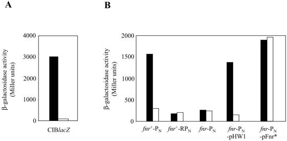
To determine whether oxygen controls the expression of the genes involved in the central pathway for anaerobic catabolism of aromatic compounds in Azoarcus sp. strain CIB, we checked the activity of the PN promoter driving the expression of the bzd catabolic genes when the cells were cultivated in the presence or absence of oxygen. To this end, we determined the β-galactosidase activity in Azoarcus sp. strain CIB lacZ, which harbors the PN::lacZ translational fusion stably inserted into the chromosome of Azoarcus sp. strain CIB (Table 1), after 48 h of anaerobic or aerobic growth on 3 mM benzoate. As shown in Fig. 2A, the levels of β-galactosidase were 1 order of magnitude higher when oxygen was absent than when it was present in the growth medium, and similar results were obtained along the growth curve (data not shown). These results suggest that indeed oxygen plays a major role in the expression of the bzd genes by inhibiting the activity of the PN promoter.
FIG. 2.
β-Galactosidase activities of E. coli and Azoarcus sp. strain CIB harboring PN::lacZ translational fusions. (A) Azoarcus sp. strain CIB lacZ (PN::lacZ) cells were grown for 48 h in MC medium containing 3 mM benzoate either aerobically (empty bar) or anaerobically in the presence of 10 mM nitrate (filled bar). (B) E. coli M182 cells (fnr+) carrying plasmid pSJ3PN (PN) (PN::lacZ) or plasmid pSJ3RPN (RPN) (bzdR-PN::lacZ) and E. coli JRG1728 cells (fnr) carrying plasmid pSJ3PN (PN), pHW1, or pQE60-His6Fnr* (pFnr*) were grown anaerobically (filled bars) or aerobically (empty bars) in glucose-containing LB medium until they reached stationary phase. β-Galactosidase activity was measured as described in Materials and Methods. The results of one experiment are shown, and the values were reproducible in three separate experiments with standard deviations of <10%.
The effect of oxygen on the activity of the PN promoter was also analyzed in a heterologous host, such as E. coli. Thus, whereas E. coli M182 cells harboring plasmid pSJ3PN (PN::lacZ) showed β-galactosidase activity along the growth curve when they grew anaerobically, the β-galactosidase levels of the same cells growing aerobically were significantly reduced and similar to those of E. coli M182 cells harboring plasmid pSJ3RPN (PR-bzdR/PN::lacZ), which expresses the BzdR repressor that inhibits the PN promoter (4) (Fig. 2B). Therefore, these data confirm the negative effect of oxygen on the transcription of the bzd catabolic genes.
The role of oxygen in repressing the expression of genes involved in aromatic catabolic pathways has been reported before. Thus, the expression of the badDEFG operon of R. palustris, encoding the four subunits of benzoyl-CoA reductase, is dramatically decreased under aerobic conditions (18). Strong down-regulation of the synthesis of benzoyl-CoA reductase of T. aromatica was found in response to oxygen, since the protein was immunologically detected only in trace amounts in aerobically grown cells. The bss genes encoding the benzylsuccinate synthase involved in toluene degradation in M. magnetotacticum TS-6 were transcribed only in anaerobically toluene-grown cells. However, there are some reports showing that genes encoding oxygen-sensitive enzymes, such as benzoyl-CoA reductase (bcr genes) from M. magnetotacticum TS-6 (47) and benzylsuccinate synthase (bss genes) from Thauera strain DNT-1 (47), are transcribed not only under anaerobic conditions, but also in cells growing aerobically in benzoate and toluene, respectively. Therefore, it appears that each organism has evolved a particular regulatory strategy for expression of the genes involved in the anaerobic catabolism of aromatic compounds.
Role of Fnr in the activity of the PN promoter.
An exhaustive analysis of the PN promoter region revealed the sequence 5′-TTGACTTAGATCAA-3′ centered at position −41.5 from the transcription start point (Fig. 3C). This sequence is almost identical to the consensus sequence, TTGAT-N4-ATCAA (where N is any of the four bases), for binding to Fnr from E. coli (50). This observation suggested that a protein of the Fnr/Crp superfamily could be involved in the regulation of the PN promoter by binding to its cognate sequence in response to oxygen deprivation. To investigate the potential role of the E. coli Fnr protein in the activity of the PN promoter, we measured the β-galactosidase activity from plasmid pSJ3PN (PN::lacZ) in E. coli JRG1728, an fnr derivative from E. coli strain M182. As shown in Fig. 2B, E. coli cells lacking Fnr did not show activity of the PN promoter when they were growing either in the presence or in the absence of oxygen. However, E. coli JRG1728(pSJ3PN) cells harboring pHW1, a plasmid that constitutively expresses the fnr gene in trans (Table 1), showed significant β-galactosidase activity similar to that of the fnr+ E. coli strain M182(pSJ3PN) when the cells were grown in the absence of oxygen (Fig. 2B). These data strongly suggest that Fnr is required for the anaerobic induction of the PN promoter in E. coli. To further confirm this assumption, we checked the expression of the PN::lacZ fusion in the presence of Fnr*, a constitutively active Fnr mutant protein that carries a D154A substitution able to form a dimer and to bind DNA in the presence of oxygen (26, 34, 62). As shown in Fig. 2B, the Fnr* protein expressed from plasmid pQE60-His6Fnr* (60) allowed high activity of the PN promoter when E. coli JRG1728(pSJ3PN, pQE60-His6Fnr*) cells were grown either in the presence or in the absence of oxygen. All these results taken together indicate that Fnr is needed for PN activity in E. coli and that the oxygen-dependent repression of the bzd genes could be bypassed by the expression of a constitutively active Fnr* protein.
FIG. 3. In vitro binding of His6-Fnr* to the PN promoter. (A) Gel retardation analyses were performed as indicated in Materials and Methods. Lane 1 shows the free PN probe; lanes 2 to 5 show retardation assays containing 1, 2.5, 5, and 10 nM of purified His6-Fnr* protein, respectively. (B) DNase I footprinting analysis of the interaction of purified His6-Fnr* and RNAP with the PN promoter region. The DNase I footprinting experiments were carried out using the PN probe labeled as indicated in Materials and Methods. Lane 1 shows a footprinting assay in the absence of proteins. Lanes 2 to 4 show footprinting assays containing 50, 100, and 150 nM purified His6-Fnr*, respectively. Lanes 5 to 7 show footprinting assays containing 50, 100, and 150 nM purified E. coli RNAP, respectively. Lanes 8 to 10 show footprinting assays containing 50 nM of His6-Fnr* and 50, 100, and 150 nM of RNAP, respectively. Lanes AG show the A+G Maxam and Gilbert sequencing reactions. An expanded view of the promoter region protected by purified His6-Fnr* (Fnr operator) is shown at the right, and the phosphodiester bonds hypersensitive to DNase I cleavage are indicated with asterisks. The two regions flanking the Fnr operator that are protected by RNAP are marked by brackets. The −10 and −35 boxes and the transcription initiation site (+1) of the PN promoter are also shown. (C) Expanded view of the PN promoter. The sequence from positions −174 to +79 is indicated. The transcription start site (+1) and the inferred −10 and −35 boxes of the PN are indicated. The ribosome binding site (RBS) and the ATG start codon of the bzdN gene are also shown in italics and boldface, respectively. The BzdR-binding regions I, II, and III (operators) and the Fnr-binding site (FNR) are boxed. The inverted repeats of the consensus Fnr-binding sequence are marked with convergent arrows above the sequence.
Fnr binding to the PN promoter.
To study the in vitro interaction of the Fnr* protein with the PN promoter, we purified the E. coli Fnr* mutant protein that retains activity in the presence of oxygen (26, 62). The purification of His-tagged Fnr* protein was carried out from E. coli cell extracts by using nickel affinity chromatography as described previously (4, 60) (see Materials and Methods). To demonstrate the interaction of Fnr with the PN promoter, gel retardation assays were performed by using purified His6-Fnr* protein and a 376-bp DNA fragment that carries the PN promoter region from position −293 to +83 (PN probe). The His6-Fnr* protein was able to retard the migration of the PN probe in a protein concentration-dependent manner (Fig. 3A). Binding of His6-Fnr* to the PN promoter was specific because it was inhibited by adding unlabeled PN probe to the retardation assays and it was not affected by adding an unlabeled heterologous probe (data not shown).
The Fnr-binding site (operator) in the PN promoter was identified by using DNase I footprinting assays. As shown in Fig. 3B, the His6-Fnr* protein protected a DNA region spanning from position −26 to −57 with respect to the transcription start site of the PN promoter (Fig. 3C). The protected region contains the palindromic sequence (TTGACTTAGATCAA) reported above, which is almost identical to the consensus Fnr-binding sequence (50), confirming such a region as the Fnr operator at the PN promoter. The location of the Fnr-binding site centered at position −41.5 from the transcription start site and overlapping the −35 box fits perfectly with PN being a typical class II Fnr-dependent promoter (24, 42). Although putative Fnr consensus binding sequences centered at positions −39.5 and −42.5 were postulated to drive the expression of the badDEFG operon (18) and of the hbaR gene (19) in R. palustris, our results provide the first experimental demonstration that an Fnr-binding site is involved in the activation of a promoter running the expression of genes for the anaerobic catabolism of aromatic compounds.
Fnr-mediated transcriptional activation of the PN promoter.
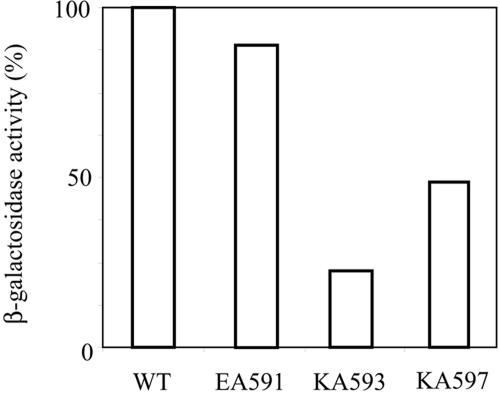
In most cases, transcription activation in class II-dependent promoters is thought to be stimulated through direct protein-protein interaction with RNAP (14, 42). DNase I footprinting assays revealed that binding of RNAP to the PN promoter is significantly increased in the presence of the His6-Fnr* protein (Fig. 3B). In this sense, the slightly long distance (19 nucleotides) between the −10 (TAACAT) and −35 (TCAACA) boxes typical of σ70-dependent promoters might explain the requirement for Fnr to facilitate the formation of the RNAP-PN closed complex. Increasing concentrations of RNAP result in increasing protection both upstream and downstream of the Fnr-binding site at the PN promoter (Fig. 3B), suggesting that Fnr interacts with RNAP. The upstream protection could be attributed to the αCTD of RNAP, since it has been suggested that in transcriptionally active complexes at class II Fnr-dependent promoters, one αCTD subunit of RNAP binds to the minor groove near position −61, immediately upstream of the bound Fnr homodimer, and makes an interaction that contributes to its activation (3, 14, 27, 60). On the other hand, the observed protection downstream of the Fnr-binding site could be due to the σ70 subunit of RNAP. Previous studies identified a small region (amino acid residues 590 to 603) in the C-terminal domain of the σ70 subunit of RNAP that is required for Fnr-dependent activation of some promoters (35). This C-terminal region of the σ70 subunit contains one acidic residue (E591) and six basic residues (K593, R596, K597, R599, H600, and R603), and amino acid substitutions within this region decrease Fnr-dependent transcription activation (7, 56). To address whether activation at the PN promoter is directed through direct contact between the C-terminal domain of the σ70-RNAP and Fnr, PN::lacZ expression was measured from anaerobically growing E. coli cells containing either wild-type σ70 or one of the previously characterized σ70 mutants [σ70(EA591), σ70(KA593), or σ70(KA597)] (see Materials and Methods). The mutant strains tested here grew at the same rate in liquid medium and formed colonies comparable in size to those of the wild-type strain, as has been previously reported (35). We observed that under anaerobic conditions, the alanine substitutions at positions 593 (KA593) and 597 (KA597) of σ70 reduced the activity of the PN promoter in vivo, whereas the EA591 mutation had no significant effect on PN activity (Fig. 4). Similar results were obtained previously at the Fnr-dependent E. coli pnarG and pdmsA promoters (35), suggesting that the Fnr-σ70-RNAP contact plays a crucial role in activation of the class II Fnr-dependent PN promoter.
FIG. 4.
Effects of wild-type σ70 and some σ70 mutants on the Fnr-mediated expression of the PN::lacZ fusion in E. coli. E. coli PK3300 cells harboring plasmid pBBR5PN (PN::lacZ) and either plasmid pGEX-2T σ70 (WT), pGEX-2Tσ70(EA591)(EA591), pGEX-2Tσ70(KA593)(KA593), or pGEX-2Tσ70(KA597)KA597) (Table 1) were grown anaerobically for 10 h in M63 minimal medium containing 20 mM glycerol in the presence of 20 μg/ml tryptophan to an A600 of 0.4. β-Galactosidase activity is shown as the percentage of β-galactosidase activity with respect to that of cells containing wild-type σ70 (370 Miller units). The results of one experiment are shown, and the values were reproducible in three independent experiments with standard deviations of <10%.
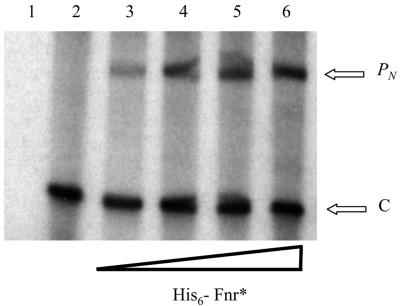
To further study the role of the Fnr protein as a transcriptional activator of the PN promoter, we performed in vitro transcription assays using purified His6-Fnr* protein, the E. coli RNAP, and the plasmid pJCD-PN, which contains the PN promoter, as a supercoiled DNA template. As shown in Fig. 5, formation of the expected 184-nucleotide transcript due to the activity of the PN promoter was stimulated by increasing concentrations of the His6-Fnr* protein in the transcription mixtures. In the absence of His6-Fnr*, the 105-nucleotide control mRNA was the only transcript observed (Fig. 5), even using RNAP at a concentration at which RNAP binds to PN alone (Fig. 3B, lanes 5 to 7). Therefore, although Fnr increases the affinity of the RNAP for the PN promoter (Fig. 3B, lanes 8 to 10), the in vitro transcription experiments revealed that Fnr is essential in transcription initiation at some step after RNAP-PN closed-complex formation. In this sense, it has been shown that Fnr, as well as some other members of the Fnr/Crp superfamily, such as Crp, activate transcription in some class II Fnr-dependent promoters by promoting isomerization from the transcriptionally inactive closed complex to the transcriptionally active open complex (51, 60).
FIG. 5.
Effect of His6-Fnr* on in vitro transcription from PN. Multiple-round transcription reactions were carried out by using pJCD-PN, a template that produces a control mRNA of 105 nucleotides (C) and an mRNA from PN of 184 nucleotides (PN). Lanes 2 to 6, in vitro transcription reactions performed with 100 nM E. coli RNAP and 0, 1, 2.5, 5, and 10 nM of His6-Fnr*, respectively. Lane 1 shows a reaction without RNAP.
The in vitro results correlate perfectly with the in vivo transcription experiments (see above), demonstrating that Fnr is strictly required for an efficient activation of the PN promoter and suggesting that the lack of expression from the PN promoter under aerobic conditions is due to the failure of wild-type Fnr protein to interact with the target promoter. To determine the mechanism of activation of PN, further studies are needed to identify the transcription initiation step facilitated by Fnr.
The AcpR protein is involved in the activation of the PN promoter in Azoarcus sp. strain CIB.
Once we had demonstrated the role of the Fnr protein from E. coli as a transcriptional activator of the heterologous PN promoter, we wanted to identify the Fnr ortholog controlling the expression of the bzd genes in the homologous system, Azoarcus sp. strain CIB. A recent amino acid sequence comparison analysis revealed the existence of a putative fnr ortholog (ebA5149) in the genome of Azoarcus sp. strain EbN1, the sole Azoarcus strain whose genome is known so far. The fnr ortholog is located between the hemN gene (ebA5151), which codes for a putative oxygen-independent coprophorphyrinogen III oxidase that is also regulated by Fnr in Pseudomonas, and the hypothetical ebA5146 gene (41). Based on the Azoarcus sp. strain EbN1 sequence, two oligonucleotides were designed at the 3′ end of ebA5146 and the 5′ end of hemN, respectively, to PCR amplify a sequence of 1,220 bp from the genome of Azoarcus sp. strain CIB. The amplified sequence contains the predicted 3′ and 5′ ends of the orthologous ebA5146 and hemN genes flanking an fnr gene (hereafter referred as the acpR gene) in Azoarcus sp. strain CIB. Interestingly, the association of fnr with hemN has also been reported in other organisms (28). The acpR gene codes for a protein of 248 amino acids that shows a significant amino acid sequence similarity to members of the Fnr/Crp superfamily of transcriptional regulators, such as the putative Fnr orthologs (EbA5149) from Azoarcus sp. strain EbN1 (accession number CAI09052; 94% identity), Dechloromonas aromatica strain RCB (accession number ZP_00150505; 75% identity), Rubrivirax gelatinosus strain PM1 (accession number AAW66138; 68% identity), Polaromonas sp. strain JS666 (accession number ZP_00364405; 58% identity), and Ralstonia solanacearum (accession number CAD14985; 54% identity).
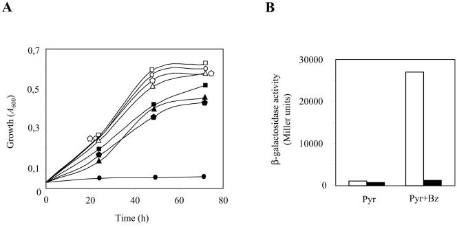
To check the role of the AcpR protein in the expression of the bzd cluster in Azoarcus sp. strain CIB, we constructed an Azoarcus sp. strain CIB mutant strain harboring a disrupted acpR gene, as detailed in Materials and Methods. We observed that the Azoarcus sp. strain CIBdacpR mutant strain was unable to grow anaerobically on aromatic compounds, such as benzoate (Fig. 6A), phenylacetate, or 4-hydroxybenzoate, that are mineralized through the bzd-encoded central pathway. On the other hand, aerobic growth on aromatic compounds was not affected in the mutant strain (data not shown). Anaerobic growth of Azoarcus sp. strain CIBdacpR on aromatic compounds was restored when the mutant cells harbored the plasmid pBBR1MCS-5acpR, which expresses the acpR gene under the control of the Plac promoter (Table 1) (Fig. 6A). These results suggest, therefore, that AcpR is essential for the anaerobic expression of the bzd cluster.
FIG. 6.
AcpR and Fnr* control the expression of the bzd genes in Azoarcus sp. strain CIB. (A) Growth curves of Azoarcus sp. strain CIB (square), Azoarcus sp. strain CIBdacpR (circle), Azoarcus sp. strain CIBdacpR carrying plasmid pBBR1MCS-5acpR (triangle), and Azoarcus sp. strain CIBdacpR carrying plasmid pIZ-FNR* (pentagon) growing anaerobically in MC medium containing succinate (empty symbols) or benzoate (filled symbols). (B) Azoarcus sp. strain CIB (open blocks) and Azoarcus sp. strain CIBdacpR (filled blocks) carrying plasmid pBBR5PN (PN::lacZ) were grown anaerobically for 48 h in MC medium containing 0.4% (wt/vol) pyruvate (Pyr) or 0.4% (wt/vol) pyruvate plus 3 mM benzoate (Pyr+Bz). β-Galactosidase activity was measured as described in Materials and Methods. The results of one experiment are shown, and values were reproducible in three separate experiments with standard deviations of <10%.
To determine whether the acpR gene product is indeed needed for the activity of the PN promoter, Azoarcus sp. strain CIB and Azoarcus sp. strain CIBdacpR were transformed with pBBR5PN, a promiscuous plasmid that harbors the PN::lacZ translational fusion. Whereas significant β-galactosidase activity was observed when Azoarcus sp. strain CIB(pBBR5PN) cells were grown anaerobically in benzoate-containing minimal medium, β-galactosidase activity in the Azoarcus sp. strain CIB dacpR(pBBR5PN) mutant strain growing either in the presence or the absence of benzoate reached only the basal levels observed when Azoarcus sp. strain CIB uses nonaromatic compounds, such as pyruvate, as the sole carbon source (Fig. 6B). These data strongly suggest that AcpR is a transcriptional activator required for the activity of the PN promoter when Azoarcus sp. strain CIB grows on benzoate under anaerobic conditions. In this sense, the physiological role of AcpR in Azoarcus sp. strain CIB in regulating aromatic-compound degradation in response to oxygen appears to be equivalent to that carried out by AadR, another member of the Fnr/Crp superfamily, in R. palustris (17, 18, 19).
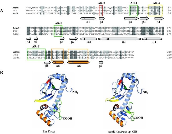
A phylogenetic analysis based on a multiple amino acid sequence alignment of AcpR with other members of the Fnr/Crp superfamily revealed that AcpR branches within the Fnr group (data not shown) that contains the well-characterized E. coli Fnr protein (43% amino acid sequence identity). Interestingly, although AcpR and AadR may play similar physiological roles in the cell, they show only 34% amino acid sequence identity, and in fact, AadR has been classified within a different group (FnrN) of the Fnr/Crp superfamily (28). In the E. coli Fnr protein, four cysteine residues, Cys-20, -23, -29, and -122, contribute to the formation of an iron-sulfur [4Fe-4S]2+ cluster that is essential for activation (dimerization) of Fnr under anaerobic conditions (23). Whereas these four cysteine residues, Cys-18, -21, -27, and -120, are perfectly conserved in AcpR from Azoarcus, in AadR only two cysteine residues, Cys-20 and -116, are conserved (Fig. 7A). The activation regions AR-1, AR-2, and AR-3 that are involved in the interaction of the E. coli Fnr protein with the αCTD, αNTD, and σ70-CTD subunits of the RNAP, respectively (16, 32, 55, 58), are also conserved in AcpR and AadR, although in the latter there is low amino acid sequence conservation in the AR-1 region (Fig. 7A). By using the three-dimensional structure of the E. coli CRP protein as a template, a three-dimensional model of AcpR from Azoarcus sp. strain CIB was generated (Fig. 7B). By comparing the model of AcpR with that previously obtained for the E. coli Fnr protein (31), we could determine the location of the putative AR-1, AR-2, and AR-3 regions of AcpR and the winged helix-turn-helix (HTH) motif (2) implicated in DNA recognition (Fig. 7B).
FIG.7.
Amino acid sequence alignment and three-dimensional model of AcpR. (A) Amino acid sequence alignment of AcpR, Fnr, and AadR. The accession numbers of the sequences are as follows: AcpR from Azoarcus sp. strain CIB, AY996130; Fnr from E. coli, AY629341; and AadR of R. palustris, CAE29675. The amino acid residues of each sequence are numbered on the right. Sequences were aligned using the multiple sequence alignment program Clustal (53). Amino acids are indicated by their standard one-letter codes. Dark gray represents identical residues in the three sequences. Light gray indicates identical residues in two of the three sequences. The AR-1, AR-2, and AR-3 regions are boxed in green, red, and yellow, respectively. Secondary-structure elements predicted from the AcpR three-dimensional model (B) are drawn at the bottom of the alignment. (B) Three-dimensional model of the E. coli Fnr and Azoarcus sp. strain CIB AcpR proteins; ribbon diagram showing the six α-helices (ribbons) and the nine β-strands (arrows). In green, red, and yellow are indicated the corresponding surfaces of the AR-1, AR-2, and AR-3 regions, respectively. The core helix-turn-helix fold of the winged HTH motif (2) is indicated in orange. The corresponding N and C termini are labeled NH2 and COOH, respectively.
Despite the relevance of the predicted structural similarity between Fnr and AcpR, the two proteins do not have the same regulatory functions within the cell. Thus, in E. coli, the lack of the Fnr protein has a pleiotropic effect on the expression of a moderate number of genes, including the incapacity of the mutant strain to grow using nitrate or fumarate as a final electron acceptor (54). In contrast, the lack of AcpR in Azoarcus did not affect anaerobic growth using nitrate as a final electron acceptor or growth on nonaromatic carbon sources, such as succinate (Fig. 6A), acetate, or malate, but altered the ability to catabolize aromatic compounds through the benzoyl-CoA pathway. Therefore, since the physiological role of AcpR appears to focus on the transcriptional control of the bzd cluster rather than being a global regulatory protein controlling the expression of important gene programs, such as other members of the Fnr group (28), AcpR can be considered an aromatic central pathway regulator for the anaerobic catabolism of aromatic compounds in Azoarcus. Since AadR from R. palustris plays a similar specialized role in controlling the expression of genes involved in anaerobic catabolism of aromatic compounds (17, 18, 19), such specialization appears to be present in different groups within the Fnr/Crp superfamily, and it might be a general principle in aromatic degraders, such as Azoarcus (41) and R. palustris (33), which contain a significant array of multiple Crp/Fnr regulators. Thus, in the genome of Azoarcus sp. strain EbN1 there are seven genes encoding putative transcriptional regulators of the Crp/Fnr family, i.e., one Fnr-like protein (AcpR), one Crp-like protein, three Dnr-like proteins, and two Nnr-like proteins (41). A multiple amino acid sequence alignment of the HTH motifs of these seven regulators revealed significant differences among them (data not shown), which agrees with the assumption that each individual member becomes adapted to fulfill a particular physiological role.
E. coli Fnr* behaves as an aromatic central-pathway regulator in Azoarcus sp. strain CIB.
As indicated above, Fnr from E. coli is a highly versatile global regulator that controls a significant number of cellular processes, and we have shown here that it also acts as a transcriptional activator of the bzd genes when expressed in E. coli. To check whether Fnr replaces the function of AcpR in Azoarcus sp. strain CIB, we cloned and expressed the FNR* gene under the Ptac promoter in plasmid pIZ-FNR* (Table 1). Whereas Azoarcus sp. strain CIB dacpR is unable to grow anaerobically using benzoate as a carbon source, cells containing plasmid pIZ-FNR* show a growth curve on benzoate similar to that of the wild-type Azoarcus sp. strain CIB (Fig. 6A), indicating that Fnr* is able to efficiently complement the lack of AcpR, and therefore, it also behaves as an aromatic central-pathway regulator in Azoarcus. This result is in agreement with the observed similarity between the HTH motifs of Fnr and AcpR (Fig. 7A). Moreover, when plasmid pIZ-FNR* was expressed in Azoarcus sp. strain CIB lacZ (Table 1) and the β-galactosidase levels of the resulting strain were analyzed, we did not observe significant activity when the cells were grown in pyruvate, but a fivefold induction of the PN::lacZ expression was detected when the cells were grown aerobically in the presence of pyruvate and benzoate (data not shown). These data show that E. coli Fnr* allows the expression of the bzd genes when Azoarcus grows aerobically in the presence of benzoate, and they constitute the first example of how the expression of an anaerobic pathway for the catabolism of aromatic compounds can be switched to aerobic conditions just by changing a key regulatory protein.
In summary, in this work we have shown that the expression of the bzd cluster for the anaerobic catabolism of aromatic compounds of Azoarcus sp. strain CIB is under the control of a complex regulatory system that involves not only the reported BzdR specific transcriptional repressor (4), but also a devoted transcriptional activator, the AcpR protein, that belongs to the Fnr/Crp superfamily of regulators and whose activity can be replaced by the Fnr protein from E. coli. Whereas benzoyl-CoA is the inducer molecule that prevents BzdR-mediated repression at the PN promoter, the absence of oxygen must be the environmental signal that triggers activation of PN by the AcpR protein and allows expression of the bzd genes when Azoarcus sp. strain CIB grows anaerobically in the presence of benzoate. The molecular mechanisms underlying these two different control levels at the PN promoter are under study.
Acknowledgments
This work was supported by Comunidad Autónoma de Madrid Grant 07 M/0076/2002, Proyecto Intramural of CSIC 2004-20E073, and Comisión Interministerial de Ciencia y Tecnología Grants BIO2003-01482 and VEM2003-20075-CO2-02. G. Durante-Rodríguez is the recipient of a predoctoral fellowship from the Plan Nacional de Formación de Personal Investigador-MEC. M. Carmona is a holder of the Ramón y Cajal Program of the Spanish Ministerio de Educación y Ciencia.
The technical work of I. Alonso and M. Zazo is greatly appreciated. The help of A. Díaz, S. Carbajo, M. Cayuela, and G. Porras with sequencing is gratefully acknowledged. We are indebted to S. Busby and C. Gross for kindly providing some strains and plasmids. We are indebted to R. Rabus and J. Heider for providing us the Azoarcus sp. strain EbN1 sequence before publication.
REFERENCES
- 1.Altschul, S. F., W. Gish, W. Miller, E. W. Myers, and D. J. Lipman. 1990. Basic local alignment search tool. J. Mol. Biol. 215:403-410. [DOI] [PubMed] [Google Scholar]
- 2.Aravind, L., V. Anantharaman, S. Balaji, M. M. Babu, and L. M. Iyer. 2005. The many faces of the helix-turn-helix domain: transcription regulation and beyond. FEMS Microbiol. Rev. 29:231-262. [DOI] [PubMed] [Google Scholar]
- 3.Attey, A., T. Belyaeva, N. Savery, J. Hoggett, N. Fujita, A. Ishihama, and S. Busby. 1994. Interactions between the cyclic AMP receptor protein and the alpha subunit of RNA polymerase at the Escherichia coli galactose operon P1 promoter. Nucleic Acids Res. 21:4375-4380. [DOI] [PMC free article] [PubMed] [Google Scholar]
- 4.Barragán, M. J. L., B. Blázquez, M. T. Zamarro, J. M. Mancheño, J. L. García, E. Díaz, and M. Carmona. 2005. BzdR, a repressor that controls the anaerobic catabolism of benzoate in Azoarcus sp. CIB, is the first member of a new subfamily of transcriptional regulators. J. Biol. Chem. 280:10683-10694. [DOI] [PubMed] [Google Scholar]
- 5.Barragán, M. J. L., M. Carmona, M. T. Zamarro, B. Thiele, M. Boll, G. Fuchs, J. L. García, and E. Díaz. 2004. The bzd gene cluster, coding for anaerobic benzoate catabolism, in Azoarcus sp. strain CIB. J. Bacteriol. 186:5762-5774. [DOI] [PMC free article] [PubMed] [Google Scholar]
- 6.Barragán, M. J. L., E. Díaz, J. L. García, and M. Carmona. 2004. Genetic clues on the evolution of anaerobic catabolism of aromatic compounds. Microbiology 150:2018-2021. [DOI] [PubMed] [Google Scholar]
- 7.Bell, A., and S. Busby. 1994. Location and orientation of an activating region in the Escherichia coli transcription factor FNR. Mol. Microbiol. 11:383-390. [DOI] [PubMed] [Google Scholar]
- 8.Bell, A., J. Cole, and S. Busby. 1990. Molecular genetic analysis of an FNR-dependent anaerobically inducible Escherichia coli promoter. Mol. Microbiol. 4:1753-1763. [DOI] [PubMed] [Google Scholar]
- 9.Blake, T., A. Barnanrd, S. Busby, and J. Green. 2002. Transcription activation by FNR: evidence for a functional activating region 2. J. Bacteriol. 184:5855-5861. [DOI] [PMC free article] [PubMed] [Google Scholar]
- 10.Boll, M., and G. Fuchs. 1995. Benzoyl-coenzyme A reductase (dearomatizing), a key enzyme of anaerobic aromatic metabolism. ATP dependence of the reaction, purification and some properties of the enzyme from Thauera aromatica strain K172. Eur. J. Biochem. 234:921-933. [DOI] [PubMed] [Google Scholar]
- 11.Bradford, M. M. 1976. A rapid and sensitive method for the quantitation of microgram quantities of protein utilizing the principle of protein-dye binding. Anal. Biochem. 72:248-254. [DOI] [PubMed] [Google Scholar]
- 12.Brodsky, L. I., V. V. Ivanov, Y. L. Kalaidzidis, A. M. Leontovich, V. K. Nikolaev, S. I. Feranchuck, and V. A. Drachev. 1995. GeneBee-NET: Internet-based server for analyzing biopolymer structure. Biochemistry 60:923-928. [PubMed] [Google Scholar]
- 13.Browning, D., D. Lee, and S. Busby. 2002. Secrets of bacterial transcription initiation taught by the Escherichia coli FNR protein, p. 127-142. In D. Hodgson and C. Thomas (ed.), Signals, switches, regulons, and cascades. Cambridge University Press, Cambridge, United Kingdom.
- 14.Busby, S., and R. H. Ebright. 1999. Transcription activation by catabolite activator protein (CAP). J. Mol. Biol. 293:199-213. [DOI] [PubMed] [Google Scholar]
- 15.Carmona, M., and B. Magasanik. 1996. Activation of transcription at sigma 54-dependent promoters on linear templates requires intrinsic or induced bending of the DNA. J. Mol. Biol. 261:348-356. [DOI] [PubMed] [Google Scholar]
- 16.de Lorenzo, V., and K. N. Timmis. 1994. Analysis and construction of stable phenotypes in gram-negative bacteria with Tn5- and Tn10-derived minitransposons. Methods Enzymol. 235:386-405. [DOI] [PubMed] [Google Scholar]
- 17.Dispensa, M., C. T. Thomas, M. K. Kim, J. A. Perrotta, J. Gibson, and C. S. Harwood. 1992. Anaerobic growth of Rhodopseudomonas palustris on 4-hydroxybenzoate is dependent on AadR, a member of the cyclic AMP receptor protein family of transcriptional regulators. J. Bacteriol. 174:5803-5813. [DOI] [PMC free article] [PubMed] [Google Scholar]
- 18.Egland, P. G., and C. Harwood. 1999. BadR, a new MarR family member, regulates anaerobic benzoate degradation by Rhodopseudomonas palustris in concert with AadR, an Fnr family member. J. Bacteriol. 181:2102-2109. [DOI] [PMC free article] [PubMed] [Google Scholar]
- 19.Egland, P. G., and C. Harwood. 2000. HbaR, a 4-hydroxybenzoate sensor and FNR-CRP superfamily member, regulates anaerobic 4-hydroxybenzoate degradation by Rhodopseudomonas palustris. J. Bacteriol. 182:100-106. [DOI] [PMC free article] [PubMed] [Google Scholar]
- 20.Felsenstein, J. 1993. PHYLIP (Phylogenetic Interface Package), version 3.5.1. University of Washington, Seattle.
- 21.Ferrández, A., B. Miñambres, B. García, E. R. Olivera, J. M. Luengo, J. L. García, and E. Díaz. 1998. Catabolism of phenylacetic acid in Escherichia coli. Characterization of a new aerobic hybrid pathway. J. Biol. Chem. 273:25974-25986. [DOI] [PubMed] [Google Scholar]
- 22.Gibson, J., and C. S. Harwood. 2002. Metabolic diversity in aromatic compound utilization by anaerobic microbes. Annu. Rev. Microbiol. 56:345-369. [DOI] [PubMed] [Google Scholar]
- 23.Green, J., S. Irvine, W. Meng, and J. R. Guest. 1996. FNR-DNA interactions at natural and semi-synthetic promoters. Mol. Microbiol. 19:125-137. [DOI] [PubMed] [Google Scholar]
- 24.Guest, J. R., J. Green, A. S. Irvine, and S. Spiro. 1996. The FNR modulation and FNR-regulated gene expression, p. 317-342. In E. C. C. Lin and A. S. Lynch (ed.), Regulation of gene expression in Escherichia coli. R. G. Landes Company, Austin, Tex.
- 25.Harwood, C. S., G. Burchhardt, H. Herrmann, and G. Fuchs. 1999. Genetic clues on the evolution of anaerobic catabolism of aromatic compounds. FEMS Microbiol. Rev. 22:439-458. [Google Scholar]
- 26.Kiley, P. J., and W. S. Reznikoff. 1991. Fnr mutants that activate gene expression in the presence of oxygen. J. Bacteriol. 173:16-22. [DOI] [PMC free article] [PubMed] [Google Scholar]
- 27.Kolb, A., S. Busby, H. Buc, S. Garges, and S. Adhya. 1993. Transcriptional regulation by cAMP and its receptor protein. Annu. Rev. Biochem. 62:749-795. [DOI] [PubMed] [Google Scholar]
- 28.Körner, H., H. J. Sofia, and W. G. Zumft. 2003. Phylogeny of the bacterial superfamily of Crp-Fnr transcription regulators: exploiting the metabolic spectrum by controlling alternative gene programs. FEMS Microbiol. Rev. 27:559-592. [DOI] [PubMed] [Google Scholar]
- 29.Kovach, M. E., P. H. Elzer, D. S. Hills, G. T. Robertson, M. A. Farris, R. M. Roop II, and K. M. Peterson. 1995. Four new derivatives of the broad-host-range cloning vector pBBR1MCS, carrying different antibiotic-resistance cassettes. Gene 166:175-176. [DOI] [PubMed] [Google Scholar]
- 30.Laemmli, U. K. 1970. Cleavage of structural proteins during the assembly of the head of bacteriophage T4. Nature 227:680-685. [DOI] [PubMed] [Google Scholar]
- 31.Lamberg, K. E., and P. J. Kiley. 2000. FNR-dependent activation of the class II dmsA and narG promoters of Escherichia coli requires FNR-activating regions 1 and 3. Mol. Microbiol. 38:817-827. [DOI] [PubMed] [Google Scholar]
- 32.Lamberg, K. E., C. Luther, K. D. Weber, and P. J. Kiley. 2002. Characterization of activating region 3 from Escherichia coli FNR. J. Mol. Biol. 315:275-283. [DOI] [PubMed] [Google Scholar]
- 33.Larimer, F. W., P. Chain, L. Hauser, J. Lamerdin, S. Malfatti, L. Do, M. L. Land, D. A. Pelletier, J. T. Beatty, A. S. Lang, F. R. Tabita, J. L. Gibson, T. E. Hanson, C. Bobst, J. L. Torres, C. Peres, F. H. Harrison, J. Gibson, and C. S. Harwood. 2004. Complete genome sequence of the metabolically versatile photosynthetic bacterium Rhodopseudomonas palustris. Nat. Biotechnol. 22:55-61. [DOI] [PubMed] [Google Scholar]
- 34.Lazazzera, B. A., D. M. Bates, and P. J. Kiley. 1993. The activity of the Escherichia coli transcription factor FNR is regulated by a change in oligomeric state. Genes Dev. 7:1993-2005. [DOI] [PubMed] [Google Scholar]
- 35.Lonetto, M. A., V. Rhodius, K. Lamberg, P. Kiley, S. Busby, and C. Gross. 1998. Identification of a contact site for different transcription activators in region 4 of the Escherichia coli RNA polymerase σ70 subunit. J. Mol. Biol. 284:1353-1365. [DOI] [PubMed] [Google Scholar]
- 36.Lovley, D. R. 2003. Cleaning up with genomics: applying molecular biology to bioremediation. Nat. Rev. 1:35-44. [DOI] [PubMed] [Google Scholar]
- 37.Marschall, C., V. Labrousse, M. Kreimer, D. Weichart, A. Kolb, and R. Hengge-Aronis. 1998. Molecular analysis of the regulation of csiD, a carbon starvation-inducible gene in Escherichia coli that is exclusively dependent on sigma s and requires activation by cAMP-CRP. J. Mol. Biol. 276:339-353. [DOI] [PubMed] [Google Scholar]
- 38.Martínez-Pérez, O., E. Moreno-Ruiz, B. Floriano, and E. Santero. 2004. Regulation of tetralin biodegradation and identification of genes essential for expression of thn operons. J. Bacteriol. 186:6101-6109. [DOI] [PMC free article] [PubMed] [Google Scholar]
- 39.Maxam, A. M., and W. Gilbert. 1980. Sequencing end-labeled DNA with base-specific chemical cleavages. Methods Enzymol. 65:499-560. [DOI] [PubMed] [Google Scholar]
- 40.Miller, J. H. 1972. Experiments in molecular genetics. Cold Spring Harbor Laboratory Press, Cold Spring Harbor, N.Y.
- 41.Rabus, R., M. Kube, J. Heider, A. Beck, K. Heitmann, F. Widdel, and R. Reinhardt. 2005. The genome sequence of an anaerobic aromatic-degrading denitrifying bacterium, strain EbN1. Arch. Microbiol. 183:27-36. [DOI] [PubMed] [Google Scholar]
- 42.Rhodius, V. A., and S. J. W. Busby. 1998. Positive activation of gene expression. Curr. Opin. Microbiol. 1:151-159. [DOI] [PubMed] [Google Scholar]
- 43.Sambrook, J., and D. W. Russell. 2001. Molecular cloning: a laboratory manual, 3rd ed. Cold Spring Harbor Laboratory Press, Cold Spring Harbor, N.Y.
- 44.Sambrook, J., E. F. Fritsch, and T. Maniatis. 1989. Molecular cloning: a laboratory manual, 2nd ed. Cold Spring Harbor Laboratory Press. Cold Spring Harbor, N.Y.
- 45.Sawers, G. 1999. The aerobic/anaerobic interface. Curr. Opin. Microbiol. 2:181-187. [DOI] [PubMed] [Google Scholar]
- 46.Schäfer, A., A. Tauch, W. Jäger, J. Kalinowski, G. Thierbach, and A. Pühler. 1994. Small mobilizable multi-purpose cloning vectors derived from the Escherichia coli plasmids pK18 and pK19: selection of defined deletions in the chromosome of Corynebacterium glutamicum. Gene 145:69-73. [DOI] [PubMed] [Google Scholar]
- 47.Shinoda, Y., J. Akagi, Y. Uchihashi, A. Hiraishi, H. Yukawa, H. Yurimoto, Y. Sakai, and N. Kato. 2005. Anaerobic degradation of aromatic compounds by magnetospirillum strains: isolation and degradation genes. Biosci. Biotechnol. Biochem. 69:1483-1491. [DOI] [PubMed] [Google Scholar]
- 48.Smith, D. B., and K. S. Johnson. 1988. Single-step purification of polypeptides expressed in Escherichia coli as fusions with glutathione S-transferase. Gene 67:31-40. [DOI] [PubMed] [Google Scholar]
- 49.Spiro, S., and J. R. Guest. 1988. Inactivation of the FNR protein of Escherichia coli by targeted mutagenesis in the N-terminal region. Mol. Microbiol. 2:701-707. [DOI] [PubMed] [Google Scholar]
- 50.Spiro, S., and J. R. Guest. 1990. FNR and its role in oxygen-regulated gene expression in Escherichia coli. FEMS Microbiol. Rev. 75:399-428. [DOI] [PubMed] [Google Scholar]
- 51.Tagami, H., and H. Aiba. 1998. A common role of CRP in transcription activation: CRP acts transiently to stimulate events leading to open complex formation at a diverse set of promoters. EMBO J. 17:1759-1767. [DOI] [PMC free article] [PubMed] [Google Scholar]
- 52.Teodorescu, O., T. Galor, J. Pillardy, and R. Elber. 2004. Enriching the sequence substitution matrix by structural information. Proteins 54:41-48. [DOI] [PubMed] [Google Scholar]
- 53.Thompson, J. D., D. G. Higgins, and T. J. Gibson. 1994. CLUSTAL W: improving the sensitivity of progressive multiple sequence alignment through sequence weighting, position-specific gap penalties and weight matrix choice. Nucleic Acids Res. 22:4673-4680. [DOI] [PMC free article] [PubMed] [Google Scholar]
- 54.Unden, G., S. Becker, J. Bongaerts, G. Hollighaus, J. Schirawsky, and S. Six. 1995. O2-sensing and O2-dependent gene regulation in facultatively anaerobic bacteria. Arch. Microbiol. 164:81-90. [PubMed] [Google Scholar]
- 55.Weber, K. D., O. D. Vincent, and P. J. Kiley. 2005. Additional determinants within Escherichia coli FNR activating region 1 and RNA polymerase alpha subunit required for transcription activation. J. Bacteriol. 187:1724-1731. [DOI] [PMC free article] [PubMed] [Google Scholar]
- 56.West, D., R. Williams, V. Rhodius, A. Bell, N. Sharma, C. Zou, N. Fujita, A. Ishihama, and S. Busby. 1993. Interactions between the Escherichia coli cyclic AMP receptor protein and RNA polymerase at class II promoters. Mol. Microbiol. 10:789-797. [DOI] [PubMed] [Google Scholar]
- 57.Widdel, F., and R. Rabus. 2001. Anaerobic biodegradation of saturated and aromatic hydrocarbons. Curr. Opin. Biotechnol. 12:259-276. [DOI] [PubMed] [Google Scholar]
- 58.William, S. M., N. J. Savery, S. J. Busby, and H. J. Wing. 1997. Transcription activation at class I FNR-dependent promoters: identification of the activating surface of FNR and the corresponding contact site in the C-terminal domain of the RNA polymerase alpha subunit. Nucleic Acids Res. 25:4028-4034. [DOI] [PMC free article] [PubMed] [Google Scholar]
- 59.Williams, R., A. Bell, G. Sims, and S. Busby. 1991. The role of two surface exposed loops in transcription activation by the Escherichia coli CRP and FNR proteins. Nucleic Acids Res. 19:6705-6712. [DOI] [PMC free article] [PubMed] [Google Scholar]
- 60.Wing, H. J., J. Green, J. R. Guest, and S. J. Busby. 2000. Role of activating region 1 of Escherichia coli FNR protein in transcription activation at class II promoters. J. Biol. Chem. 275:29061-29065. [DOI] [PubMed] [Google Scholar]
- 61.Wing, H. J., S. M. Williams, and S. Busby. 1995. Spacing requirements for transcription activation by Escherichia coli FNR protein. J. Bacteriol. 177:6704-6710. [DOI] [PMC free article] [PubMed] [Google Scholar]
- 62.Ziegelhoffer, E. C., and P. J. Kiley. 1995. In vitro analysis of a constitutively active mutant form of the Escherichia coli global transcription factor FNR. J. Mol. Biol. 245:351-361. [DOI] [PubMed] [Google Scholar]


