

Abstract
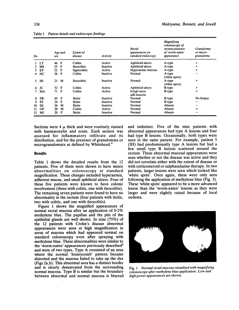
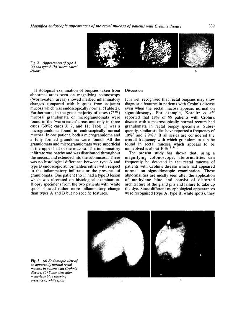
The magnified endoscopic appearances of the rectum are described in 12 patients with Crohn's disease with apparent rectal sparing on sigmoidoscopy. Five of them had minor abnormalities on colonoscopy but the remaining seven had a normal rectum. After the application of 0.2% methylene blue, examination using a magnifying endoscope (Olympus CF-HM) revealed characteristic 'worm-eaten' appearances in 75% of the patients regardless of the activity of their disease. Histological examination of biopsy specimens from these lesions showed marked inflammatory changes, and granulomas or microgranulomas were found in 75%. Inflammatory changes were not seen in mucosa which appeared normal on magnifying colonoscopy although microgranuloma were found in three cases. These observations confirm the focal nature of Crohn's disease and may suggest that the early lesions are mucosal and frequently contain granulomata.
Full text
PDF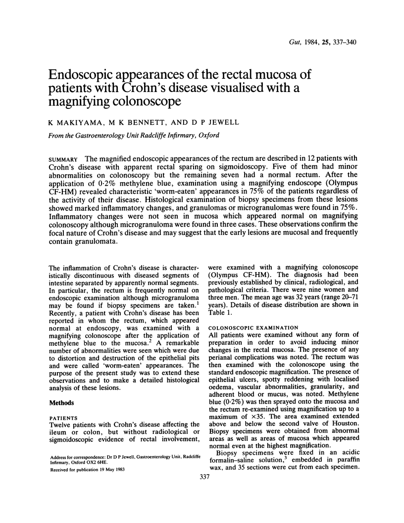
Images in this article
Selected References
These references are in PubMed. This may not be the complete list of references from this article.
- Curran R. C., Gregory J. Effects of fixation and processing on immunohistochemical demonstration of immunoglobulin in paraffin sections of tonsil and bone marrow. J Clin Pathol. 1980 Nov;33(11):1047–1057. doi: 10.1136/jcp.33.11.1047. [DOI] [PMC free article] [PubMed] [Google Scholar]
- Dunne W. T., Cooke W. T., Allan R. N. Enzymatic and morphometric evidence for Crohn's disease as a diffuse lesion of the gastrointestinal tract. Gut. 1977 Apr;18(4):290–294. doi: 10.1136/gut.18.4.290. [DOI] [PMC free article] [PubMed] [Google Scholar]
- Gear E. V., Jr Dobbins WO 3d,+DOBBINS WO III: Rectal biopsy. A review of its diagnostic usefulness. Gastroenterology. 1968 Oct;55(4):522–544. [PubMed] [Google Scholar]
- Goodman M. J., Skinner J. M., Truelove S. C. Abnormalities in the apparently normal bowel mucosa in Crohn's disease. Lancet. 1976 Feb 7;1(7954):275–278. doi: 10.1016/s0140-6736(76)91404-5. [DOI] [PubMed] [Google Scholar]
- Hill R. B., Kent T. H., Hansen R. N. Clinical usefulness of rectal biopsy in Crohn's disease. Gastroenterology. 1979 Oct;77(4 Pt 2):938–944. [PubMed] [Google Scholar]
- Korelitz B. I., Sommers S. C. Rectal biopsy in patients with Crohn's disease. Normal mucosa on sigmoidoscopic examination. JAMA. 1977 Jun 20;237(25):2742–2744. [PubMed] [Google Scholar]
- Morson B. C. Rectal and colonic biopsy in inflammatory bowel disease. Am J Gastroenterol. 1977 May;67(5):417–426. [PubMed] [Google Scholar]
- Rotterdam H., Korelitz B. I., Sommers S. C. Microgranulomas in grossly normal rectal mucosa in Crohn's disease. Am J Clin Pathol. 1977 Jun;67(6):550–554. doi: 10.1093/ajcp/67.6.550. [DOI] [PubMed] [Google Scholar]
- Surawicz C. M., Meisel J. L., Ylvisaker T., Saunders D. R., Rubin C. E. Rectal biopsy in the diagnosis of Crohn's disease: value of multiple biopsies and serial sectioning. Gastroenterology. 1981 Jan;80(1):66–71. [PubMed] [Google Scholar]