

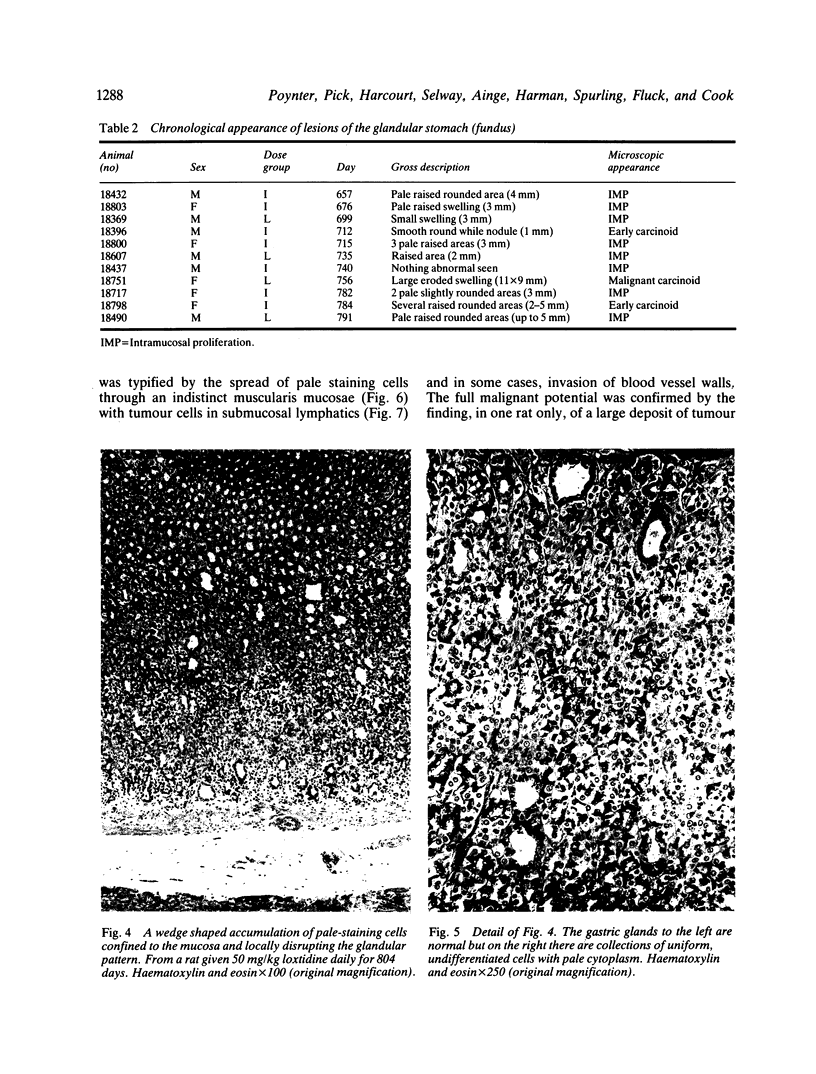
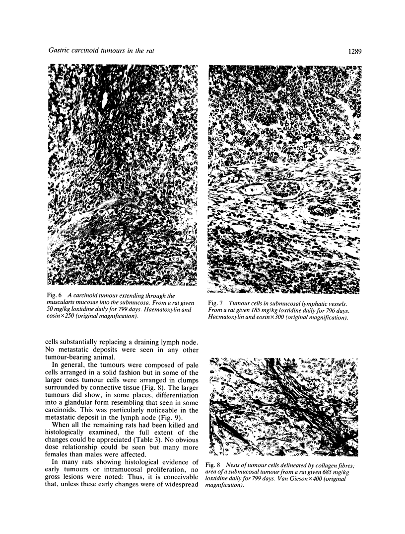
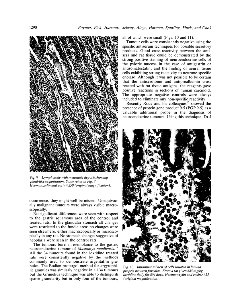
Abstract
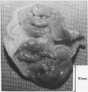
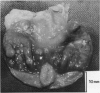
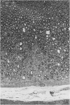
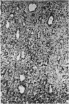
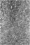
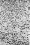
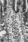
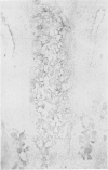
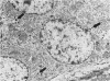
The oral administration of loxtidine, a potent histamine H2-antagonist, to a total of 378 rats at doses of 50, 185, or 685 mg/kg/day for 116 weeks resulted in the late formation of carcinoid tumours of the gastric fundus. The first such tumour was detected after 712 days of treatment. There was no dose related response; 11 rats at the low level of treatment were affected, 12 at the intermediate and 11 at the high. Twenty seven females but only seven males were affected. No gastric tumours were found in the 228 controls. There is no evidence that loxtidine acts as a direct carcinogen and it is suggested that the tumours were the result of prolonged achlorhydria produced by a potent unsurmountable histamine H2 receptor antagonist.
Full text
PDF
Images in this article
Selected References
These references are in PubMed. This may not be the complete list of references from this article.
- Beems R. B., Spit B. J., Koëter H. B., Feron V. J. Nature and histogenesis of sulfite-induced gastric lesions in rats. Exp Mol Pathol. 1982 Jun;36(3):316–325. doi: 10.1016/0014-4800(82)90061-2. [DOI] [PubMed] [Google Scholar]
- Betton G. R., Salmon G. K. Pathology of the forestomach in rats treated for 1 year with a new histamine H2-receptor antagonist, SK&F 93479 trihydrochloride. Scand J Gastroenterol Suppl. 1984;101:103–108. [PubMed] [Google Scholar]
- Capella C., Polak J. M., Timson C. M., Frigerio B., Solcia E. Gastric carcinoids of argyrophil ECL cells. Ultrastruct Pathol. 1980 Jul-Sep;1(3):411–418. doi: 10.3109/01913128009141444. [DOI] [PubMed] [Google Scholar]
- Capella C., Solcia E., Snell K. C. Ultrastructure of endocrine cells and argyrophil carcinoids of the stomach of Praomys (Mastomys) natalensis. J Natl Cancer Inst. 1973 Jun;50(6):1471–1485. doi: 10.1093/jnci/50.6.1471. [DOI] [PubMed] [Google Scholar]
- Feron V. J., Wensvoort P. Gastric lesions in rats after the feeding of sulphite. Pathol Eur. 1972;7(2):103–111. [PubMed] [Google Scholar]
- Grimelius L. A silver nitrate stain for alpha-2 cells in human pancreatic islets. Acta Soc Med Ups. 1968;73(5-6):243–270. [PubMed] [Google Scholar]
- Heyderman E. Immunoperoxidase technique in histopathology: applications, methods, and controls. J Clin Pathol. 1979 Oct;32(10):971–978. doi: 10.1136/jcp.32.10.971. [DOI] [PMC free article] [PubMed] [Google Scholar]
- Häkanson R., Hedenbro J., Liedberg G., Rehfeld J. F., Stadil F. Activation of histidine decarboxylase by H2-receptor blockade: mechanism of action. Br J Pharmacol. 1975 Jan;53(1):127–130. doi: 10.1111/j.1476-5381.1975.tb07339.x. [DOI] [PMC free article] [PubMed] [Google Scholar]
- KAHLSON G., ROSENGREN E., SVAHN D., THUNBERG R. MOBILIZATION AND FORMATION OF HISTAMINE IN THE GASTRIC MUCOSA AS RELATED TO ACID SECRETION. J Physiol. 1964 Nov;174:400–416. doi: 10.1113/jphysiol.1964.sp007494. [DOI] [PMC free article] [PubMed] [Google Scholar]
- Kroes R., Garbis-Berkvens J. M., de Vries T., van Nesselrooy H. J. Histopathological profile of a Wistar rat stock including a survey of the literature. J Gerontol. 1981 May;36(3):259–279. doi: 10.1093/geronj/36.3.259. [DOI] [PubMed] [Google Scholar]
- Kunze E., Schauer A., Eder M., Seefeldt C. Early sequential lesions during development of experimental gastric cancer with special reference to dysplasias. J Cancer Res Clin Oncol. 1979;95(3):247–264. doi: 10.1007/BF00410646. [DOI] [PubMed] [Google Scholar]
- Rode J., Dhillon A. P., Doran J. F., Jackson P., Thompson R. J. PGP 9.5, a new marker for human neuroendocrine tumours. Histopathology. 1985 Feb;9(2):147–158. doi: 10.1111/j.1365-2559.1985.tb02431.x. [DOI] [PubMed] [Google Scholar]
- Sher S. P. Tumors in control hamsters, rats, and mice: literature tabulation. Crit Rev Toxicol. 1982 Mar;10(1):49–79. doi: 10.3109/10408448209033631. [DOI] [PubMed] [Google Scholar]
- Soga J., Koro T., Tazawa K., Kanahara H., Sano M. Argyrophil cell microneoplasia in the Mastomys' stomach--an observation on early carcinoid formation. J Natl Cancer Inst. 1975 Oct;55(4):1001–1006. doi: 10.1093/jnci/55.4.1001. [DOI] [PubMed] [Google Scholar]
- Solcia E., Capella C., Buffa R., Usellini L., Frigerio B., Fontana P. Endocrine cells of the gastrointestinal tract and related tumors. Pathobiol Annu. 1979;9:163–204. [PubMed] [Google Scholar]
- Streett C. S., Cimprich R. E., Robertson J. L. Pathologic findings in the stomachs of rats treated with the H2-receptor antagonist tiotidine. Scand J Gastroenterol Suppl. 1984;101:109–117. [PubMed] [Google Scholar]
- Sugimura T., Fujimura S. Tumour production in glandular stomach of rat by N-methyl-N'-nitro-N-nitrosoguanidine. Nature. 1967 Dec 2;216(5118):943–944. doi: 10.1038/216943a0. [DOI] [PubMed] [Google Scholar]
- Tahara E., Ito H., Nakagami K., Shimamoto F. Induction of carcinoids in the glandular stomach of rats by N-methyl-N'-nitro-N-nitrosoguanidine. J Cancer Res Clin Oncol. 1981;100(1):1–12. doi: 10.1007/BF00405896. [DOI] [PubMed] [Google Scholar]
- Wilander E., El-Salhy M., Pitkänen P. Histopathology of gastric carcinoids: a survey of 42 cases. Histopathology. 1984 Mar;8(2):183–193. doi: 10.1111/j.1365-2559.1984.tb02335.x. [DOI] [PubMed] [Google Scholar]