Abstract
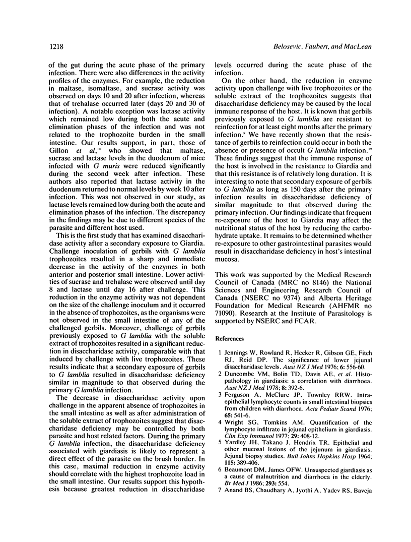
The sequence of changes in the activity of six disaccharidases in the small intestine of gerbils during primary and secondary G lamblia infections was examined. The primary G lamblia infection induced a transient reduction in disaccharidase activity which was related to the highest trophozoite burden in the small intestine. During the primary exposure, a 30% to 85% decrease in the activity of enzymes was observed on days 10 and 20 after infection. Secondary exposure of gerbils to G lamblia caused a sharp decrease in disaccharidase activity as early as 24 h after challenge. The reduction in the enzyme activity was not influenced by the size of the challenge inoculum and occurred even when there were no live trophozoites in the small intestine. Disaccharidase deficiency could also be induced by challenge with the soluble extract of the trophozoites. Multiple challenge administrations of G lamblia trophozoites to gerbils induced a persistent disaccharidase deficiency. The results indicate that disaccharidase deficiency associated with the primary G lamblia infection probably represents a direct effect of the parasite on the brush border of the small intestine. On the other hand, the observed disaccharidase deficiency in the secondary G lamblia infection appears to be induced by the local immune responses of the host.
Full text
PDF


Selected References
These references are in PubMed. This may not be the complete list of references from this article.
- Anand B. S., Chaudhary R., Jyothi A., Yadev R. S., Baveja U. K. Experimental examination of the direct damaging effects of Giardia lamblia on intestinal mucosal scrapings of mice. Trans R Soc Trop Med Hyg. 1985;79(5):613–617. doi: 10.1016/0035-9203(85)90167-1. [DOI] [PubMed] [Google Scholar]
- Beaumont D. M., James O. F. Unsuspected giardiasis as a cause of malnutrition and diarrhoea in the elderly. Br Med J (Clin Res Ed) 1986 Aug 30;293(6546):554–555. doi: 10.1136/bmj.293.6546.554. [DOI] [PMC free article] [PubMed] [Google Scholar]
- Belosevic M., Dick T. A. Trichinella spiralis: comparison with an Arctic isolate. Exp Parasitol. 1980 Apr;49(2):266–276. doi: 10.1016/0014-4894(80)90123-x. [DOI] [PubMed] [Google Scholar]
- Belosevic M., Faubert G. M. Giardia muris: correlation between oral dosage, course of infection, and trophozoite distribution in the mouse small intestine. Exp Parasitol. 1983 Aug;56(1):93–100. doi: 10.1016/0014-4894(83)90100-5. [DOI] [PubMed] [Google Scholar]
- Belosevic M., Faubert G. M., MacLean J. D., Law C., Croll N. A. Giardia lamblia infections in Mongolian gerbils: an animal model. J Infect Dis. 1983 Feb;147(2):222–226. doi: 10.1093/infdis/147.2.222. [DOI] [PubMed] [Google Scholar]
- Belosevic M., Faubert G. M., Skamene E., MacLean J. D. Susceptibility and resistance of inbred mice to Giardia muris. Infect Immun. 1984 May;44(2):282–286. doi: 10.1128/iai.44.2.282-286.1984. [DOI] [PMC free article] [PubMed] [Google Scholar]
- Dahlqvist A. Assay of intestinal disaccharidases. Anal Biochem. 1968 Jan;22(1):99–107. doi: 10.1016/0003-2697(68)90263-7. [DOI] [PubMed] [Google Scholar]
- Duncombe V. M., Bolin T. D., Davis A. E., Cummins A. G., Crouch R. L. Histopathology in giardiasis: a correlation with diarrhoea. Aust N Z J Med. 1978 Aug;8(4):392–396. doi: 10.1111/j.1445-5994.1978.tb04908.x. [DOI] [PubMed] [Google Scholar]
- Erlandsen S. L., Chase D. G. Morphological alterations in the microvillous border of villous epithelial cells produced by intestinal microorganisms. Am J Clin Nutr. 1974 Nov;27(11):1277–1286. doi: 10.1093/ajcn/27.11.1277. [DOI] [PubMed] [Google Scholar]
- Ferguson A., McClure J. P., Townley R. R. Intraepithelial lymphocyte counts in small intestinal biopsies from children with diarrhoea. Acta Paediatr Scand. 1976 Sep;65(5):541–546. doi: 10.1111/j.1651-2227.1976.tb04929.x. [DOI] [PubMed] [Google Scholar]
- Gillon J., Al Thamery D., Ferguson A. Features of small intestinal pathology (epithelial cell kinetics, intraepithelial lymphocytes, disaccharidases) in a primary Giardia muris infection. Gut. 1982 Jun;23(6):498–506. doi: 10.1136/gut.23.6.498. [DOI] [PMC free article] [PubMed] [Google Scholar]
- Jennings W., Rowland R., Hecker R., Gibson G. E., Fitch R. J., Reid D. P. The significance of lowered jejunal disaccharidase levels. Aust N Z J Med. 1976 Dec;6(6):556–560. doi: 10.1111/j.1445-5994.1976.tb03994.x. [DOI] [PubMed] [Google Scholar]
- Lewis P. D., Jr, Belosevic M., Faubert G. M., Curthoys L., MacLean J. D. Cortisone-induced recrudescence of Giardia lamblia infections in gerbils. Am J Trop Med Hyg. 1987 Jan;36(1):33–40. doi: 10.4269/ajtmh.1987.36.33. [DOI] [PubMed] [Google Scholar]
- Roberts-Thomson I. C., Stevens D. P., Mahmoud A. A., Warren K. S. Giardiasis in the mouse: an animal model. Gastroenterology. 1976 Jul;71(1):57–61. [PubMed] [Google Scholar]
- Smith P. D., Gillin F. D., Spira W. M., Nash T. E. Chronic giardiasis: studies on drug sensitivity, toxin production, and host immune response. Gastroenterology. 1982 Oct;83(4):797–803. [PubMed] [Google Scholar]
- Wright S. G., Tomkins A. M. Quantification of the lymphocytic infiltrate in jejunal epithelium in giardiasis. Clin Exp Immunol. 1977 Sep;29(3):408–412. [PMC free article] [PubMed] [Google Scholar]
- Wright S. G., Tomkins A. M., Ridley D. S. Giardiasis: clinical and therapeutic aspects. Gut. 1977 May;18(5):343–350. doi: 10.1136/gut.18.5.343. [DOI] [PMC free article] [PubMed] [Google Scholar]
- YARDLEYJHTAKANO J., HENDRIX T. R. EPITHELIAL AND OTHER MUCOSAL LESIONS OF THE JEJUNUM IN GIARDIASIS. JEJUNAL BIOPSY STUDIES. Bull Johns Hopkins Hosp. 1964 Nov;115:389–406. [PubMed] [Google Scholar]


