

Abstract
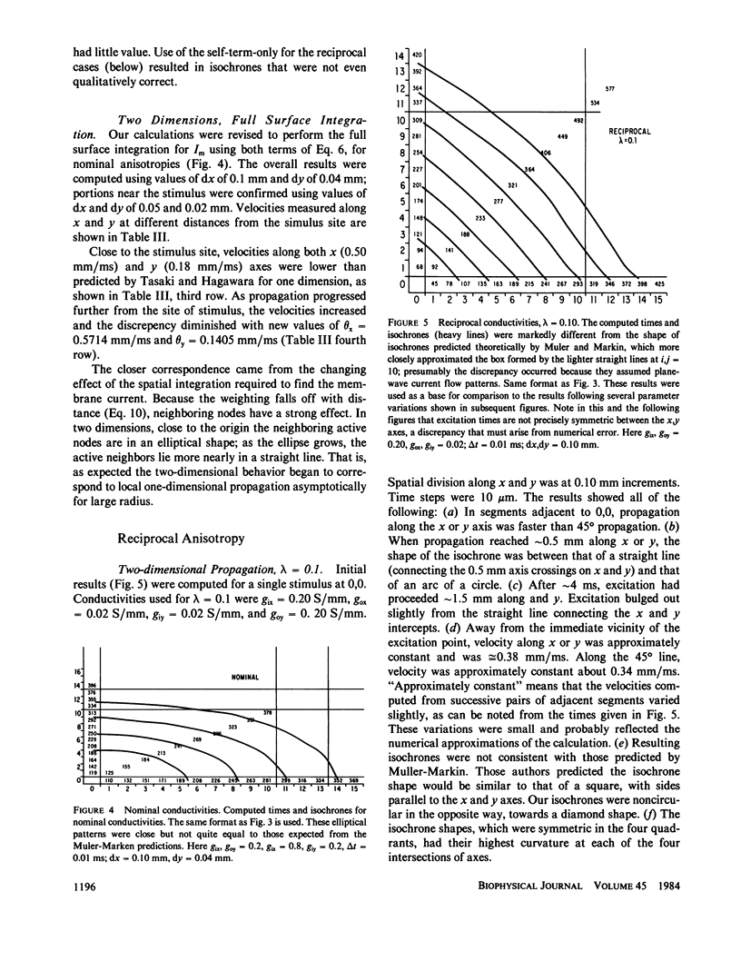
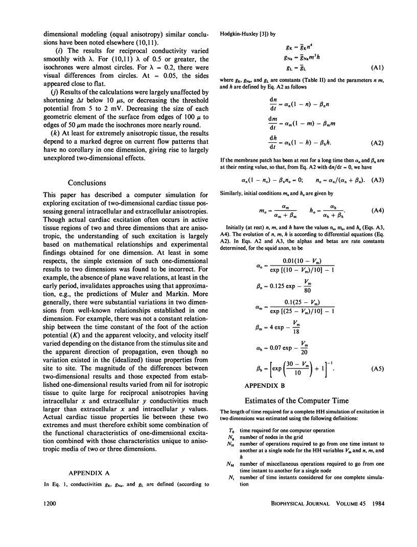
This paper reports on a simulation of propagation for anisotropic two-dimensional cardiac tissue. The tissue structure assumed was that of a Hodgin-Huxley membrane separating inside and outside anisotropic media, obeying Ohm's law in each case. Membrane current was found by an integral expression involving partial spatial derivatives of Vm weighted by a function of distance. Numerical solutions for transmembrane voltage as a function of time following excitation at a single central site were computed using an algorithm that examined only the portion of the tissue undergoing excitation at each moment; thereby, the number of calculations required was reduced to a large but achievable number. Results are shown for several combinations of the four conductivity values: With isotropic tissue, excitation spread in circles, as expected. With tissue having nominally normal ventricular conductivities, excitation spread in patterns close to ellipses. With reciprocal conductivities, isochrones approximated a diamond shape, and were in conflict with the theoretical predictions of Muler and Markin; the time constant of the foot of the action potentials, as computed, varied between sites along axes as compared with sites along the diagonals, even though membrane properties were identical everywhere. Velocity of propagation changed for several milliseconds following the stimulus. Patterns that would have been expected from well-known studies in one dimension did not always occur in two dimensions, with the magnitude of the difference varying from nil for isotropic conductivities to quite large for reciprocal conductivities.
Full text
PDF


Selected References
These references are in PubMed. This may not be the complete list of references from this article.
- Ebihara L., Shigeto N., Lieberman M., Johnson E. A. The initial inward current in spherical clusters of chick embryonic heart cells. J Gen Physiol. 1980 Apr;75(4):437–456. doi: 10.1085/jgp.75.4.437. [DOI] [PMC free article] [PubMed] [Google Scholar]
- Geselowitz D. B., Barr R. C., Spach M. S., Miller W. T., 3rd The impact of adjacent isotropic fluids on electrograms from anisotropic cardiac muscle. A modeling study. Circ Res. 1982 Nov;51(5):602–613. doi: 10.1161/01.res.51.5.602. [DOI] [PubMed] [Google Scholar]
- HODGKIN A. L., HUXLEY A. F. A quantitative description of membrane current and its application to conduction and excitation in nerve. J Physiol. 1952 Aug;117(4):500–544. doi: 10.1113/jphysiol.1952.sp004764. [DOI] [PMC free article] [PubMed] [Google Scholar]
- Joyner R. W., Ramón F., Morre J. W. Simulation of action potential propagation in an inhomogeneous sheet of coupled excitable cells. Circ Res. 1975 May;36(5):654–661. doi: 10.1161/01.res.36.5.654. [DOI] [PubMed] [Google Scholar]
- Kootsey J. M., Johnson E. A. The origin of the T-wave. Crit Rev Bioeng. 1980;4(3):233–270. [PubMed] [Google Scholar]
- Plonsey R., Barr R. C. Current flow patterns in two-dimensional anisotropic bisyncytia with normal and extreme conductivities. Biophys J. 1984 Mar;45(3):557–571. doi: 10.1016/S0006-3495(84)84193-4. [DOI] [PMC free article] [PubMed] [Google Scholar]
- Plonsey R., Rudy Y. Electrocardiogram sources in a 2-dimensional anisotropic activation model. Med Biol Eng Comput. 1980 Jan;18(1):87–94. doi: 10.1007/BF02442485. [DOI] [PubMed] [Google Scholar]
- Spach M. S., Miller W. T., 3rd, Miller-Jones E., Warren R. B., Barr R. C. Extracellular potentials related to intracellular action potentials during impulse conduction in anisotropic canine cardiac muscle. Circ Res. 1979 Aug;45(2):188–204. doi: 10.1161/01.res.45.2.188. [DOI] [PubMed] [Google Scholar]
- TASAKI I., HAGIWARA S. Capacity of muscle fiber membrane. Am J Physiol. 1957 Mar;188(3):423–429. doi: 10.1152/ajplegacy.1957.188.3.423. [DOI] [PubMed] [Google Scholar]