Abstract
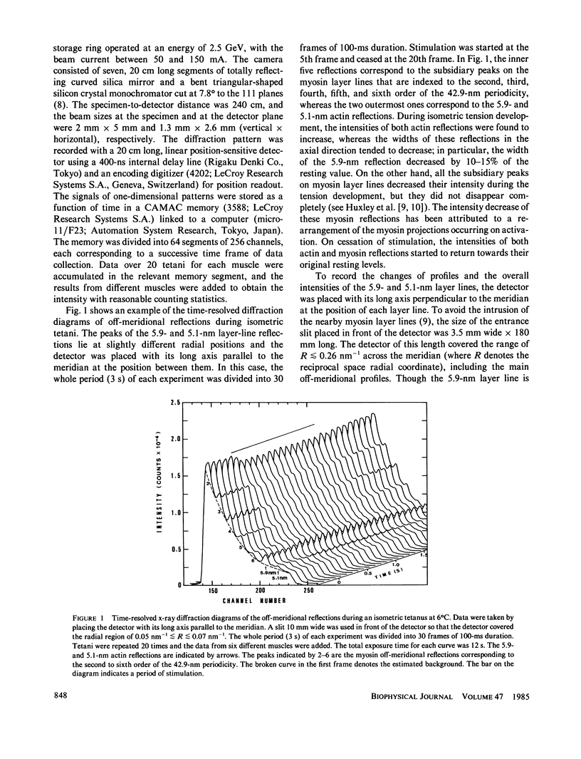
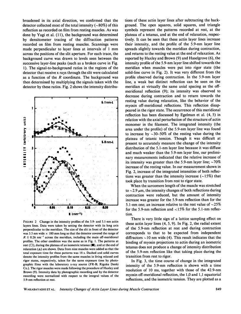
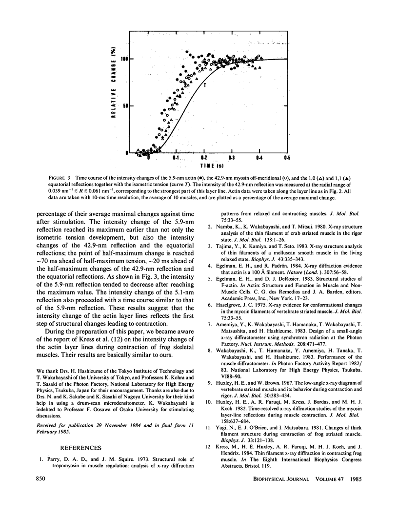
Time-resolved x-ray diffraction studies have been made on the 5.9- and 5.1-nm actin layer lines from frog skeletal muscles during an isometric tetanus at 6 degrees C, using synchrotron radiation. The integrated intensities of these actin layer lines were found to increase during a tetanus by 30-50% for the 5.9-nm reflection and approximately 70% for the 5.1-nm reflection of the resting values. The intensity increase of both reflections was greater than that taking place in the transition from rest to rigor state. The intensity change of the 5.9-nm reflection preceded those of the myosin 42.9-nm off-meridional reflection and of the equatorial reflections, as well as the isometric tension development. The intensity profile of the 5.9-nm layer line during contraction was found to be different from that observed in the rigor state.
Full text
PDF


Selected References
These references are in PubMed. This may not be the complete list of references from this article.
- Huxley H. E., Brown W. The low-angle x-ray diagram of vertebrate striated muscle and its behaviour during contraction and rigor. J Mol Biol. 1967 Dec 14;30(2):383–434. doi: 10.1016/s0022-2836(67)80046-9. [DOI] [PubMed] [Google Scholar]
- Huxley H. E., Faruqi A. R., Kress M., Bordas J., Koch M. H. Time-resolved X-ray diffraction studies of the myosin layer-line reflections during muscle contraction. J Mol Biol. 1982 Jul 15;158(4):637–684. doi: 10.1016/0022-2836(82)90253-4. [DOI] [PubMed] [Google Scholar]
- Namba K., Wakabayashi K., Mitsui T. X-ray structure analysis of the thin filament of crab striated muscle in the rigor state. J Mol Biol. 1980 Mar 25;138(1):1–26. doi: 10.1016/s0022-2836(80)80002-7. [DOI] [PubMed] [Google Scholar]
- Parry D. A., Squire J. M. Structural role of tropomyosin in muscle regulation: analysis of the x-ray diffraction patterns from relaxed and contracting muscles. J Mol Biol. 1973 Mar 25;75(1):33–55. doi: 10.1016/0022-2836(73)90527-5. [DOI] [PubMed] [Google Scholar]
- Tajima Y., Kamiya K., Seto T. X-ray structure analysis of thin filaments of a molluscan smooth muscle in the living relaxed state. Biophys J. 1983 Sep;43(3):335–343. doi: 10.1016/S0006-3495(83)84357-4. [DOI] [PMC free article] [PubMed] [Google Scholar]
- Yagi N., O'Brien E. J., Matsubara I. Changes of thick filament structure during contraction of frog striated muscle. Biophys J. 1981 Jan;33(1):121–137. doi: 10.1016/S0006-3495(81)84876-X. [DOI] [PMC free article] [PubMed] [Google Scholar]


