Abstract
Porcine reproductive and respiratory syndrome virus (PRRSV) glycoprotein 5 (GP5) is the most abundant envelope glycoprotein and a major inducer of neutralizing antibodies in vivo. Three putative N-linked glycosylation sites (N34, N44, and N51) are located on the GP5 ectodomain, where a major neutralization epitope also exists. To determine which of these putative sites are used for glycosylation and the role of the glycan moieties in the neutralizing antibody response, we generated a panel of GP5 mutants containing amino acid substitutions at these sites. Biochemical studies with expressed wild-type (wt) and mutant proteins revealed that the mature GP5 contains high-mannose-type sugar moieties at all three sites. These mutations were subsequently incorporated into a full-length cDNA clone. Our data demonstrate that mutations involving residue N44 did not result in infectious progeny production, indicating that N44 is the most critical amino acid residue for infectivity. Viruses carrying mutations at N34, N51, and N34/51 grew to lower titers than the wt PRRSV. In serum neutralization assays, the mutant viruses exhibited enhanced sensitivity to neutralization by wt PRRSV-specific antibodies. Furthermore, inoculation of pigs with the mutant viruses induced significantly higher levels of neutralizing antibodies against the mutant as well as the wt PRRSV, suggesting that the loss of glycan residues in the ectodomain of GP5 enhances both the sensitivity of these viruses to in vitro neutralization and the immunogenicity of the nearby neutralization epitope. These results should have great significance for development of PRRSV vaccines of enhanced protective efficacy.
Porcine reproductive and respiratory syndrome virus (PRRSV) belongs to the family Arteriviridae within the order Nidovirales which also includes equine arteritis virus (EAV), lactate dehydrogenase-elevating virus (LDV), and simian hemorrhagic fever virus. The viral genome is a linear, positive-stranded RNA molecule of approximately 15.0 kb in length and possesses a cap structure at the 5′ end and a poly(A) tail at the 3′ end. Eight open reading frames (ORFs) are in the viral genome (9, 34). The first two open reading frames (ORF1a and ORF1ab) encode viral nonstructural (NS) polyproteins that are involved in polyprotein processing and genome transcription and replication (47). The viral structural proteins, encoded in ORFs 2 to 7, are expressed from six subgenomic capped and polyadenylated mRNAs that are synthesized as a 3′-coterminal nested set of mRNAs with a common leader sequence at the 5′ end.
The major viral envelope protein is glycoprotein 5 (GP5), which is encoded in ORF5 of the viral genome (29, 35, 36). GP5 is a glycosylated transmembrane protein of approximately 25 kDa (10, 16, 35). It has a putative N-terminal signal peptide and possesses three potential N-linked glycosylation sites which are located in a small ectodomain comprising the first 40 residues of the mature protein (28, 35). In EAV and LDV, the major envelope glycoprotein forms a disulfide-linked heterodimer with the ORF6 gene product, the viral matrix (M) protein (13, 15, 45). Similar interaction between PRRSV GP5 and M protein has been observed but the mode of interaction has not been defined yet (12, 28).
It has been postulated that formation of heterodimers of GP5 and M proteins may play a critical role in assembly of infectious PRRSV. In addition to its role in virus assembly, GP5 appears to be involved in entry of the virus into susceptible host cells. GP5 is presumed to interact with the host cell receptor sialoadhesin (11) for entry into porcine alveolar macrophages, the in vivo target cells for PRRSV. The role of GP5 in receptor recognition is supported by the presence of a major neutralization epitope in the N-terminal ectodomain (38), implying a central role for the GP5 ectodomain in the infection process.
The N-linked glycans of the GP5 ectodomain may be critical for proper functioning of the protein. N-linked glycosylation, in general, is important for correct folding, targeting, and biological activity of proteins (17-19, 51, 55). In many enveloped viruses, the envelope proteins are modified by the addition of sugar moieties and the N-linked glycosylation of envelope protein plays diverse functions in viral glycoproteins such as receptor binding, membrane fusion, penetration into cells, and virus budding (6, 14). Recent studies have demonstrated the role of N-linked glycosylation of Hantaan virus glycoprotein in protein folding and intracellular trafficking (43) as well as in the biological activity and antigenicity of influenza virus hemagglutinin (HA) protein (1). Furthermore, it has become evident that glycosylation of viral envelope proteins is a major mechanism for viral immune evasion and persistence used by several different enveloped viruses to escape, block, or minimize the virus-neutralizing antibody response. Examples of this effect have been reported for simian immunodeficiency virus (40) and human immunodeficiency virus type 1 (50), hepatitis B virus (25), and influenza virus (44) and more importantly, in the case of the arterivirus LDV (8).
Recently the development of reverse genetic systems for PRRSV has been reported from several laboratories, including ours (33, 37, 48, 49). Evidently, mutational studies with infectious clones have led to a better understanding of the mechanisms of transcription and replication of the viral genome of arteriviruses (46). Thus, in order to examine the importance of N-linked glycosylation in the biological activity of GP5 of PRRSV in generating infectious virus or eliciting neutralizing antibodies in vivo, we have constructed a series of mutant GP5 proteins in which each of the potential N-linked glycosylation sites has been mutated either individually or in various combinations. The resulting mutant proteins were examined for their glycosylation pattern and role in infectious virus recovery and in cross neutralization by antibodies raised, through experimental inoculations, against the wild-type (wt) PRRSV and against the mutant viruses. Our data show that all three putative glycosylation sites are used for glycosylation with high-mannose-type glycans and glycosylation of GP5 protein at residue 44 is critical for recovery of infectious PRRSV. Very importantly, our data from neutralization and antibody response studies indicate that natural infection with PRRSV may involve an immune evasion based on glycan-shielding mechanisms as was previously described for other viruses, helping to explain the rather ineffective protective humoral immune response that is observed in PRRSV-infected animals (31, 38).
MATERIALS AND METHODS
Cells, media, and antibodies.
MARC-145 (23) cells were propagated in Dulbecco's modified Eagle's medium (DMEM) containing 10% fetal bovine serum (FBS) and 100 units of penicillin, 20 units of streptomycin, and 20 units of kanamycin per ml of growth medium. These cells were used for RNA electroporation, virus infection, viral growth, and plaque assays. The baby hamster kidney (BHK-21) cells were maintained in minimal essential medium (MEM) with Earl's salt containing 5% FBS and the above-mentioned antibiotics. BHK-21 cells were used for transient expression of GP5 followed by either immunofluorescence assays or radiolabeling and immunoprecipitation experiments. All cells were maintained at 37°C and 5% CO2. Rabbit polyclonal antibodies to the PRRSV GP5 and M proteins were kindly provided by Carl A. Gagnon (University of Quebec, Montreal, Canada). The monoclonal antibody (SDOW17) against nucleocapsid protein (N) was purchased from National Veterinary Services Laboratories (Ames, IA). The anti-mouse Alexa-488-labeled antibody was obtained from Molecular Probes (Eugene, OR).
Genetic manipulation of plasmids encoding GP5 and PRRSV infectious clone.
The full-length PRRSV infectious cDNA clone (FL-12) in pBR322 (49) was digested with EcoRV and BstZ17I restriction enzymes and the ∼4.9-kbp fragment encompassing the majority of ORF2, complete ORFs 3 to 7, and the entire 3′ untranslated region (UTR) of PRRSV was cloned into pBR322 using the same enzyme sites. This intermediate plasmid served as the template for mutagenesis to introduce mutations at the potential N-linked glycosylation sites (N34, N44, and N51) within GP5 (Fig. 2).
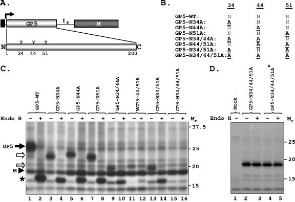
FIG. 2.
Glycosylation analysis of wt GP5 and its mutants in transfected cells. (A) Schematic of the bicistronic vector and PRRSV GP5 with the three putative glycosylation sites at amino acid positions 34, 44, and 51 shown. (B) Various mutants used in the present study. (C) Expression of wt and mutant GP5 and their sensitivity to Endo H. The experiment was performed as described in the legend to Fig. 1; proteins were immunoprecipitated with anti-GP5 antibody, digested with Endo H (+) or left undigested (−), and analyzed by electrophoresis. Mutant GP5 proteins are shown by open and shaded arrows. The protein band (∼17 kDa) identified by the asterisk is generated by Endo H digestion of the wt and mutant GP5 proteins. (D) Expression of the triple mutant GP5 N34/44/51A from plasmids with and without M. Cells were mock transfected (lane 1) or transfected with plasmids encoding the triple mutant-containing M coding region (GP5-N34/44/51A, lanes 2 and 3) or without the M coding region (GP5-N34/44/51A*, lanes 4 and 5). Proteins were radiolabeled and detected with anti-GP5 antibody as described above. The mobility of proteins is shown in kilodaltons on the right.
Mutagenesis was carried out using overlap extension PCR with synthetic primers (Table 1), Pfu polymerase (Stratagene), and standard techniques (4, 20, 42). The PCR product was digested with BsrGI and BstEII restriction enzymes and replaced back in the intermediate plasmid. Clones containing the desired mutations were identified and confirmed by sequencing. The entire coding region of GP5 was sequenced to ensure that additional mutations were not present in the clones. The EcoRV-PacI fragment from the intermediate plasmid containing mutations in the GP5 coding region was moved back into the full-length cDNA clone using the same restriction enzyme sites. The GP5 coding region in the full-length clones was again sequenced with PRRSV-specific internal primers to confirm the presence of the mutations.
TABLE 1.
Primers used in this studya
| Primer | Nucleotide sequence |
|---|---|
| GP5-For | 5′ GCCGGAATTCGGAGCCGCCGCCACCATGTTGGGGAGATGCTTGAC 3′ |
| GP5-Rev | 5′ CCCCGAATTCCTAAAGACGACCCCATTGTTC 3′ |
| M-For | 5′ ATATATGGATCCGCCACCATGGGGTCGTCTTTAGACGAC 3′ |
| M-Rev | 5′ ATATATGCATGCTTATTTGGCATATTTGACAAGG 3′ |
| GP5-N34A-For | 5′ GCCAACAGCGCCAGCAGCTCTC 3′ |
| GP5-N44A-For | 5′ GTTGATTTACGCCTTGACGCTATG 3′ |
| GP5-N51A-For | 5′ GTGAGCTGGCTGGCACAGATTG 3′ |
| GP5-N34/44A-For | 5′ GCCAACAGCGCCAGCAGCTCTCATCTTCAGTTGATTTACGCCTTGACGCTATG 3′ |
| GP5-N44/51A-For | 5′ GTTGATTTACGCCTTGACGCTATGTGAGCTGGCTGGCACAGATTG 3′ |
| GP5-N34/51A-For | 5′ GCCAACAGCGCCAGCAGCTCTCATCTTCAGTTGATTTACAACTTGACGCTATGTGAG CTGGCTGGCACAGATTG 3′ |
| GP5-N34/44/51A-For | 5′ GCCAACAGCGCCAGCAGCTCTCATCTTCAGTTGATTTACGCCTTGACGCTATGTGAG CTGGCTGGCACAGATTG 3′ |
| PRRSV-13177-For | 5′ CTACCAACATCAGGTCGATGGCGG 3′ |
| PRRSV-14473-Rev | 5′ GTCGGCCGCGACTTACCTTTAGAG 3′ |
Underlining indicates mutations. Restriction enzyme sites incorporated in the primers are shown in bold; the initiation codons for the GP5 and M proteins are shown in bold italics.
The wt GP5 and individual mutants were cloned in a bicistronic vector (GP5-IE-M) where GP5 is the first cistron followed by the encephalomyocarditis virus (EMCV) internal ribosome entry site (IRES) (21) and M coding sequences (Fig. 1A). To construct this plasmid, a DNA fragment encompassing the entire IRES element of EMCV (IE) was released from the pIRES2-EGFP (Clontech) vector using BamHI and NcoI restriction enzymes. The fragment was treated with mung bean nuclease (New England BioLabs) and cloned in the pGEM3 (Promega) vector digested with SmaI to obtain the vector pGEM3-IE. The full-length GP5 coding region was PCR amplified using a forward primer (GP5-For) that contained an EcoRI enzyme site and Kozak sequence followed by ATG and GP5 sequences and a reverse primer (GP5-Rev) (Table 1) containing an EcoRI enzyme site. The PCR fragment was cloned at the unique EcoRI site in the pGEM3-IE vector just upstream of IE so that the coding sequence of GP5 is under the control of the T7 RNA polymerase promoter.
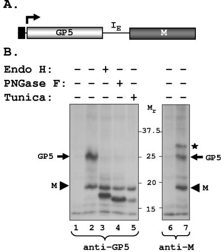
FIG. 1.
Transient expression of PRRSV GP5 and M protein. (A) Schematic of the bicistronic construct showing the GP5 and M coding regions flanking the IRES from EMCV (IE). The coding regions are under the control of the T7 RNA polymerase promoter (black rectangle) present immediately upstream of the GP5 coding region. The bent arrow shows the position and direction of transcription by T7 RNA polymerase from the vector. (B) Expression of GP5 and M proteins in cells transfected with the bicistronic vector. Mock-transfected (lane 1) or plasmid-transfected cells (lanes 2 to 7) were radiolabeled as described in Materials and Methods and immunoprecipitated with anti-GP5 antibody (lanes 1 to 5) or anti-M antibody (lanes 6 and 7). Immunoprecipitated proteins were left untreated (−) (lanes 1, 2, 6, and 7) or treated (+) with Endo H (lane 3) or PNGase F (lane 4) and analyzed by electrophoresis. Lane 5 contains immunoprecipitated proteins from transfected cells treated or not with tunicamycin. The mobility of proteins is shown in kilodaltons. The asterisk identifies a cellular protein that coimmunoprecipitates with anti-M antibody.
The resulting plasmid (pGEM3-GP5-IE) was then used to clone the coding region of the M protein. To this end, the M coding region was PCR amplified using primer pairs (M-For and M-Rev) (Table 1) containing BamHI and SphI restriction enzyme sites and cloned in pGEM3-GP5-IE at the corresponding sites. The GP5 and M coding region in the resulting plasmid (GP5-IE-M) was sequence confirmed. The IE and M coding region was deleted from GP5-IE-M to generate plasmid pGEM3-GP5, which expresses only GP5 protein in transfected cells. The full-length GP5 coding region was also cloned in a cytomegalovirus promoter-driven vector (pcDNA 3.0; Clontech) for complementation studies.
In vitro transcription and electroporation.
The full-length plasmids were digested with AclI and linearized DNA was used as the template to generate capped RNA transcripts using the mMESSAGE mMACHINE Ultra T7 kit as per the manufacturer's (Ambion, Austin, TX) recommendations and as described earlier (48, 49). The reaction mixture was treated with DNase I to digest the DNA template and extracted with phenol and chloroform and finally precipitated with isopropanol. The integrity of the in vitro transcripts was analyzed by glyoxal-agarose gel electrophoresis followed by ethidium bromide staining.
MARC-145 cells were electroporated with approximately 5.0 μg of in vitro transcripts along with 5.0 μg of total RNA isolated from MARC-145 cells. About 2 × 10⁶ cells in 400 μl of DMEM containing 1.25% dimethyl sulfoxide were pulsed once using a Bio-Rad Gene Pulser Xcell at 250 V and 950 μF in a 4.0-mm cuvette. The cells were diluted in normal growth medium and plated in a 60-mm cell culture plate. A small portion of the electroporated cells was plated in a 24-well plate to examine expression of N protein at 48 h postelectroporation, which would indicate genome replication and transcription. Once expression of N protein is confirmed using an indirect immunofluorescence assay, the supernatant from bulk of the electroporated cells in the 60-mm plate was collected at 48 h postelectroporation, clarified, and passed onto naïve MARC-145 cells. The infected cells were observed for cytopathic effect along with the expression of N protein using immunofluorescence assay. The supernatants from infected cells showing both cytopathic effect and positive fluorescence were considered to contain infectious virus. After confirmation, the virus stock was grown and frozen at −80°C in small aliquots for further studies. In all the experiments, FL-12, containing the wt PRRSV genome, and FL-12pol−, containing the polymerase-defective PRRSV genome (48, 49), were used as controls.
Metabolic radiolabeling and analyses of proteins.
BHK-21 cells in six-well plates were infected with recombinant vaccinia virus (vTF7-3) at a multiplicity of infection (MOI) of 3.0 and subsequently transfected with the bicistronic plasmid DNA encoding wt or various mutant GP5s under the T7 RNA polymerase promoter. DNA transfection was carried out using Lipofectamine2000 as per the manufacturer's protocol (Life Technologies). At 16 h posttransfection, cells were washed twice with phosphate-buffered saline (PBS) and starved in methionine- and cysteine-free DMEM for 1 hour and radiolabeled with 0.6 ml of methionine- and cysteine-free DMEM containing 100 μCi of Expre35S35S protein labeling mix (NEN Life Sciences, MA) per ml of medium for 3 hours. Following radiolabeling, the cells were washed in cold PBS three times and cell extracts were prepared in 300 μl of radioimmunoprecipitation assay (RIPA) buffer (10 mM Tris-HCl, pH 8.0, 150 mM NaCl, 1% Triton X-100, 0.1% sodium dodecyl sulfate [SDS], 1% sodium deoxycholate, and 1× protease inhibitor). The clarified cell extracts were incubated overnight at 4°C with rabbit anti-GP5 or anti-M protein antibody. A slurry of approximately 4.0 mg of protein A-Sepharose (Pharmacia, Uppsala, Sweden) in 100 μl RIPA buffer was added and further incubated for 2 h. The immunoprecipitated complexes were washed three times with 500 μl of RIPA buffer and used for further analysis.
For pulse-chase analysis, infected and transfected cells were labeled for 2 h as described above and either harvested immediately or washed twice with warm PBS and maintained in DMEM containing a 100-fold excess of unlabeled methionine for 2 or 4 h before harvesting. In PRRSV-infected infected cells, the pulse-chase experiment was performed similarly at 48 h postinfection.
For endoglycosidase H (Endo H) treatment, the immunoprecipitated complexes were resuspended in 20 μl of 1× denaturing buffer (0.5% SDS, 1.0% β-mercaptoethanol) and boiled for 10 min. The supernatant was collected, adjusted to 1× G5 buffer (0.05 M sodium citrate, pH 5.5), and incubated for 16 h at 37°C with 100 units of Endo H (New England Biolabs). The undigested control samples were processed similarly but no Endo H was added. Following Endo H digestion, the samples were mixed with an equal volume of 2× SDS-polyacrylamide gel electrophoresis (PAGE) sample buffer, boiled for 5 min, and resolved by SDS-12% PAGE under denaturing conditions along with protein marker (Protein Plus Precision Standard; Bio-Rad). The gel was fixed with 10% acetic acid for 15 min, washed three times with water, treated with 0.5 M sodium salicylate for 30 min, dried, and finally exposed to X-ray film at −70°C. For peptide N-glycosidase F (PNGase F) (New England Biolabs) digestion, immunoprecipitated complexes were resuspended in 1× G7 buffer (0.05 M sodium phosphate, pH 7.5, 1.0% NP-40) and digestion was performed by incubating for 16 h at 37°C with 2 units of the enzyme. To examine synthesis of GP5 in the presence of tunicamycin (Sigma), transfected cells were treated with 2.0 μg of tunicamycin per ml of medium for 1 hour and radiolabeling was performed in the presence of the drug for 3 h as above.
For obtaining radiolabeled extracellular virions or intracellular virus-expressed GP5, MARC-145 cells were infected with wt or mutant PRRSVs. At 48 h postinfection, the cells were starved for 1 hour and radiolabeled with 100 μCi of Expre35S35S protein labeling mix per ml of medium containing 90% methionine- and cysteine-free DMEM and 10% regular DMEM for 24 h. Following labeling, the culture supernatant was harvested and cleared of cell debris and the extracellular virions were pelleted at 100,000 × g for 3 h at 4°C. The viral pellets were resuspended in 200 μl of RIPA buffer and immunoprecipitated with anti-GP5 antibody and the proteins were examined with or without Endo H treatment. For immunoprecipitation of intracellular virus-expressed GP5, infection was carried out as described above and at 24 h postinfection, the cells were starved for 1 hour and radiolabeled as described above for 2 h prior to preparing cell extracts.
Viral growth kinetics and plaque assay.
MARC-145 cells were infected with mutant or wt PRRSV at an MOI of 3.0 PFU per cell and incubated at 37°C in an incubator. At various time points postinfection, aliquots of culture supernatants from infected cells were collected and the virus titer in the supernatants was determined and expressed as 50% tissue culture infectious dose per ml (TCID50/ml). To examine the plaque morphology of mutant viruses, the plaque assay was performed using MARC-145 cells. Cells were infected with 10-fold serial dilutions of individual viruses for 1 hour at 37°C. The infected cell monolayer was washed with PBS and overlaid with DMEM-5% FBS containing 0.8% SeaPlaque agarose (FMC Bioproducts, ME). After 96 h, the agarose plugs were removed and the cell monolayer was incubated with staining solution (20% formaldehyde, 9.0% ethanol, and 0.1% crystal violet) for 30 min at room temperature. The cells were gently washed with water to remove excess dye and air dried to examine and count the plaques.
Complementation of virus recovery by expressing wt GP5 in trans.
BHK-21 cells were transfected with pcDNA-GP5. At 40 h posttransfection, the cells were harvested and electroporated with capped in vitro transcripts derived from full-length PRRSV cDNA encoding mutant GP5. The electroporated cells were diluted with fresh medium and plated in a six-well plate. The supernatant from the electroporated cells was collected at 48 h postelectroporation, centrifuged to remove cell debris, and used to infect naïve MARC-145 cells. The infected MARC-145 cells were examined at 48 h postinfection for expression of N protein by immunofluorescence assay as described above. The number of positive cells was counted to assign the number of pseudoparticles produced in the supernatant. The average number of positive cells was calculated from three independent experiments and is presented as the number of pseudoparticles produced per microgram of in vitro-transcribed RNA transfected into the cells.
Serum neutralization assays.
The titer of PRRSV-neutralizing antibodies in a serum sample was determined using the fluorescence focus neutralization assay described previously (54). Serial dilutions of test sera were incubated for 60 min at 37°C in the presence of 200 TCID50 of the challenge virus, which consisted of either FL12 (wt PRRSV) or any of the GP5 mutant-encoding viruses FL-N34A, FL-N51A, and FL-N34/51A in DMEM containing 5% FBS. The mixtures were added to 96-well microtitration plates containing confluent MARC-145 cells which had been seeded 48 h earlier. After incubation for 24 h at 37°C in a humidified atmosphere containing 5% CO2, the cells were fixed for 10 min with a solution of 50% methanol and 50% acetone. After extensive washing with PBS, the expression of N protein of PRRSV was detected with monoclonal antibody SDOW17 (36) using a 1:500 dilution, followed by incubation with fluorescein isothiocyanate-conjugated goat anti-mouse immunoglobulin G (Sigma) at a 1:100 dilution. Neutralization titers were expressed as the reciprocal of the highest dilution that inhibited 90% of the foci present in the control wells.
Experimental inoculation of pigs with GP5 mutants and wt PRRSV.
High-titer stocks (obtained through three passages in MARC-145 cells) of the GP5 mutant viruses (FL-N34A, FL-N51A, and FL-N34/51A) and FL-12 (wt PRRSV) were used to infect young pigs. Twenty-one-day-old, recently weaned pigs were purchased from a specific-pathogen-free herd with a certified record of absence of PRRSV infection. All animals were negative for anti-PRRSV antibodies as tested by enzyme-linked immunosorbent assay (Iddex Labs, Portland, ME). Three pigs per group were infected with either FL-12 wt PRRSV or mutants FL-N34A, FL-N51A, and FL-N34/51A. In all cases, the inoculum consisted of 105 TCID50 diluted in 2 ml and was administered intramuscularly in the neck. The rectal temperatures of the inoculated animals were monitored for 15 days postinoculation. Viremia was measured by regular isolation on MARC-145 cells at days 4, 7, and 14 postinoculation. Serum samples were drawn weekly for a total of 48 days postinoculation. The serum samples were used to detect homologous and heterologous cross-neutralization titers for each of the mutants and wt PRRSV.
RESULTS
Expression and characterization of PRRSV GP5.
GP5 of PRRSV strain 97-7895 (49) has three putative N-linked glycosylation sites (N34, N44, and N51). To examine the glycosylation pattern of GP5, we first generated a bicistronic vector in which the coding regions of the GP5 and M proteins flanking the EMCV IRES were placed under the control of the T7 RNA polymerase promoter (Fig. 1A). The rationale for constructing the bicistronic vector is that the GP5 and M proteins are known (for LDV and EAV) or postulated (for PRRSV) to interact with each other (32) and that such interactions may be important for protein folding, glycosylation, intracellular transport, and/or other biological activities of GP5 (45).
Transient expression of GP5 and M by transfection of the bicistronic plasmid followed by radiolabeling and immunoprecipitation with anti-GP5 antibody revealed two major protein species. The protein species migrating with a mass of ∼25.5 kDa is the fully glycosylated form of GP5 (Fig. 1B, lane 2). Since each N-linked glycosylation adds ∼2.5 kDa of molecular mass to a protein (24), this indicates that all three potential glycosylation sites may be used for glycosylation of GP5. The ∼19.0-kDa protein species is the viral M protein, since it was also was specifically immunoprecipitated with anti-M antibody (Fig. 1B, lane 7). The results indicate that the GP5 and M proteins interact with each other in cells expressing both proteins, as both proteins were coimmunoprecipitated with anti-GP5 antibody as well as anti-M antibody.
Upon treatment with Endo H, an enzyme that removes high-mannose-type oligosaccharide chains, the size of GP5 was reduced to ∼17 kDa, whereas the size of the M protein remained unchanged (Fig. 1B, lane 3). Treatment of GP5 with PNGase F (Fig. 1B, lane 4), an enzyme that removes all types of sugars from the protein backbone, or synthesis of GP5 in the presence of tunicamycin (Fig. 1B, lane 5) resulted in a protein that migrated with slightly faster electrophoretic mobility than the protein with Endo H treatment. This is expected, since tunicamycin treatment or digestion with PNGase F would generate unglycosylated proteins, whereas Endo H treatment would result in proteins that retain N-acetylglucosamine residues at each of the N-linked glycosylation sites. No difference in the Endo H sensitivity of GP in the presence or absence of M protein was observed (data not shown).
Furthermore, in cells transfected with the plasmid encoding GP5 alone, in addition to the ∼25.5-kDa and ∼17-kDa protein species, a GP5 molecule with a molecular mass of ∼19 kDa was also detected (data not shown). We reason that the ∼19-kDa protein, which comigrates with the M protein, is the unglycosylated form of full-length GP5 and that the ∼17-kDa species is the amino-terminal signal-cleaved unglycosylated form of the protein. It is of note that a prominent protein species of ∼30-kDa molecular mass (identified by the asterisk in Fig. 1B) was detected with anti-M antibody. The identity of this protein is not known but it could be a cellular protein that interacts with the M protein.
The results from the above studies suggest that the unglycosylated and fully glycosylated forms of GP5 possess apparent molecular sizes of ∼19 kDa and ∼25.5 kDa, respectively. The ∼17-kDa protein species is likely generated by cleavage of the amino-terminal signal sequence. It appears that all three potential glycosylation sites are used to generate the fully glycosylated form of GP5. The glycan moieties added to these sites are of the high-mannose type since they are sensitive to digestion by Endo H. In addition, the results indicate that both unglycosylated and fully glycosylated forms of GP5 appear to interact with the M protein.
Analysis of N-linked glycosylation sites used for glycosylation of GP5.
To more precisely determine whether all or some of the potential N-linked glycosylation sites in GP5 are used for addition of sugar moieties, a panel of mutants in which all three potential glycosylation sites, N34, N44, and N51 (Fig. 2A), were altered to alanines either individually or in various combinations were generated in the bicistronic plasmid (Fig. 2B). In plasmid-transfected cells, the proteins were radiolabeled and immunoprecipitated with anti-GP5 antibody. The immune complexes were either left untreated or treated with Endo H and examined by SDS-PAGE.
As can be seen from the data presented in Fig. 2C, mutant GP5 proteins carrying single mutations (N34A, N44A, or N51A) migrated as ∼23.0-kDa protein species (lanes 3, 5, and 7, open arrow), which, upon treatment with Endo H, migrated as ∼17-kDa protein species (Fig. 2C, lanes 4, 6, and 8, asterisk), similar to the wt GP5 after Endo H treatment (Fig. 2C, lane 2). The minor differences in electrophoretic mobility of the proteins are most likely reflective of the fact that the wt protein would retain all three N-acetylglucosamine residues following Endo H treatment compared to the single mutants, which would contain two such residues. The double mutants (N34/44A, N44/51A, and N34/51A) produced protein species that migrated close to ∼20.5 kDa (Fig. 2C, lanes 9, 11, and 13, gray arrow) and upon Endo H treatment, the size of the proteins was reduced to ∼17 kDa (Fig. 2C, lanes 10, 12, and 14). The triple mutant (N34/44/51A) generated a protein that migrated as ∼19.0-kDa protein (Fig. 2C, lane 15) and was resistant to Endo H digestion (Fig. 2C, lane 16). To unequivocally ascertain that the triple mutant protein was synthesized, we examined expression of the triple mutant GP5 in the absence of M protein. As can be seen from the results in Fig. 2D, the triple mutant was synthesized in transfected cells (lane 4) as a ∼19-kDa protein, which was resistant to Endo H (lane 5).
Thus, from the above mutational studies, it is clear that all the three potential glycosylation sites are used for glycosylation to generate fully mature PRRSV GP5. It appears that all three glycosylation sites are modified by high-mannose-type glycan moieties.
Recovery of infectious PRRSV with GP5 mutants.
To assess the importance of N-linked glycosylation in the generation of infectious PRRSV, the coding regions of the mutant GP5 proteins were inserted into the full-length cDNA clone (49). Capped in vitro transcripts produced from the clones were electroporated into MARC-145 cells and generation of infectious PRRSV was examined. Our results showed that infectious virus was readily recovered from the cells electroporated with full-length transcripts containing mutations at N34, N51, and N34/51. However, under similar conditions for virus recovery, repeated attempts to recover other mutant viruses were unsuccessful.
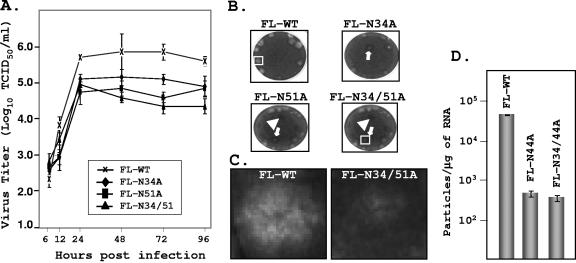
Although the growth kinetics of the recovered viruses were similar to that of the wt virus, the overall yield of viruses FL-N34A and FL-N51A, containing mutations at N34 and N51, respectively, was approximately one log less in MARC-145 cells, while that of FL-N34/51A with the double mutation (N34/51A) was almost 1.5 log less than that of the wt PRRSV (Fig. 3A). Reverse transcription-PCR amplification of RNA from infected cells followed by nucleotide sequencing indicated that these viruses are stable and contained the desired mutations and no other mutations were detected in the entire GP5 region (data not shown).
FIG. 3.
Characterization of mutant viruses encoding mutant GP5. (A) Single-step growth kinetics of wt (FL-12) and various mutant PRRSVs in MARC-145 cells. Cells in six-well plates were infected with PRRSV at an MOI of 3, culture supernatants were collected at the indicated times after infection, and virus titers were determined. Average titers with standard deviations (error bars) from three independent experiments are shown. (B) Plaque morphology of mutant viruses. Arrows and arrowheads show plaques that are less clear. Boxes show representative plaques that have been magnified in succeeding panels. (C) Magnified images of representative plaques from wt PRRSV (FL-WT) and one mutant virus (FL-N34/51A). (D) trans-Complementation to recover mutant PRRSVs; quantitative analysis of mutant virus recovery from cells expressing wt GP5 protein. The average yields of viruses from three independent experiments with standard deviations (represented by error bars) are shown.
The viral plaque assay was performed on MARC-145 cells to monitor the plaque phenotype of the mutant viruses. The majority of the plaques generated by wt PRRSV were clear and distinct, while the mutant viruses produced plaques that had different phenotypes. The FL-N34A, FL-N51A, and FL-N34/51A viruses generated some plaques that were less distinct and many of the cells within these plaques appeared normal. In addition, the mutant viruses also produced some plaques in which the viruses failed to clear the cell monolayer (Fig. 3B, solid arrow). Higher-magnification images (Fig. 3C) of representative plaques show that the plaques produced by wt PRRSV are much bigger than those from the mutant viruses and that the mutant viruses fail to lyse the cell monolayer in the plaque. These data indicate that the recovered mutant viruses are indeed less cytopathic than wt PRRSV.
Since we were unable to recover infectious PRRSV with mutant templates FL-N44A, FL-N31/44A, FL-N44/51A, and FL-N31/44/51A, it is possible that mutations in the GP5 coding region may have affected some other function(s) of the RNA templates, such as packaging of the genomic RNA into particles. To address this, we examined whether cells expressing wt GP5 in trans could support packaging of mutant RNA templates that are otherwise defective in generating infectious PRRSV. BHK-21 cells transfected with pcDNA-GP5 were electroporated with in vitro transcripts and at 48 h postelectroporation, the culture supernatants were collected and used to infect naïve MARC-145 cells to determine the production of PRRSV pseudoparticles.
If pseudoparticles are generated, one would then expect to observe expression of the N protein in these infected MARC-145 cells. The expression of N is only possible when naïve MARC-145 cells receive full-length encapsidated mutant RNA genome that sets up replication following entry of the pseudoparticles into cells. Of all the mutants that could not be recovered previously, we were able to recover pseudoparticles containing two mutant full-length genomes (FL-N44A and FL-N34/44A). Multiple attempts to recover pseudoinfectious particles with the other mutant templates (FL-N44/51A and FL-N31/44/51A) were unsuccessful.
A quantitative estimation of the number of infectious pseudoparticles produced from these experiments suggests that approximately 1,000 particles are produced per microgram of mutant RNA electroporated into the cells (Fig. 3D). This is approximately 100-fold less than that obtained with RNA encoding wt GP5. The production of such low levels of infectious pseudoparticles could be due to the fact that only about 5 to 10% of cells that expressed wt GP5 received the full-length transcripts, as seen by the expression of the N protein in these cells. It is also possible that low levels of expression of wt GP5 in the transfected cells may have contributed to the low levels of production of these pseudovirions.
Examination of GP5 incorporated into mutant viruses and those expressed in infected and transfected cells.
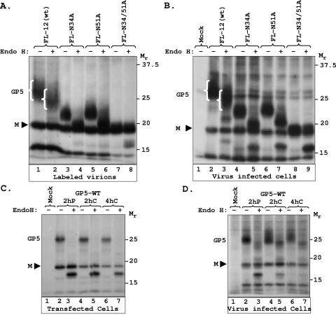
To determine the nature of the GP5 protein incorporated into infectious virions produced from transfected cells, we generated radiolabeled PRRSV from cells infected with the wt and mutant viruses. The extracellular virions present in the culture supernatant were pelleted by ultracentrifugation and GP5 present in these virions was examined by immunoprecipitation using anti-GP5 antibody and subsequent electrophoretic analysis. The results show that the wt GP5 incorporated into virions migrated as a broadly diffuse band of ∼25- to 27-kDa protein species (Fig. 4A, lane 1), which is partially resistant to Endo H digestion (lane 2). Mutant GP5 (N34A and N51A) incorporated into virions were sensitive to Endo H. Based on the sizes of the products generated following Endo H digestion, it appears that only one glycan moiety in these single-site mutants is sensitive, while the other is resistant. In contrast, the double mutant GP5 (N34/51A) was partially resistant to Endo H. Furthermore, Endo H digestion of GP5 from mutant viruses also produced various amounts of GP5 protein backbone, indicating that these viruses incorporate GP5 proteins that contain Endo H-resistant as well as Endo H-sensitive glycan moieties.
FIG. 4.
Examination of GP5 incorporated into mutant virions and synthesized in mutant virus-infected cells or in transfected cells. (A) Radiolabeled virions from culture supernatants of infected cells were pelleted, GP5 protein was immunoprecipitated, treated with Endo H (+) or not (−), and analyzed by electrophoresis. The positions of wt GP5 without and with Endo H digestion (lanes 1 and 2, respectively) are shown by white brackets. (B) Cells infected with various mutant viruses were radiolabeled, GP5 was immunoprecipitated, treated with Endo H (+) or not (−), and analyzed by electrophoresis. The positions of wt GP5 without and with Endo H digestion (lanes 2 and 3, respectively) are shown by white brackets. (C) Pulse-chase analysis and Endo H sensitivity of GP5 expressed in transfected cells. Cells were transfected with GP5-IE-M, pulse-labeled for 2 h (2hrP), and subsequently chased for 2 h (2hrC) or 4 h (4hrC). Proteins were immunoprecipitated with anti-GP5 antibody, digested with Endo H (+) or not (−), and analyzed by electrophoresis as described in Materials and Methods. (D) Pulse-chase analysis and Endo H sensitivity of GP5 in cells infected with wt PRRSV. The experiment was performed as in panel C. The mobility of proteins is shown in kilodaltons on the right side of each panel.
Since in cells transfected with the bicistronic vector, the wt as well as the mutant GP proteins were completely Endo H sensitive (Fig. 1 and 2), we were surprised by the observation that GP5 on PRRSV virions contained largely Endo H-resistant forms. To examine if the Endo H-resistant forms of the protein are also synthesized in infected cells, MARC-145 cells infected with wt or mutant PRRSV were radiolabeled, and GP5 protein was immunoprecipitated with anti-GP5 antibody and analyzed by electrophoresis with or without Endo H digestion. The results of such an experiment are shown in Fig. 4B.
The majority of wt GP5 contained Endo H-resistant glycans at all three sites (lanes 2 and 3), whereas the two single mutants contained Endo H-resistant glycans at only one site (lanes 4 to 7). Some of the glycan moieties in the double mutant are resistant, while others are sensitive to Endo H (Fig. 4B, lanes 8 and 9). Although the pattern of Endo H resistance is similar to what is observed for virion-associated GP5, it is different from that observed in cells expressing both GP5 and M proteins (Fig. 1 and 2).
To further determine if the GP5 protein synthesized in transfected cells is different from that synthesized in PRRSV-infected cells, we examined the Endo H resistance of GP5 by pulse-chase experiments. In transfected cells, GP5 synthesized during a 2-h pulse was completely sensitive to Endo H and remained fully sensitive to the enzyme even after a 4-h chase (Fig. 4C), indicating that GP5 remains in the endoplasmic reticulum or cis-Golgi compartment. However, in PRRSV-infected cells, GP5 was partially sensitive to Endo H following a 2-h pulse and the Endo H sensitivity of the protein decreased with increasing chase times (Fig. 4D). These results indicate that other viral proteins present in PRRSV-infected cells may play a role in further modification of glycans on GP5.
Influence of hypoglycosylation of GP5 on PRRSV's ability to be neutralized by specific antibodies.
The level of glycosylation of viral glycoproteins that are involved in the interaction with viral receptors is known to affect the ability of virions to react with virus-neutralizing antibodies (2, 41). To test whether this phenomenon occurs in the case of PRRSV, the PRRSV GP5 mutants with altered glycosylation patterns (FL-N34A, FL-N51A, and FL-N34/51A) were compared with wt PRRSV (FL-12) in their ability to be neutralized by convalescent antisera.
Towards this end, we used convalescent antisera (47 days postinfection) from four animals that had been infected with wt PRRSV. Similar doses (2,000 TCID50) of infectious PRRSV GP5 mutants (FL-N34A, FL-N51A and FL-N34/51A) as well as of the infectious clone-derived wt PRRSV (FL-12) were used as the challenge virus in serum neutralization assays following our standard assay protocol and the set of four anti-wt PRRSV (FL-12) sera used as references. Table 2 shows the different endpoint neutralizing titers obtained.
TABLE 2.
Effect of alteration of glycosylation pattern of PRRSV GP5 on the ability of the infectious virion to react with neutralizing antibodiesa
| Animal no. | Endpoint with PRRSV strain:
|
|||
|---|---|---|---|---|
| Wt PRRSV (FL-12) | FL-N34A | FL-N51A | FL-N34/51A | |
| 11404 | 1:32 | 1:256 | 1:256 | 1:32,768 |
| 11346 | 1:8 | 1:64 | 1:256 | 1:16,384 |
| 11457 | 1:16 | NDb | 1:128 | 1:2,048 |
| 11407 | 1:8 | ND | 1:64 | 1:2,048 |
Data are inverse endpoint dilutions showing neutralization. Animals were infected with 105 TCID50 of wt PRRSV FL12, and serum samples were taken 47 days postinfection.
ND, not determined.
Normally, a wt PRRSV convalescent-phase serum sample collected at 47 to 54 days postinfection contains moderate levels of wt PRRSV neutralizing activity (1:8 to 1:32) (Tables 2 and 3), reflecting the relatively weak and sluggish character of the neutralizing antibody response that is typical of infections with wt PRRSV (31). However, the use of hypoglycosylated PRRSV mutants (which lack one or two glycan moieties on the GP5 ectodomain) as the challenge virus in the serum neutralization assays seems to have significantly enhanced the endpoint of the reference sera, with endpoint titer enhancement ranging from 6- to 22-fold (Table 2). This observation clearly suggests that the removal of one or particularly two of the glycan moieties increases the accessibility of the neutralizing epitope to specific antibodies. These results appear to indicate the presence of significant amount of PRRSV-neutralizing antibodies in the wt PRRSV-infected convalescent-phase sera that would otherwise have been undetectable because of the use of wt PRRSV containing fully glycosylated GP5 in typical serum neutralization assays.
TABLE 3.
Effect of altered GP5 glycosylation pattern on ability of PRRSV strains to induce neutralizing antibodies to wt PRRSV
| Strain | Geometric mean endpoint at indicated day postinfection
|
|||
|---|---|---|---|---|
| 0 | 14 | 21 | 48 | |
| Wt PRRSV (FL-12) | 1.0 | 1.0 | 3.2 | 25.4 |
| FL-N34A | 1.0 | 1.0 | 4.0 | 128.0 |
| FL-N51A | 1.0 | 1.0 | 4.0 | 128.0 |
| FL-N34/51A | 1.0 | 1.0 | 5.0 | 161.3 |
Influence of hypoglycosylation of GP5 on PRRSV's ability to induce neutralizing antibodies in vivo.
One remarkable effect that has been reported in the literature is that carbohydrate removal from a viral envelope glycoprotein leads to production of high titers of neutralizing antibodies against the mutant virus when this mutant is used for in vivo inoculation of the host, in some cases also inducing higher titers of antibody to the wt virus than the wt virus itself (40). We infected groups of pigs with identical doses of either wt PRRSV (FL-12) or one of the mutants with altered glycosylation patterns.
Interestingly, clinical and virological assessment of the infection by evaluation of rectal temperature and evaluation of viremia at days 4, 7, and 10 postinfection indicated a similar pattern of infection in all groups, as previously described for FL-12 (49) without evidence of virulence attenuation or exacerbation for either of the mutants (data not shown). However, sequential sampling of serum from these animals for 48 days indicated pronounced differences between wt PRRSV and the mutants in the kinetics of induction of a PRRSV-neutralizing antibody response (Tables 3 and 4). The mutants developed early and more robust homologous neutralizing antibody responses than that developed by wt PRRSV, to the point where, in the case of the mutants, the characteristically weak and sluggish nature of the PRRSV-neutralizing antibody response appears to have been corrected (Table 4).
TABLE 4.
Effect of altered GP5 glycosylation pattern on ability of PRRSV strain to induce neutralizing antibodies to a homologous strain
| Strain | Geometric mean endpoint at indicated day postinfection
|
|||
|---|---|---|---|---|
| 0 | 14 | 21 | 48 | |
| Wt PRRSV (FL-12) | 1.0 | 1.0 | 3.2 | 25.4 |
| FL-N34A | 1.0 | 4.0 | 128 | 8,192 |
| FL-N51A | 1.0 | 4.0 | 128 | 2,048 |
| FL-N34/51A | 1.0 | 4.0 | 64 | 4,096 |
The kinetics of the appearance of mutant-homologous neutralizing antibodies (Table 4) indicates a more regular neutralizing antibody seroconversion consistent with that described for other viral infections such as influenza virus or pseudorabies virus but not for PRRSV (31). Of utmost importance is the fact that the infection with GP5 glycosylation mutants induced a wt PRRSV-specific neutralizing antibody response that is significantly higher than the response with wt PRRSV itself. The mutant viruses FL-N34A and FL-N51A induced fivefold-higher (P < 0.05) levels of neutralizing antibody titer against wt PRRSV than did wt PRRSV itself, while mutant FL-N34/51A induced a sixfold-higher (P < 0.01) titer of wt PRRSV-neutralizing antibodies than did wt PRRSV itself (Table 3).
DISCUSSION
In the present study, we examined the influence of glycosylation of GP5 of PRRSV on recovery of infectious virus and its role in the ability of the mutant viruses to be neutralized by antibodies and in inducing neutralizing antibodies in vivo. We have found that all three potential glycosylation sites (N34, N44, and N51) in GP5 are used for the addition of glycan moieties. Our results reveal that glycan addition at the N44 site is most critical for the recovery of infectious virus. Furthermore, our results show that PRRSVs containing hypoglycosylated forms of GP5 are exquisitely sensitive to neutralization by antibodies and that the mutant viruses induce significantly higher levels of neutralizing antibodies not only to the homologous mutant viruses but also to wt PRRSV.
Confirmation that all three potential N-linked glycosylation sites are used for glycan addition in GP5 was provided by using mutants with alterations at single or multiple sites (Fig. 2). Biochemical studies showed that the PRRSV GP5 protein, when coexpressed with M protein in transfected cells, contains Endo H-sensitive high-mannose-type glycans. The observation that the majority of GP5 incorporated into virions is resistant to Endo H (Fig. 4A) whereas GP5 expressed in the presence of M protein in transfected cells is fully Endo H sensitive is intriguing. It is possible that GP5 expressed in the presence of M protein in transfected cells accumulates mostly in the endoplasmic reticulum or in the cis-Golgi region and therefore remains Endo H sensitive. However, in PRRSV-infected cells, GP5 may interact with additional viral proteins and the transport of GP5 beyond the endoplasmic reticulum or cis-Golgi apparatus is facilitated through the formation of complexes with other viral proteins. Consistent with this interpretation, we have observed that in wt or mutant PRRSV-infected cells, GP5 protein is also resistant to Endo H.
We suggest that GP5, which is synthesized in the endoplasmic reticulum in infected cells, is transported to the medial and/or trans-Golgi region where the majority of GP5 molecules acquire Endo H resistance prior to being incorporated into PRRSV virions. Several studies with arteriviruses, including PRRSV, suggest that GP5 and M protein form a heterodimer, which may play a key role in viral infectivity (13, 15, 28). In EAV and LDV, direct interaction of GP5 and M protein through the formation of disulfide bridges has been demonstrated (13, 15). Such interactions may occur prior to further processing of N-linked oligosaccharide side chains, presumably before GP5 is transported out of the endoplasmic reticulum or the cis-Golgi compartment.
It is interesting that the pattern of Endo H resistance of GP5 incorporated into wt and mutant virions is different. While the majority of GP5 molecules in wt PRRSV were Endo H resistant, most of the GP5 molecules in the single-site mutant virions (FL-N34A and FL-N51A) were Endo H sensitive (Fig. 4A). Furthermore, of the two glycan moieties in these mutants, only one was sensitive, while the other was resistant. The double mutant (FL-N34/51A) virion also incorporated GP5 that contained glycans, some of which were also sensitive to Endo H. These data are consistent with the interpretation that wt as well as mutant PRRSV virions incorporate a mixed population of GP5 molecules that contain different glycan moieties at different sites. Previous studies demonstrating incorporation of differentially glycosylated forms of GP5 into wt PRRSV virions (28) further strengthen our interpretation.
From the pattern of Endo H sensitivity of GP5 incorporated into the virions, it is tempting to speculate that the N44 site may contain the Endo H-resistant glycans, although some GP5 molecules with Endo H-sensitive glycans at this site were incorporated into virions. Whether this unusual pattern of glycans at various sites in GP5 and incorporation of various forms of GP5 into virions has any relevance to the pattern of immune response seen in PRRSV-infected animals remains to be investigated.
In a recent study, it was shown that of the two N-linked glycosylation sites (N46 and N53) in GP5 of Lelystad PRRSV, glycosylation of N46 residue was strongly required for virus particle production. Infectious virus yield was reduced by approximately 100-fold by a mutation at N46 (53). Our results suggest that glycan addition at N44 (for North American PRRSV) is absolutely essential for recovery of infectious PRRSV. It is possible that the European and North American isolates of PRRSV differ somewhat in their requirements for N-linked glycosylation for production of infectious viruses. In this regard, it is of note that the Lelystad virus contains only two N-linked glycosylation sites, whereas the North American isolates we have used in this study contain three such sites.
GP5 is the most important glycoprotein of PRRSV involved in the generation of PRRSV-neutralizing antibodies and protective immunity. Our results reveal that the absence of glycans at residues 34 and 51 in the GP5 ectodomain, while generating viable PRRSV mutants, enhances both the sensitivity of these mutants to neutralization by antibodies and the immunogenicity of the nearby neutralization epitope. The immediate effect of the absence of glycans in GP5 of mutant PRRSVs has been increased sensitivity of the viruses to neutralization by convalescent-phase sera from pigs infected with wt PRRSV (Table 2).
Studies with human immunodeficiency virus type 1 and simian immunodeficiency virus have shown that acquisition or removal of glycans in the variable loops of gp160 modifies their sensitivity to neutralization (5, 7, 30). Therefore, it has been postulated (41) that glycans play at least two types of essential roles during biosynthesis of viral envelope glycoproteins. In one case, lack of glycans entails defects of the glycoprotein and thus in the overall viability of the viral strain. We postulate that the glycans at N44 of PRRSV GP5 serve a similar role. In the second case, the glycans potentially serve to shield viral proteins against neutralization by antibodies (41). For PRRSV GP5, glycans at N34 and N51 may have a similar role. In the case of human immunodeficiency virus, “glycan shielding” is postulated to be a primary mechanism to explain evasion from neutralizing immune response, ensuring in vivo persistence of human immunodeficiency virus (50).
This invites us to draw some parallels with PRRSV. Infection with PRRSV, which is known to persist for several months in individual animals (3, 52), presents an unusual behavior in terms of induction of virus-specific neutralizing immune response (31). It is well documented that animals infected with PRRSV usually take longer than normal to establish a detectable PRRSV-neutralizing antibody response. Once established, this PRRSV-neutralizing response is weak, and varies significantly from animal to animal (31, 38). The delay in neutralizing antibody response has been postulated to be due to the presence of a nearby immunodominant decoy epitope (amino acid positions 27 to 30), which evokes a robust, early, nonprotective immune response that masks and/or slows the response to the neutralizing epitope (amino acid positions 37 to 45) (26, 38). While this is a plausible explanation for the atypical character of the PRRSV-neutralizing antibody response, it remains to be tested. In our laboratory, deletion of the decoy epitope has consistently proven lethal to the recovery of infectious PRRSV (I. H. Ansari et al., unpublished results), making it difficult to test this hypothesis.
It is possible that an alternative or complementary mechanism to explain the peculiar nature of the PRRSV-neutralizing response could be envisioned by the glycan-shielding phenomenon proposed for the human and simian immunodeficiency viruses (22, 50). The use of mutant PRRSVs lacking one or two glycan moieties in our studies provides evidence for the first time of the presence of large amounts of PRRSV-neutralizing antibodies in the sera of wt PRRSV-infected animals that were otherwise undetectable because of the use of wt PRRSV in the serum neutralization assays. The PRRSV-neutralizing antibodies, while present in the host's response, are unable to react with the infecting wt PRRSV virions due to blocking or shielding of the neutralizing epitope by the glycan moieties on GP5.
One important precedent for neutralization escape by glycosylation of glycoproteins in arteriviruses has been described for LDV. It is highly resistant to antibody neutralization due to the heavy glycan shielding of its major glycoprotein, VP-3 (8). However, certain naturally occurring strains of LDV are highly susceptible to neutralization due to loss of two glycosylation sites on the ectodomain of VP-3. Interestingly, this neutralization-sensitive phenotype correlates with a high degree of neurotropism in the host acquired by these easily neutralizable LDV strains. Such enhancement of neuropathogenicity probably reflects the facilitation of interaction of the viral glycoproteins with receptors in neural cells, possibly due to the absence of glycan shielding (8).
In the young-pig model that we used for inoculation with PRRSV, we were not able to detect pathogenic differences between any of the mutant PRRSVs and wt PRRSV, although we limited our observations to temperature and viremia measurements. It is possible that under different experimental conditions (i.e., in a pregnant-sow model), some alterations in the pathogenicity of these mutant PRRSVs might be observed. It is not known whether naturally occurring hypoglycosylated PRRSV strains are common, although previous reports have suggested their presence (27, 39).
A remarkable observation in our experiments has been that the GP5 mutants, when infecting pigs in vivo, can outperform wt PRRSV in their ability to mount a sizable wt PRRSV-neutralizing response at late phases of infection (Table 3). Our results seem to mimic one of the most dramatic effects of viral glycoprotein carbohydrate removal so far reported in the literature by Reitter et al. (40). These authors observed that rhesus monkeys infected with simian immunodeficiency virus mutants lacking selected glycans moieties mount higher neutralization responses not only against the mutant virus but also against wt simian immunodeficiency virus than those caused by the wt virus itself. In a parallel scenario, we have observed not only higher neutralizing titers against homologous PRRSV mutants but also sizable titers against wt PRRSV (Table 3). In addition, the response occurred earlier, with neutralizing titers detectable at 14 days postinfection, an observation not typically noted with wt PRRSV infection (Table 4).
The increased neutralization of wt PRRSV by sera from pigs infected with the PRRSV mutants suggests that glycans were masking a neutralizing epitope(s) that does not induce neutralizing antibodies when glycans are present. This observation has great significance for the design of better, more efficacious PRRSV vaccines, suggesting that new, rationally designed vaccines should carry modifications in the glycosylation pattern of GP5 in order to enhance the production of neutralizing antibodies. In addition, it will be important to study the effects that this removal of carbohydrates from immunologically prominent glycoproteins of PRRSV may have on increasing serum neutralization titers not only to the homologous immunizing strain, but also to diverse unrelated PRRSV strains.
Acknowledgments
This research was supported in part by NRI CGP grant 2004-01576 from the United States Department of Agriculture, by National Pork Board project 04-112, and by project P20RR15636 from the COBRE program of the National Center for Research Resources, NIH.
We thank Carl A. Gagnon, University of Quebec, Canada, for providing polyclonal antibodies against PRRSV GP5 and Dan Rock (University of Illinois at Urbana) for comments on the manuscript. We also thank Debasis Nayak and Monica Brito for technical help and other members of our laboratories for valuable suggestions during this study.
REFERENCES
- 1.Abe, Y., E. Takashita, K. Sugawara, Y. Matsuzaki, Y. Muraki, and S. Hongo. 2004. Effect of the addition of oligosaccharides on the biological activities and antigenicity of influenza A/H3N2 virus hemagglutinin. J. Virol. 78:9605-9611. [DOI] [PMC free article] [PubMed] [Google Scholar]
- 2.Alexander, S., and J. H. Elder. 1984. Carbohydrate dramatically influences immune reactivity of antisera to viral glycoprotein antigens. Science 226:1328-1330. [DOI] [PubMed] [Google Scholar]
- 3.Allende, R., W. W. Laegreid, G. F. Kutish, J. A. Galeota, R. W. Wills, and F. A. Osorio. 2000. Porcine reproductive and respiratory syndrome virus: description of persistence in individual pigs upon experimental infection. J. Virol. 74:10834-10837. [DOI] [PMC free article] [PubMed] [Google Scholar]
- 4.Ausubel, F. M., R. Brent, R. E. Kingston, D. D. Moore, J. D. Seidman, J. A. Smith, and K. Struhl (ed.). 2001. Current protocols in molecular biology, 2nd ed. John Wiley and Sons, New York, N.Y.
- 5.Bolmstedt, A., S. Sjolander, J. E. Hansen, L. Akerblom, A. Hemming, S. L. Hu, B. Morein, and S. Olofsson. 1996. Influence of N-linked glycans in V4-V5 region of human immunodeficiency virus type 1 glycoprotein gp160 on induction of a virus-neutralizing humoral response. J. Acquir. Immune Defic. Syndr. Hum. Retrovir. 12:213-220. [DOI] [PubMed] [Google Scholar]
- 6.Braakman, I., and E. van Anken. 2000. Folding of viral envelope glycoproteins in the endoplasmic reticulum. Traffic 1:533-539. [DOI] [PMC free article] [PubMed] [Google Scholar]
- 7.Chackerian, B., L. M. Rudensey, and J. Overbaugh. 1997. Specific N-linked and O-linked glycosylation modifications in the envelope V1 domain of simian immunodeficiency virus variants that evolve in the host alter recognition by neutralizing antibodies. J. Virol. 71:7719-7727. [DOI] [PMC free article] [PubMed] [Google Scholar]
- 8.Chen, Z., K. Li, and P. G. Plagemann. 2000. Neuropathogenicity and sensitivity to antibody neutralization of lactate dehydrogenase-elevating virus are determined by polylactosaminoglycan chains on the primary envelope glycoprotein. Virology 266:88-98. [DOI] [PubMed] [Google Scholar]
- 9.Conzelmann, K. K., N. Visser, P. Van Woensel, and H. J. Thiel. 1993. Molecular characterization of porcine reproductive and respiratory syndrome virus, a member of the arterivirus group. Virology 193:329-339. [DOI] [PMC free article] [PubMed] [Google Scholar]
- 10.Dea, S., C. A. Gagnon, H. Mardassi, B. Pirzadeh, and D. Rogan. 2000. Current knowledge on the structural proteins of porcine reproductive and respiratory syndrome (PRRS) virus: comparison of the North American and European isolates. Arch. Virol. 145:659-688. [DOI] [PMC free article] [PubMed] [Google Scholar]
- 11.Delputte, P. L., and H. J. Nauwynck. 2004. Porcine arterivirus infection of alveolar macrophages is mediated by sialic acid on the virus. J. Virol. 78:8094-8101. [DOI] [PMC free article] [PubMed] [Google Scholar]
- 12.Delputte, P. L., N. Vanderheijden, H. J. Nauwynck, and M. B. Pensaert. 2002. Involvement of the matrix protein in attachment of porcine reproductive and respiratory syndrome virus to a heparinlike receptor on porcine alveolar macrophages. J. Virol. 76:4312-4320. [DOI] [PMC free article] [PubMed] [Google Scholar]
- 13.de Vries, A. A., S. M. Post, M. J. Raamsman, M. C. Horzinek, and P. J. Rottier. 1995. The two major envelope proteins of equine arteritis virus associate into disulfide-linked heterodimers. J. Virol. 69:4668-4674. [DOI] [PMC free article] [PubMed] [Google Scholar]
- 14.Doms, R. W., R. A. Lamb, J. K. Rose, and A. Helenius. 1993. Folding and assembly of viral membrane proteins. Virology 193:545-562. [DOI] [PubMed] [Google Scholar]
- 15.Faaberg, K. S., and P. G. Plagemann. 1995. The envelope proteins of lactate dehydrogenase-elevating virus and their membrane topography. Virology 212:512-525. [DOI] [PubMed] [Google Scholar]
- 16.Gagnon, C. A., G. Lachapelle, Y. Langelier, B. Massie, and S. Dea. 2003. Adenoviral-expressed GP5 of porcine respiratory and reproductive syndrome virus differs in its cellular maturation from the authentic viral protein but maintains known biological functions. Arch. Virol. 148:951-972. [DOI] [PMC free article] [PubMed] [Google Scholar]
- 17.Helenius, A. 1994. How N-linked oligosaccharides affect glycoprotein folding in the endoplasmic reticulum. Mol. Biol. Cell 5:253-265. [DOI] [PMC free article] [PubMed] [Google Scholar]
- 18.Helenius, A., and M. Aebi. 2001. Intracellular functions of N-linked glycans. Science 291:2364-2369. [DOI] [PubMed] [Google Scholar]
- 19.Helenius, A., and M. Aebi. 2004. Roles of N-linked glycans in the endoplasmic reticulum. Annu. Rev. Biochem. 73:1019-1049. [DOI] [PubMed] [Google Scholar]
- 20.Horton, R. M., Z. L. Cai, S. N. Ho, and L. R. Pease. 1990. Gene splicing by overlap extension: tailor-made genes using the polymerase chain reaction. BioTechniques 8:528-535. [PubMed] [Google Scholar]
- 21.Jang, S. K., M. V. Davies, R. J. Kaufman, and E. Wimmer. 1989. Initiation of protein synthesis by internal entry of ribosomes into the 5′ nontranslated region of encephalomyocarditis virus RNA in vivo. J. Virol. 63:1651-1660. [DOI] [PMC free article] [PubMed] [Google Scholar]
- 22.Johnson, W. E., H. Sanford, L. Schwall, D. R. Burton, P. W. Parren, J. E. Robinson, and R. C. Desrosiers. 2003. Assorted mutations in the envelope gene of simian immunodeficiency virus lead to loss of neutralization resistance against antibodies representing a broad spectrum of specificities. J. Virol. 77:9993-10003. [DOI] [PMC free article] [PubMed] [Google Scholar]
- 23.Kim, H. S., J. Kwang, I. J. Yoon, H. S. Joo, and M. L. Frey. 1993. Enhanced replication of porcine reproductive and respiratory syndrome (PRRS) virus in a homogeneous subpopulation of MA-104 cell line. Arch. Virol. 133:477-483. [DOI] [PubMed] [Google Scholar]
- 24.Kornfeld, R., and S. Kornfeld. 1985. Assembly of asparagine-linked oligosaccharides. Annu. Rev. Biochem. 54:631-664. [DOI] [PubMed] [Google Scholar]
- 25.Lee, J., J. S. Park, J. Y. Moon, K. Y. Kim, and H. M. Moon. 2003. The influence of glycosylation on secretion, stability, and immunogenicity of recombinant HBV pre-S antigen synthesized in Saccharomyces cerevisiae. Biochem. Biophys. Res. Commun. 303:427-432. [DOI] [PubMed] [Google Scholar]
- 26.Lopez, O. J., and F. A. Osorio. 2004. Role of neutralizing antibodies in PRRSV protective immunity. Vet. Immunol. Immunopathol. 102:155-163. [DOI] [PubMed] [Google Scholar]
- 27.Magar, R., R. Larochelle, E. A. Nelson, and C. Charreyre. 1997. Differential reactivity of a monoclonal antibody directed to the membrane protein of porcine reproductive and respiratory syndrome virus. Can. J. Vet. Res. 61:69-71. [PMC free article] [PubMed] [Google Scholar]
- 28.Mardassi, H., B. Massie, and S. Dea. 1996. Intracellular synthesis, processing, and transport of proteins encoded by ORFs 5 to 7 of porcine reproductive and respiratory syndrome virus. Virology 221:98-112. [DOI] [PubMed] [Google Scholar]
- 29.Mardassi, H., S. Mounir, and S. Dea. 1995. Structural gene analysis of a Quebec reference strain or porcine reproductive and respiratory syndrome virus (PRRSV). Adv. Exp. Med. Biol. 380:277-281. [DOI] [PubMed] [Google Scholar]
- 30.Matsushita, S., M. Robert-Guroff, J. Rusche, A. Koito, T. Hattori, H. Hoshino, K. Javaherian, K. Takatsuki, and S. Putney. 1988. Characterization of a human immunodeficiency virus neutralizing monoclonal antibody and mapping of the neutralizing epitope. J. Virol. 62:2107-2114. [DOI] [PMC free article] [PubMed] [Google Scholar]
- 31.Meier, W. A., J. Galeota, F. A. Osorio, R. J. Husmann, W. M. Schnitzlein, and F. A. Zuckermann. 2003. Gradual development of the interferon-gamma response of swine to porcine reproductive and respiratory syndrome virus infection or vaccination. Virology 309:18-31. [DOI] [PubMed] [Google Scholar]
- 32.Meulenberg, J. J. 2000. PRRSV, the virus. Vet. Res. 31:11-21. [DOI] [PubMed] [Google Scholar]
- 33.Meulenberg, J. J., J. N. Bos-de Ruijter, R. van de Graaf, G. Wensvoort, and R. J. Moormann. 1998. Infectious transcripts from cloned genome-length cDNA of porcine reproductive and respiratory syndrome virus. J. Virol. 72:380-387. [DOI] [PMC free article] [PubMed] [Google Scholar]
- 34.Meulenberg, J. J., M. M. Hulst, E. J. de Meijer, P. L. Moonen, A. den Besten, E. P. de Kluyver, G. Wensvoort, and R. J. Moormann. 1993. Lelystad virus, the causative agent of porcine epidemic abortion and respiratory syndrome (PEARS), is related to LDV and EAV. Virology 192:62-72. [DOI] [PMC free article] [PubMed] [Google Scholar]
- 35.Meulenberg, J. J., A. Petersen-den Besten, E. P. De Kluyver, R. J. Moormann, W. M. Schaaper, and G. Wensvoort. 1995. Characterization of proteins encoded by ORFs 2 to 7 of Lelystad virus. Virology 206:155-163. [DOI] [PMC free article] [PubMed] [Google Scholar]
- 36.Nelson, E. A., J. Christopher-Hennings, T. Drew, G. Wensvoort, J. E. Collins, and D. A. Benfield. 1993. Differentiation of U.S. and European isolates of porcine reproductive and respiratory syndrome virus by monoclonal antibodies. J. Clin. Microbiol. 31:3184-3189. [DOI] [PMC free article] [PubMed] [Google Scholar]
- 37.Nielsen, H. S., G. Liu, J. Nielsen, M. B. Oleksiewicz, A. Botner, T. Storgaard, and K. S. Faaberg. 2003. Generation of an infectious clone of VR-2332, a highly virulent North American-type isolate of porcine reproductive and respiratory syndrome virus. J. Virol. 77:3702-3711. [DOI] [PMC free article] [PubMed] [Google Scholar]
- 38.Ostrowski, M., J. A. Galeota, A. M. Jar, K. B. Platt, F. A. Osorio, and O. J. Lopez. 2002. Identification of neutralizing and nonneutralizing epitopes in the porcine reproductive and respiratory syndrome virus GP5 ectodomain. J. Virol. 76:4241-4250. [DOI] [PMC free article] [PubMed] [Google Scholar]
- 39.Pirzadeh, B., C. A. Gagnon, and S. Dea. 1998. Genomic and antigenic variations of porcine reproductive and respiratory syndrome virus major envelope GP5 glycoprotein. Can. J. Vet. Res. 62:170-177. [PMC free article] [PubMed] [Google Scholar]
- 40.Reitter, J. N., R. E. Means, and R. C. Desrosiers. 1998. A role for carbohydrates in immune evasion in AIDS. Nat. Med. 4:679-684. [DOI] [PubMed] [Google Scholar]
- 41.Reynard, F., A. Fatmi, B. Verrier, and F. Bedin. 2004. HIV-1 acute infection env glycomutants designed from 3D model: effects on processing, antigenicity, and neutralization sensitivity. Virology 324:90-102. [DOI] [PubMed] [Google Scholar]
- 42.Sarkar, G., and S. S. Sommer. 1990. The “megaprimer” method of site-directed mutagenesis. BioTechniques 8:404-407. [PubMed] [Google Scholar]
- 43.Shi, X., and R. M. Elliott. 2004. Analysis of N-linked glycosylation of Hantaan virus glycoproteins and the role of oligosaccharide side chains in protein folding and intracellular trafficking. J. Virol. 78:5414-5422. [DOI] [PMC free article] [PubMed] [Google Scholar]
- 44.Skehel, J. J., D. J. Stevens, R. S. Daniels, A. R. Douglas, M. Knossow, I. A. Wilson, and D. C. Wiley. 1984. A carbohydrate side chain on hemagglutinins of Hong Kong influenza viruses inhibits recognition by a monoclonal antibody. Proc. Natl. Acad. Sci. USA 81:1779-1783. [DOI] [PMC free article] [PubMed] [Google Scholar]
- 45.Snijder, E. J., J. C. Dobbe, and W. J. Spaan. 2003. Heterodimerization of the two major envelope proteins is essential for arterivirus infectivity. J. Virol. 77:97-104. [DOI] [PMC free article] [PubMed] [Google Scholar]
- 46.Snijder, E. J., and J. J. Meulenberg. 1998. The molecular biology of arteriviruses. J. Gen. Virol. 79:961-979. [DOI] [PubMed] [Google Scholar]
- 47.Snijder, E. J., and J. M. Meulenberg. 2001. Arteriviruses, p. 1205-1220. In D. Knipe et al. (ed.), Fields virology, 4th ed. Lippincott Williams & Wilkins, Philadelphia, Pa.
- 48.Truong, H., Z. Lu, G. Kutish, F. Osorio, and A. Pattnaik. 2004. Infectious PRRSV from transcripts of a full-length cDNA clone. Proc. 23rd Annu. Mtg. Am. Soc. Virol., Quebec, Canada, abstr. W17-10, p. 109, 2004.
- 49.Truong, H. M., Z. Lu, G. F. Kutish, J. Galeota, F. A. Osorio, and A. K. Pattnaik. 2004. A highly pathogenic porcine reproductive and respiratory syndrome virus generated from an infectious cDNA clone retains the in vivo virulence and transmissibility properties of the parental virus. Virology 325:308-319. [DOI] [PMC free article] [PubMed] [Google Scholar]
- 50.Wei, X., J. M. Decker, S. Wang, H. Hui, J. C. Kappes, X. Wu, J. F. Salazar-Gonzalez, M. G. Salazar, J. M. Kilby, M. S. Saag, N. L. Komarova, M. A. Nowak, B. H. Hahn, P. D. Kwong, and G. M. Shaw. 2003. Antibody neutralization and escape by HIV-1. Nature 422:307-312. [DOI] [PubMed] [Google Scholar]
- 51.Williams, D. B. 1995. Calnexin leads glycoproteins into the fold. Glycoconj. J. 12:iii-iv. [DOI] [PubMed] [Google Scholar]
- 52.Wills, R. W., J. J. Zimmerman, K. J. Yoon, S. L. Swenson, M. J. McGinley, H. T. Hill, K. B. Platt, J. Christopher-Hennings, and E. A. Nelson. 1997. Porcine reproductive and respiratory syndrome virus: a persistent infection. Vet. Microbiol. 55:231-240. [DOI] [PubMed] [Google Scholar]
- 53.Wissink, E. H., M. V. Kroese, J. G. Maneschijn-Bonsing, J. J. Meulenberg, P. A. van Rijn, F. A. Rijsewijk, and P. J. Rottier. 2004. Significance of the oligosaccharides of the porcine reproductive and respiratory syndrome virus glycoproteins GP2a and GP5 for infectious virus production. J. Gen. Virol. 85:3715-3723. [DOI] [PubMed] [Google Scholar]
- 54.Wu, W. H., Y. Fang, R. Farwell, M. Steffen-Bien, R. R. Rowland, J. Christopher-Hennings, and E. A. Nelson. 2001. A 10-kDa structural protein of porcine reproductive and respiratory syndrome virus encoded by ORF2b. Virology 287:183-191. [DOI] [PubMed] [Google Scholar]
- 55.Zhang, M., B. Gaschen, W. Blay, B. Foley, N. Haigwood, C. Kuiken, and B. Korber. 2004. Tracking global patterns of N-linked glycosylation site variation in highly variable viral glycoproteins: HIV, SIV, and HCV envelopes and influenza hemagglutinin. Glycobiology 14:1229-1246. [DOI] [PubMed] [Google Scholar]


