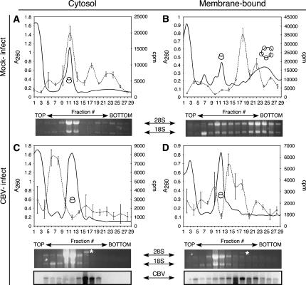
FIGURE 4.
Global mRNA translation is compartmentalized to the ER following CBV infection. HeLa cells were mock-infected (A,B) or infected with CBV (C,D) for 6 h and fractionated to yield cytosol and ER. Polyribosome profiles of cytosolic (free) (A,C) and ER (membrane-bound) (B,D) ribosomes were determined by velocity sedimentation on 15%–50% linear sucrose gradients. Gradient fractions were collected and analyzed by UV spectroscopy (solid line). Total RNA was isolated from individual gradient fractions. mRNA content was measured by quantitative incorporation of [32P]dCTP-radiolabeled nucleotide precursor into cDNA using an oligo (dT)-primed reverse transcription assay (dashed line, open circles). The sedimentation patterns of ribosomal subunits and ribosomes were determined through analysis of ribosomal RNA (indicated by 28S and 18S), and CBV virions (*) were determined by agarose denaturing gels. For C and D, RNA gels were transferred to nylon membranes and processed for Northern analysis using a probe directed against CBV RNA.

