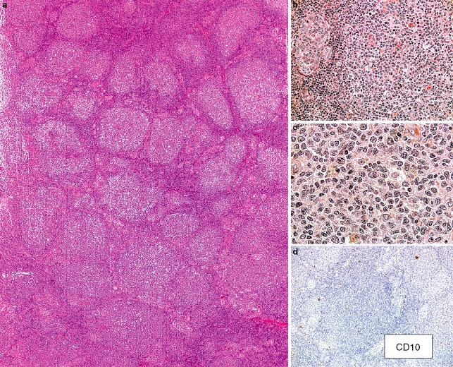
Figure 2.
Follicular lymphoma of the thyroid. a, The thyroid in this 70-year-old male is extensively infiltrated by numerous lymphoid follicles with relatively homogeneous-appearing follicular/germinal centres. b, The interfollicular regions (left) adjacent to the neoplastic-appearing follicle demonstrate many small lymphocytes and prominent lymphoepithelial lesions. c, There are numerous small angulated lymphocytes with pale cytoplasm but only infrequent centroblasts in this follicular/germinal centre. (a–c, haematoxylin and eosin.) d, There are only scattered cells staining distinctly for CD10. Weak positivity was described in the case presentation. (CD10 immunostain with haematoxylin counterstain.) Other immunophenotypic studies were reported to show the following: CD20+, IgM–, IgD–, Bcl-6+, Bcl-2– and 50% Ki67 staining in the follicular structures. Polymerase chain reaction (PCR) studies did not demonstrate a clonal IGH rearrangement and neither PCR nor cytogenetic fluorescence in situ hybridization studies showed an IGH/BCL2 translocation. Case contributed by E. Navratil, A. Dogan, P. Isaacson.

