Abstract
Two sibling species of tephritid fruit fly, Bactrocera tryoni and Bactrocera neohumeralis, are differentiated by their time of mating, which is genetically determined and requires interactions between the endogenous circadian clock and light intensity. The cryptochrome (cry) gene, a light-sensitive component of the circadian clock, was isolated in the two Bactrocera species. The putative amino acid sequence is identical in the two species. In the brain, in situ hybridization showed that cry is expressed in the lateral and dorsal regions of the central brain where PER immunostaining was also observed and in a peripheral cell cluster of the antennal lobes. Levels of cry mRNA were analyzed in whole head, brain, and antennae. In whole head, cry is abundantly and constantly expressed. However, in brain and antennae the transcript cycles in abundance, with higher levels during the day than at night, and cry transcripts are more abundant in the brain and antennae of B. neohumeralis than in that of B. tryoni. Strikingly, these results are duplicated in hybrid lines, generated by rare mating between B. tryoni and B. neohumeralis and then selected on the basis of mating time, suggesting a role for the cry gene in the mating isolation mechanism that differentiates the species.
REPRODUCTIVE isolation based on time of mating is a feature that prevents gene flow between species in sympatry. The possible role of clock genes in setting the time of mating is therefore of interest to questions of sympatric speciation. The clock genes period (per) and timeless (tim) control the circadian rhythms of female mating activity in the model organism, Drosophila melanogaster (Sakai and Ishida 2001). Moreover, gene transfer experiments implicate per in the species-specific behaviors of locomotor activity, love song rhythms, and time of mating (Petersen et al. 1988; Wheeler et al. 1991; Tauber et al. 2003). Populations of the melon fly, Bactrocera cucurbitae, showing differences in mating time, also show differences in circadian fluctuations of per mRNA (Miyatake et al. 2002).
Two Australian tephritid fruit flies provide an opportunity to study the molecular basis of a difference in mating time. The sibling species, B. tryoni and B. neohumeralis, occur sympatrically along the east coast of Queensland, with the range of B. neohumeralis contained entirely within the larger range of B. tryoni. Genetically, the two species are very closely related. In terms of DNA sequences tested, B. tryoni and B. neohumeralis are differentiated by only two single-nucleotide changes plus one trinucleotide indel in the ribosomal ITS 2 sequence (Morrow et al. 2000) and by differences in the frequency of some polymorphic microsatellite alleles (Wang et al. 2003). No fixed amino acid difference has been found in any gene tested to date and shared polymorphisms in coding and noncoding regions are common (Raphael et al. 2004). The very low level of genetic differentiation suggests that the sibling Bactrocera species are defined, not by widespread small differences throughout the genome, but by differences in the genes that determine the mating isolation mechanism.
B. tryoni and B. neohumeralis are reproductively isolated by time of mating: B. tryoni mates at dusk, in a narrow window of optimal light intensity (Tychsen and Fletcher 1971), whereas B. neohumeralis mates during the day in bright light (Smith 1979a). Hybrids resulting from forced mating between the two species are viable and fertile (Smith 1979a; Meats et al. 2003). A color difference of the humeral calli (yellow in B. tryoni, brown in B. neohumeralis) also differentiates the species and segregates with mating time in hybrid lines selected for day or dusk mating (Meats et al. 2003).
The daily rhythm of mating activity in the flies is controlled by the endogenous circadian clock (Tychsen and Fletcher 1971). The mating time difference was found to have a genetic basis by a study of the segregation of behavioral differences in hybrids (Smith 1979a), which indicated interaction between the circadian clock and light intensity. Consequently, the genes involved in circadian regulation and light response are possible candidates for species differentiation. The per homolog has been isolated from B. tryoni and B. neohumeralis (An et al. 2002), but sequence comparisons have revealed no difference in the putative amino acid sequence. The mRNA expression profiles of per in the two Bactrocera species are identical and closely follow the Drosophila pattern, in which maximum levels of expression occur during the first 4 hr of the dark phase and are maintained for 5–8 hr and expression falls to minimum levels at the end of the dark phase (Hardin et al. 1990; An et al. 2002). These results suggest that the components of the central pacemaker operate in a similar way in the two species and in Drosophila.
Entrainment of the endogenous clock to the external day-night cycle enables the organism to coordinate its activities with the external world, with light the major stimulus that sets the clock at each dawn and dusk. In Drosophila, multiple pathways contribute to photo-entrainment of the clock: the compound eyes, an extraocular pathway, and the blue-light photoreceptor cryptochrome (Helfrich-Forster et al. 2001). The Drosophila timeless protein (TIM) is also sensitive to light but this effect is mediated by binding to cryptochrome (CRY; Ceriani et al. 1999). The cryptochrome (cry) gene was first identified as encoding a blue-light photoreceptor in Arabidopsis (Ahmad and Cashmore 1993), and its role in the circadian pacemaker was revealed later to include plants, insects, and mammals (Young and Kay 2001). The cry gene shares sequence homology and a binding site for the flavin adenine dinucleotide (FAD) cofactor with other members of the cryptochrome/photolyase protein family.
The cry transcript is expressed in the lateral neurons (LNs) of Drosophila brain (Egan et al. 1999; Emery et al. 2000), believed to be the central pacemaker for behavioral rhythms. Expression has also been monitored in peripheral tissues such as the Malpighian tubules (Ivanchenko et al. 2001). In Drosophila, the cryb mutation blocks molecular cycling of the other circadian components, PER and TIM, in peripheral tissues in light-dark cycles, but PER/TIM molecular cycling is present in the central pacemaker cells (LNs) of the brain (Stanewsky et al. 1998). Although cryb flies have normal activity rhythms, they show defects in the ability to reset the clock after light pulses (Stanewsky et al. 1998) and in entrainment to a new light-dark regime, where adjustment of the evening activity peak is primarily affected (Emery et al. 2000). The cryb mutation results in loss of circadian fluctuation of olfactory function (Krishnan et al. 2001), but expression of cry in the antennae has not been directly tested in Drosophila. CRY is required in a cell autonomous fashion to maintain the molecular cycling of PER and TIM in the periphery (Emery et al. 2000), but the normal cycling of PER/TIM in the LNs of cryb flies suggests light input from the visual system for these cells (Stanewsky et al. 1998). Thus, CRY plays a major role in entrainment of the Drosophila clock to light-dark cycles (Emery et al. 1998, 2000), although a photoreceptor-independent role for CRY has been suggested in the periphery (Krishnan et al. 2001).
From studies in Drosophila and other organisms, the response to blue light is strongly dependent on the concentration of CRY protein (Lin et al. 1998; Emery et al. 2000). CRY overexpression in Drosophila results in behavioral hypersensitivity to light of low intensities (Emery et al. 1998). Since B. tryoni mates at dusk, when blue light predominates, we hypothesized that cry may have a role in the mechanism mediating the effect of light intensity on mating behavior. Therefore, we have characterized the cry homolog in B. tryoni and B. neohumeralis and studied its expression in whole head, brain, and antennae. We find that the level of cry transcript is significantly higher in the brain and antennae of B. neohumeralis compared with B. tryoni and that this difference is regenerated in hybrid lines of the two species, selected for early and late mating time.
MATERIALS AND METHODS
Culturing conditions for fruit flies:
The rearing techniques for both B. tryoni and B. neohumeralis flies have been described by Bateman (1967). The cultures were maintained at constant temperature of 25° and 60–70% relative humidity. The light-dark cycle was maintained as 14 hr light, 1 hr natural dusk, and 9 hr dark. Stock flies exhibited the typical mating behavior of each species. Adults emerged onto fresh medium and were maintained for 10–14 days to become reproductively mature.
Generation of B. neohumeralis and B. tryoni hybrids, selected for day-mating and dusk-mating behavior:
Flies were forced to mate by caging females of one species with males of the other. This parental cross was done in both directions, i.e., male B. tryoni mated with female B. neohumeralis and the reciprocal. The F1 was fertile and mated mainly at dusk. No difference was observed in the F1 of the reciprocal crosses. The F1 progeny were combined for the selection experiment. The F2 showed mating time variation, and selection was started at this point. On emergence, sexes were segregated until they were mature. The selection procedure included a filter whereby the day-mating flies were removed before dusk-mating pairs were used to start the next generation and vice versa for the selection of the day-mating line. After 6–8 generations of selection, the lines were stable (Meats et al. 2003).
Cloning of cry in B. neohumeralis and B. tryoni and sequencing analysis:
The cry transcript was isolated by a PCR-based strategy. The sequences for the primers used are indicated by arrows in Figure 1. Degenerate primers DF1 (TGGCVMGGMGGAGARACASARGC), DR1 (AAVGMVSWCGWWGAYASCCACATCC), and DR2 (CCWGCRYWBACVSWCCARTCBGC) were designed on the basis of regions of the cry gene conserved between D. melanogaster and mouse. After first-round PCR with DF1 and DR1, seminested PCR was conducted with DF1 and DR2. PCR was performed in a Hybaid OmniGene thermal cycler. The reaction mix contained 2.0 μm MgCl2, 250 μm each dNTP, 25 pmol each primer, and 1 unit Biotech Tth plus DNA polymerase. The first-round cycling conditions were 94° for 4 min; hold at 72° while the Tth enzyme mixture is added; and then 1 cycle of 46° for 1 min and 72° for 2 min; 1 cycle of 94° for 45 sec, 50° for 1 min, 72° for 2 min; and then 28 cycles of 93° for 30 sec, 56° for 45 sec, 72° for 1 min; and final extension at 72° for 5 min. Diluted PCR product (1 μl of 1:50 dilution) was used as template for the second-round PCR. The cycling conditions were 1 cycle of 94° for 2 min, 60° for 1 min, 72° for 1 min; 1 cycle of 94° for 1 min, 58° for 1 min, 72° for 1 min; the annealing temperature reduces by 2° for each following cycle until it reaches 46°; and then 25 cycles of 93° for 1 min, 46° for 1 min, 72° for 1 min; and final extension at 72° for 5 min. The amplified DNA fragments were purified using the GeneClean kit (GeneWorks) and then subcloned into EcoRV/ddT-tailed pBluescript vector (Marchuk et al. 1991), followed by sequencing with the ABI Automated DNA sequencer. The sequence revealed that the fragments from both B. neohumeralis and B. tryoni were homologs of cry. For 3′ rapid amplification of cDNA ends (RACE; Frohman 1994), forward primers (cryF1 and cryF2) were generated from the 3′ end of the cloned sequence. Total RNA (0.2–1.0 μg) from fly heads was reverse transcribed using Superscript III reverse transcriptase (Invitrogen, Carlsbad, CA) and poly(dT)-adapter primer in a 20-μl volume as recommended by the manufacturer. The first RACE-PCR, including 25 pmol cryF1 and 1/20 of the cDNA, was carried out as follows: 94° for 3 min; hold at 72° while the Tth plus is added; and then 1 cycle of 56° for 1 min and 72° for 2 min; 30 cycles of 94° for 40 sec, 56° for 1 min, 72° for 2 min; and final extension at 72° for 5 min. The nested RACE-PCR was carried out by the same procedure, using a 1-μl aliquot of the 1:50 diluted first-round PCR product as template with adapter RACE primer (Frohman 1994) and cryF2. PCR products were examined on 1% agarose gels. Specific products were purified and subcloned into pBluescript for sequence analysis.
Figure 1.—
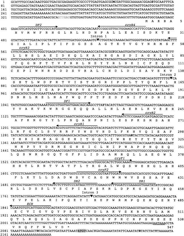
Nucleotide and deduced amino acid sequence of cryptochrome cDNA in B. tryoni. Nucleotides are numbered on the left while amino acids are numbered on the right. Primers described in the text are indicated by arrows. The positions of introns are designated by solid triangles. The end of translation is marked by an asterisk. Polymorphic sites are indicated in boldface type: R, A or G; Y, C or T; M, A or C; K, G or T; S, C or G. The HincII restriction site in the 5′-UTR used for inverse PCR is indicated in outline type. The four nucleotides (ATGT) absent from B. neohumeralis cry cDNA are shaded in the 3′-UTR.
To isolate the 5′ end of the gene, degenerate primer DF2 (GTNATHTGGTTYMGRCAYGG) was designed corresponding to the beginning of CRY translation in other species. PCR was conducted with the degenerate primer and a nested pair of nondegenerate reverse primers (cryR1 and cryR2). A 710-bp fragment was amplified and further sequencing revealed it to extend from nucleotide 405 to 1115. 5′ RACE (Frohman 1994) was then used to obtain the very 5′ end of the cry transcript. The initial reverse transcription was conducted with cryR2 and then first round PCR was conducted with primer cryR3 and second round PCR was conducted with nested primer cryR4 (Figure 1). Since the Bactrocera genome is relatively A + T rich, a poly(G) adapter primer was used in this approach. The reaction conditions were the same as for 3′ RACE. Inverse PCR on genomic DNA revealed the splicing site of intron 1. The restriction site used in this approach was HincII (Figure 1).
Samples of B. tryoni and B. neohumeralis were processed side by side. The sequence alignment of cry from the two species was done in Sequencher 3.1.1. Bioinformatic analyses were performed via Biomanager (http://biomanager.angis.org.au). From the cry DNA sequence, the open reading frame was identified and translated into amino acid sequence by the GCG Map program. The percentage identities between different protein sequences were scored by the GCG Bestfit program. The comparison of Bactrocera CRY and CRYs from other species were conducted by using Clustalw (accurate).
Tissue dissection and the generation of cDNA:
Flies of the same age and size were sampled at 09:30 and 23:00 by snap freezing. For each sample, six fly brains and six pairs of antennae were dissected in PBS buffer with RNase-free instruments and each tissue was transferred into Trizol containing 10 ng kanamycin RNA. A standard Trizol RNA extraction protocol was employed, according to the manufacturer's protocol (Invitrogen). Glycogen (20 μg) was added to each sample to improve the precipitation of RNA. Half of each RNA extract was reverse transcribed using Superscript III (Invitrogen) according to the manufacturer's protocol.
Real-time PCR and quantification of cry expression:
A time series of fly-head cDNA samples were obtained as previously described (An et al. 2002). Relative quantitative PCR was achieved using primers cryF1 and ECR, spanning a 56-bp intron (Figure 1), and double-strand DNA-binding fluorescent dye SYBR Green I (Molecular Probes, Eugene, OR). Each 20-μl reaction contained 2 mm MgCl2, 0.2 mm dNTP, 1× PCR buffer, 0.5 μm each primer, SYRB Green I (2× concentrate, the stock is 10,000× concentrate in DMSO), and cDNA template. No-template controls were run to check for contamination and determine the level of primer dimer formation. Amplification was performed in 0.2-ml tubes on a Rotor Gene 2000 real-time PCR machine (Corbett Research) supported by software Rotor-Gene 4.4. PCR parameters were an initial denature at 94° for 120 sec, followed by 40 cycles of 93° for 20 sec, 60° for 30 sec, and 72° for 45 sec. Fluorescence data were acquired using a low gain at 80°, where only specific products were assumed to be present. A melting analysis on resulting PCR products supported this for all samples measured. In addition, PCR products were run on 1.5% agarose gels to confirm that the correct band sizes were present without additional bands. Levels of cry cDNA were expressed relative to kanamycin cDNA controls in the same samples.
Statistical analysis:
A two-factor ANOVA was carried out, in which the two factors were: (1) time of day (morning vs. night expression) and (2) expression between species (B. neohumeralis vs. B. tryoni). The model tested here is one in which individual values are of the form yij = mean + Di + Gj + D × Gij + error, where Di represents the time of day (i = 1, 2), Gj represents the effect of genotype or species in this case (j = 1, 2), and D × Gij represents the interaction.
The same analysis was then carried out for the selected lines, in which the genotypes or “species” are actually hybrid lines selected for B. tryoni and B. neohumeralis mating behavior, respectively. The model is the same in this case.
A combined ANOVA can be given involving all four genotypes Gj (j = 1, 4). It is then convenient to further break down the Gj component into Sk + Tl + S × Tkl, where Sk is the “species” (k = 1, 2), and Tl (l = 1, 2) represents the type, pure species vs. hybrid line. The comparison between genotypes, having three degrees of freedom, is therefore resolved into three comparisons each with one degree of freedom. Any significant interaction term D × Gij may be broken down in a similar way. The combined analysis is then effectively a three-factor ANOVA, allowing both sets of data to be analyzed together and simultaneously allowing a comparison between pure species and hybrid lines. Results are reported in Table 1.
TABLE 1.
ANOVA ofcry expression in (A) brain and (B) antennae
| Source of variation | Sum of squares |
d.f. | F-ratio | P |
|---|---|---|---|---|
| A. Brain | ||||
| 1. Time of day (morning/night) | 0.648 | 1 | 28.34 | 0.000*** |
| 2. Genotype | 0.305 | 3 | 4.45 | 0.019* |
| 1. Time × 2. Genotype | 0.0042 | 3 | 0.062 | 0.979 |
| 2a. “Species”a | 0.228 | 1 | 10.00 | 0.006** |
| 2b. Type (pure species/hybrid line) | 0.068 | 1 | 2.98 | 0.103 |
| 2a. “Species” × 2b. Type | 0.008 | 1 | 0.37 | 0.552 |
| Error | 0.3657 | 16 | ||
| B. Antennae | ||||
| 1. Time of day (morning/night) | 2.477 | 1 | 75.18 | 0.000*** |
| 2. Genotype | 0.518 | 3 | 5.24 | 0.0104* |
| 1. Time × 2. Genotype | 0.120 | 3 | 1.21 | 0.338 |
| 2a. “Species”a | 0.469 | 1 | 14.24 | 0.002** |
| 2b. Type (pure species/hybrid line) | 0.049 | 1 | 1.48 | 0.242 |
| 2a. “Species” × 2b. Type | 0.0002 | 1 | 0.007 | 0.934 |
| Error | 0.5271 | 16 | ||
P < 0.05;
P < 0.01;
P < 0.001.
B. tryoni and B. neohumeralis in the pure species, lines selected for B. tryoni and B. neohumeralis mating behavior in the hybrid lines.
Immunostaining:
Fresh dissected fly heads were soaked in OCT mountant (BDH) for 10 min before being frozen in a cryostat. Sections (10–12 μm) were cut at −18° and placed onto gelatinized slides. After drying at room temperature for 1 hr, the sections were fixed in 4% paraformaldehyde for 2 hr at room temperature. After three washes in PBT (PBS, 0.1% Triton 100), the sections were preincubated with blocking solution (PBS, 2.5% normal goat serum, 5% BSA, 0.1% Triton 100) at room temperature for 1 hr and then incubated with rabbit anti-PER antibody (raised against the D. melanogaster PER protein, gift from J. Hall, Brandeis University) 1:500 diluted in blocking solution at 4° overnight. The sections were rinsed in PBT (six times, 10 min each) and incubated with Cy3-conjugated anti-rabbit IgG in blocking solution for 2 hr at room temperature. After washing in PBS, the sections were mounted in mounting media (Vector, Burlingame, CA). Twenty flies were sampled at 09:00, 16:00, and 23:00.
In situ hybridization:
The cry RNA probes used in this study correspond to nucleotides from 994 to 1860. Antisense and sense RNA probes were synthesized using Ambion's MEGAscript II in vitro transcription kit, in the presence of digoxigenin-UTP for labeling (Morris et al. 2004). The sensitivity of the RNA probe was assessed by detection on nylon membranes.
Dissected fly brains were fixed in freshly made 4% paraformaldehyde for 30 min at room temperature. Afterwards, the tissues went through dehydration and rehydration and were bleached with 6% hydrogen peroxide for 30 min. The tissues were then treated with 10 μg/ml Proteinase K for 10 min at room temperature, followed by another 30 min of fixation in 0.2% glutaraldehyde and 4% paraformaldehyde. Tissues were thoroughly washed with PBS between steps. Brain tissues were prehybridized for 2 hr in 1 ml of hybridization buffer (50% deionized formamide, 5× SSC, 1% SDS, 50 μg/ml heparin, and 200 μg/ml sonicated salmon sperm DNA). RNA probe (0.2 μg) was added to each sample. The hybridization reaction was carried out overnight at 70°. Following the protocol of Davidson et al. (1999), the tissues were then washed at 70° with hybridization buffer, washing solution I (2× SSC, 0.1% SDS), washing solution II (0.2× SSC, 0.1% SDS), and washing solution III (0.1× SSC, 0.5% SDS) for 20 min each, followed by anti-DIG-AP conjugated antibody detection and color development according to the manufacturer's instructions (Roche). The brains from 12 flies, both males and females, were fixed, 6 at 09:30 and 6 at 17:00.
RESULTS
Sequence of the cry transcript in B. neohumeralis and B. tryoni:
The cloning procedure for cry cDNA reveals a complete sequence of 2180 bp (GenBank accession no. AY708049). Conceptual translation of the cDNA sequence yields a 5′ untranslated region (UTR) of 386 bp and an open reading frame (ORF) of 1653 bp (from 387 to 2030), encoding 547 amino acids (Figure 1). The entire gene has an A + T content of 57.23%, while the coding region and 5′-UTR are 54.60 and 58.50% A + T, respectively. CRY belongs to the cryptochrome/photolyase protein family, so we compared the putative amino acid sequence of Bactrocera CRY with CRY from other insects (vinegar fly D. melanogaster, silk moth A. pernyi) and 6-4 photolyase from D. melanogaster. The Bactrocera protein shows only 41.3% identity with Drosophila 6-4 photolyase, but shares 73.4% identity with Drosophila CRY, allowing us to conclude that the isolated cDNA is much more likely to encode CRY than a member of the photolyase family. Bactrocera CRY shares 57.4% amino acid identity with CRY from the silk moth A. pernyi and 41.8 and 42.8% identity with mCRY1 and mCRY2 from mouse. Alignment of the Bactrocera CRY sequence with CRYs from other organisms (Figure 2) shows the high similarity among CRY proteins and the conservation of cofactor binding sites, including 5, 10-methenyltetrahydrofolate (MTHF), FAD, and cyclobutane pyrimidine dimer (CPD) binding sites. The aspartic acid residue involved in flavin binding at position 429 (Figure 2) that is mutated in the cryb mutant of Drosophila (Stanewsky et al. 1998) is conserved in all CRY proteins. In agreement with the analysis of Drosophila CRY (Emery et al. 1998), the MTHF binding sites are less conserved than the binding sites of the other two cofactors.
Figure 2.—
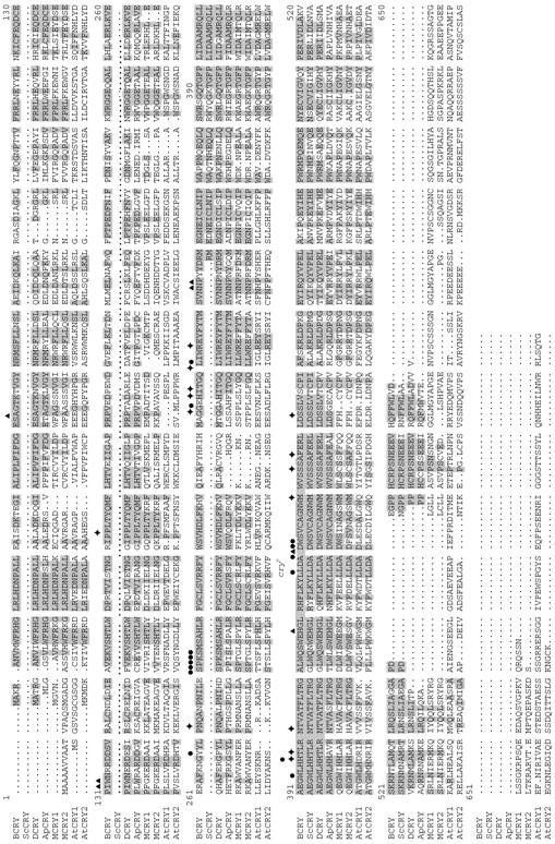
Alignment of the putative Bactrocera CRY amino acid sequences with CRY from other species. BCRY, Bactrocera CRY; ScCRY, S. crassipalpis (flesh fly) CRY; DCRY, D. melanogaster CRY; ApCRY, A. pernyi CRY; MCRY1, mouse CRY1; MCRY2, mouse CRY2; AtCRY1, Arabidopsis thaliana CRY1; AtCRY2, A. thaliana CRY2. The highlighted amino acids are conserved between Bactrocera and other species. ▴, MTHF binding sites; •, FAD binding sites; ♦, CPD binding sites (Emery et al. 1998). cryb indicates the aspartic acid residue at position 429, which is mutated in the cryb mutant of Drosophila (Stanewsky et al. 1998).
No fixed amino acid difference was detected between the CRY protein sequences from B. tryoni and B. neohumeralis. In the DNA sequence, there are several polymorphic sites shared between B. tryoni and B. neohumeralis (Figure 1), none of which alter the amino acid sequence of the translated protein. In the 3′-UTR, there are four nucleotides in a row (ATGT, 2114–2117) in B. tryoni cry cDNA absent in the parallel position of cry in B. neohumeralis. Another single nucleotide (A) at position 2076 is also deleted in B. neohumeralis. These putative fixed nucleotide differences have not been checked in large numbers of individual flies from wild populations, so we cannot rule out that they may be shared polymorphisms in wild flies.
By amplification of genomic fragments and subsequent sequencing, three introns have been identified. The splice sites are marked in Figure 1. Intron 1 appears to be very long since it failed to amplify directly from genomic DNA. The splice site was revealed by inverse PCR on genomic DNA. Intron 1 in Drosophila cry is within 3 bp of Bactrocera intron 1. Drosophila cry has introns in equivalent positions to introns 2 and 3 of Bactrocera cry. The first intron in Drosophila cry is ∼1 kb in length (http://www.fruitfly.org/sequence/), and the sizes of intron 2 (156 bp) and intron 3 (236 bp) are larger than those of Bactrocera cry (54 and 56 bp, respectively).
Constant level of cry transcripts in the whole head of B. tryoni and B. neohumeralis:
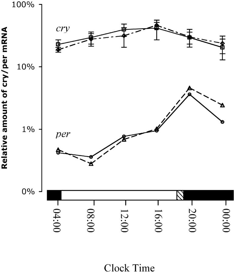
A feature shared by many clock gene transcripts is that their abundance is subject to circadian and diurnal oscillation. In the head of D. melanogaster, the cry mRNA level cycles (Emery et al. 1998; Egan et al. 1999), reaching a peak 4–6 hr after lights on and a trough 4–6 hr after dark. In the head of Bactrocera flies, semiquantitative reverse-transcriptase (RT)-PCR indicated abundant and constant expression of cry throughout a 24-hr period relative to the kanamycin reference RNA (data not shown). These samples had been used in an earlier study that showed diurnal cycling of per expression (An et al. 2002). Subsequently, a real-time PCR strategy was employed using two series of samples collected independently (Figure 3). Real-time PCR amplifications of both the kanamycin control and a per cDNA fragment were performed along with the cry amplifications. Statistical tests (ANOVA) showed no difference in cry expression levels between different time points. The expression profile of both genes is identical between B. neohumeralis and B. tryoni, as are male and female head samples. Bactrocera cry appears to be abundantly expressed in the fly head. It is present at nearly 10-fold of the peak level of the per transcript. The constant high levels of cry transcript in the whole head would mask any diurnal fluctuation in specific areas of the fly brain or other head tissues. Therefore, we identified regions of the Bactrocera brain potentially involved in central pacemaker activity and examined cry expression in dissected tissues from the head.
Figure 3.—
Diurnal expression patterns of per and cry mRNA in whole head of male flies. Similar results were obtained for female flies. The transcription levels of Bactrocera per and cry in the whole head of B. tryoni and B. neohumeralis were measured in 4-hourly cDNA samples from flies maintained in 24-hr light-dark cycles. The relative abundance of per and cry mRNA is shown as a percentage of kanamycin mRNA on a log scale. cry mRNA is the mean of three independent real-time PCR assays. Open, solid, and hatched bars are the hours of lights on, lights off, and dusk lighting, respectively. Solid line, B. neohumeralis; dashed line, B. tryoni.
PER expression reveals putative central clock cells in Bactrocera brain:
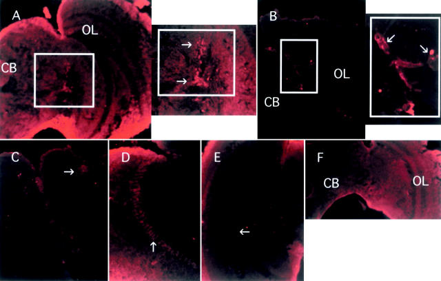
To identify central pacemaker cells, we used anti-PER antibody generated against Drosophila PER (see materials and methods) to stain Bactrocera adult brain sections. A group of cells in the lateral area were labeled (Figure 4A). The position of the cells matches that of the so-called lateral neurons, which have proved to be the central circadian pacemakers in Drosophila (Kaneko and Hall 2000). There are also some signals in the dorsal region of the brain (Figure 4C). Obvious staining was detected in the medulla of the optic lobes (Figure 4D). These are likely to be glial cells, which were also found to express PER in Drosophila (Stanewsky 2002). The signal is weaker in the dorsal region of the Bactrocera brain than in the lateral area. The staining in the lateral cell cluster is dense, but with tissue sections it is difficult to count the number of cells. The intensity of PER staining in all three brain areas—lateral area, optic lobes, and dorsal region—shows diurnal oscillation. Expression peaks at night, is lower in the morning, and reaches a trough in the afternoon (Figure 4, A, B, D, and E). The PER expression profile is similar to that of Drosophila and is consistent with the per mRNA expression tested previously (An et al. 2002). The localized expression of PER is not sex specific and no difference was found between B. neohumeralis and B. tryoni. The three groups of PER positive cells are located in the candidate regions involved in pacemaking processes of the brain.
Figure 4.—
Immunoreactivity to anti-PER antibody detected in frontal sections of male B. tryoni brain. Similar results were seen in female brain. Arrows indicate the positive signals. Staining at the edge of the section is due to background. (A) Central brain and optic lobe with framed lateral region. The enlarged lateral region is shown on the right. CB, central brain; OL, optic lobe. The fly was sampled in the afternoon at 16:00. (B) Central brain and optic lobe from a fly sampled at night (23:00) with enlarged lateral region on the right showing more intense PER staining compared to A. (C) Dorsal region. The fly was sampled in the night at 23:00. (D) Optic lobe showing staining of glial cells. The fly was sampled at 23:00. (E) Optic lobe of a fly sampled at 16:00. Note extremely weak staining in glial cells (arrow). (F) Negative control. Staining of a fly brain sampled at 23:00 without anti-PER antibody. Diffuse background fluorescence is present, but there is no evidence of cell-specific staining.
The cry transcript is expressed in the lateral area, antennal lobes, and dorsal region of the brain:
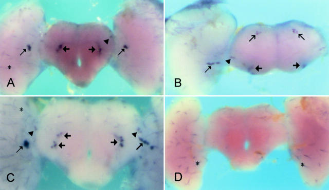
As shown in Figure 5, whole-mount in situ hybridization with antisense cry probe revealed signals in the lateral area and dorsal region of the brain. However, the staining is not evenly distributed. The signals in the lateral area are more intense than those in the dorsal region. The cells expressing cry in the lateral and dorsal areas of the Bactrocera brain are close to or coincident with the PER positive cells, which similarly showed stronger immunohistochemical staining in the lateral area than in the dorsal region. In the lateral area, cells with both large and small cell bodies expressed cry mRNA (Figure 5, A and C). We discovered that cry is also expressed in the antennal lobes of the Bactrocera brain. Rather than staining the whole antennal lobe, the signals are concentrated on cell clusters at the periphery of both antennal lobes (Figure 5, A and C). Like the lateral area, the signal in the antennal lobes is denser and more striking than that in the dorsal region. The location of the cry-expressing cells in the brain did not differ between male and female or between B. neohumeralis and B. tryoni. In several brains with some attached retinal tissue, hybridization in the retina was observed with both sense and antisense probes (not shown); hence, the signal in retina was regarded as false positive.
Figure 5.—
In situ hybridization of cry antisense (A, B, and C) and sense probes (D) to whole-mount brains (B. tryoni male). Similar results were obtained for females. Thin arrow, large lateral cells; arrowhead, small lateral cells; thick arrow, cells at the periphery of the antennal lobes; open arrow, dorsal cells; asterisk, nonspecific staining. The staining observed in adhering retinal tissue in B was also observed in control specimens. (A, C, and D) anterior view; (B) antero/dorsal view. The fly used in A was sampled at 09:30, while the other three brains were from flies sampled at 17:00.
Levels of cry transcript differ in the antennae and brain of B. tryoni, B. neohumeralis, and hybrid lines:
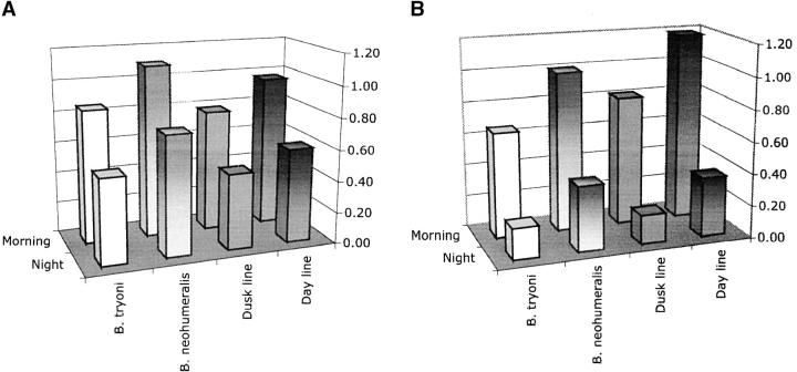
Since Drosophila cry mRNA in the head has a peak level of expression at ZT5 and a trough at ZT 17–19 (Emery et al. 1998), we chose time points 09:30 (5 hr after lights on) and 23:00 (5 hr after lights off) for testing brain tissue to see if there is any evidence of diurnal cycling of cry transcription. Brains were dissected from the pure species and from flies from hybrid lines selected for day- or dusk-mating time (see materials and methods) to test whether any variation in expression level is related to the mating time difference. The cDNA samples generated from brain tissue were tested by real-time PCR using ECF and ECR primers. The PCR was repeated three times for each sample. Data are shown in Figure 6A. The results show that the cry transcript is more abundant in the morning than at night and slightly more abundant in B. neohumeralis compared to B. tryoni. Moreover, both differences are duplicated in the day- and dusk-mating hybrids (Figure 6A).
Figure 6.—
Relative abundance of cry mRNA in the brain (A) and antennae (B) of male flies. Similar results were obtained for female flies. For each tissue cry expression is shown for the pure species (B. tryoni and B. neohumeralis) and the hybrid lines (dusk mating and day mating) at two time points (morning, 09:30; night, 23:00). The data shown are the means of three independent real-time PCR experiments. Heights of the 95% error bars are 0.19 and 0.21 for A and B, respectively.
As described in materials and methods, the data for cry expression in the brain of pure species and hybrid lines were initially analyzed separately. The similarity of results suggested the possibility of a joint analysis, which is shown in Table 1A. Time of day for cry expression is highly significant. The comparison among the four genotypes, B. tryoni, B. neohumeralis, and hybrid lines selected for B. tryoni and B. neohumeralis mating behavior, respectively, is also significant, but at a lower level. A further breakdown (Table 1A, lines 2a and 2b) shows that this significance is attributable to the “species” difference rather than to the difference between pure species and hybrid lines.
Bactrocera cry transcript was detected in the antennal lobe of the brain by in situ hybridization. Subsequently, RT-PCR revealed its presence in antennal tissue. The level of cry mRNA in the antennae was then analyzed by real-time PCR as described above at 09:30 and 23:00. As for the brain, the cry transcript is more abundant in the morning than at night for both the pure species and the day/dusk hybrids (Figure 6B). Furthermore, the level of transcript is higher in the antennae of B. neohumeralis and the day-mating hybrids than in B. tryoni and the dusk-mating hybrids at both time points (Figure 6B). ANOVA on the combined data yielded a P-value of 0.002 for “species” and <0.001 for time of day of cry expression, indicating the high significance of both aspects of the results, and there was no significant interaction between factors (Table 1B). Our results show that cry expression in the dusk-mating line behaves like B. tryoni cry, while cry expression in the day-mating line is very similar to B. neohumeralis cry and indicates that cry transcription is associated with the mating time difference in the Bactrocera species.
DISCUSSION
The sibling species B. tryoni and B. neohumeralis constitute an ideal model system to study the molecular mechanisms of behavioral differences that contribute to speciation and the maintenance of species integrity. Features of the model are: the extreme genetic similarity of the pair of sibling species that occur in sympatry, the existence of a clear, genetically determined behavior that confers premating isolation on the two species, and the capacity to generate hybrid lines that reproduce the mating isolation behavior (Meats et al. 2003). Since time of mating is a circadian behavior that is modulated by light intensity (Tychsen and Fletcher 1971; Smith 1979a), components of the central circadian pacemaker are strong candidates for a causative role in the mating isolation mechanism. A previous study (An et al. 2002) reported the isolation of the per gene in the two Bactrocera species, but failed to find any difference in amino acid sequence, expression level, or diurnal cycling phase. In this study we report that isolation of the cry gene in B. tryoni and B. neohumeralis has revealed no difference in amino acid sequence of the putative CRY protein in the two species, but has revealed a difference in gene regulation, which may function in the mating isolation mechanism that maintains species identity.
The cryptochrome gene in Bactrocera:
The cry gene belongs to the crptochrome/photolyase gene family, and cry is expressed abundantly throughout the head and body of Drosophila where it entrains cell autonomous circadian clocks. Its primary function, however, is probably to entrain the central pacemaker, located in the lateral neurons of the fly brain (Stanewsky et al. 1998; Emery et al. 2000). Drosophila 6-4 photolyase is very abundant in the ovary but hardly detectable in the head (Todo et al. 1996). The cry genes from Bactrocera were isolated from male head cDNA, but are also expressed throughout the body (data not shown). The putative amino acid sequence shows a much higher similarity to Drosophila CRY (73.4%) than to Drosophila 6-4 photolyase (41.3%), confirming that the isolation strategy has yielded a Bactrocera cry gene. Interaction with the cofactor FAD determines the light absorption spectra of CRY in mammals and plants (Lin et al. 1995), and we note that the sequence of the FAD binding site is conserved in Bactrocera cry. CRY in mammals fails to manifest the DNA repair activity of photolyases (Kobayashi et al. 1998); however, the biochemical function of insect CRYs has not been directly tested and they may possess DNA repair activity.
As in other insects studied to date, only one copy of the cry gene is found in the two Bactrocera species (Family Tephritidae), whereas there are two cry genes in the genomes of human, mouse, and Arabidopsis. Comparing Bactrocera with other insects, the dipteran D. melanogaster and the lepidopteran Antheraea pernyi, the overall percentage identities of PER are 68.4 and 45.3%, respectively (An et al. 2002), while those of CRY are 73.4 and 57.4%. It seems that CRY may be more conserved than PER during evolution.
Expression of circadian genes in the brain:
Immunohistochemistry of PER reveals staining of cells in the lateral and dorsal areas of the Bactrocera brain, similar to the localization of PER in Drosophila (Siwiki et al. 1988). In situ hybridization shows cells expressing cry mRNA in the dorsal and lateral areas of the Bactrocera brain, in a location similar to that of the PER expressing cells and of the pacemaker neurons in the Drosophila brain. These results agree with those of Egan et al. (1999), showing cry mRNA localized to lateral neurons in Drosophila brain, and with the results of Emery et al. (2000) showing cry promoter driven EGFP expression in the lateral and dorsal neurons of Drosophila. Not previously reported is the expression of cry in the antennal lobes. Specifically, these striking signals in the Bactrocera brain were localized to a cluster of cells at the periphery of the antennal lobes, a position consistent with the location of projection neurons in Drosophila, which send axons to higher brain centers such as the mushroom bodies (Jefferis et al. 2001), suggesting a function for cry in the processing of olfactory information.
Diurnal oscillation of the cry transcript:
Circadian genes in all organisms studied to date are regulated by a negative feedback loop. As for most of the circadian genes, cry mRNA oscillates in the head of Drosophila with a fivefold amplitude (Emery et al. 1998). A fourfold amplitude, with a cycling phase similar to that of Drosophila cry, is observed for mcry1 mRNA in the suprachiasmatic nuclei of mouse (Miyamoto and Sancar 1998). However, in Bactrocera, a constant high level of cry expression in whole head masked diurnal cycling of mRNA abundance in specific head tissues. A similar lack of cycling has been reported in adult head of the flesh fly Sarcophaga crassipalpis (Goto and Denlinger 2002).
Diurnal cycling of cry transcripts was revealed in dissected whole brain of B. tryoni and B. neohumeralis, but the structures of the Bactrocera head that showed most pronounced diurnal cycling were the antennae. Consistent with features of the cry oscillation in Drosophila, the cycling we observed in Bactrocera shows higher transcript levels in the morning than at night, which is almost entirely opposite to the phase of per mRNA cycling in these organisms (Hardin et al. 1990; An et al. 2002).
The lower level of cry mRNA cycling in the brain compared to antennae suggests that there may be regions of the brain that display more pronounced rhythmicity, masked by higher levels of expression in other regions. We have established that cry is expressed in the antennal lobes, which are connected to the antennae. Therefore, we hypothesize that cry mRNA oscillates with a substantial amplitude primarily in the antennal lobes of the Bactrocera brain. Western blotting and density analysis of immunohistochemical data will ultimately provide a picture of the CRY protein expression profile. If the situation in Bactrocera is similar to that in Drosophila, one would expect the protein to be abundant during the night and degrade during the day, regardless of the extent of cry mRNA cycling, as Drosophila CRY is unstable in the presence of light (Emery et al. 1998). The protein interacts with both TIM (Ceriani et al. 1999) and PER (Rosato et al. 2001) in a light-dependent manner, thus providing the light input to entrain the clock.
The proportion of cry mRNA at its maximum level in the antennae is roughly 10% of that in the whole head (data not shown). Therefore, cry must be expressed in other tissues of the head apart from brain and antennae. The photoreceptor cells of the eye are likely sites of cry expression as this is the location of other clock components in Drosophila.
Abundance of the cry transcript correlates with mating time:
We found that cry transcript levels differ in the two Bactrocera species, being higher in the day-mating B. neohumeralis than in the dusk-mating B. tryoni. Strikingly, this effect is reproduced in the hybrid lines selected for day- and dusk-mating time. Thus a higher level of cry mRNA correlates with day-mating time and suggests that regulation of cry gene expression has a role in the mating isolation mechanism. We do not know whether the difference in cry mRNA regulation between the two species is a primary effect of the cry locus or whether it is the downstream effect of another genetic locus. A third possibility is that the result in the hybrids is due to genetic linkage of the cry gene or a regulator with a major locus that controls time of mating. Given the interaction between light and mating time in the two species and the sensitivity of CRY protein to light, we favor the hypothesis that regulation of cry expression has a role in the response of the flies to light intensity. Evidence for a direct effect of cry expression on mating time will require the knockdown of cry expression in brain and antennae of day-mating flies.
The quantitative difference in cry mRNA between day- and dusk-mating flies suggests the existence of a quantitative difference in CRY protein. A number of examples illustrate the sensitivity of organisms to the quantity of CRY protein. In Arabidopsis and Drosophila, CRY overexpression increases sensitivity to light (Emery et al. 1998; Lin et al. 1998). Even quite subtle differences of CRY concentration can have a dramatic effect on phenotype (El-Din El-Assal et al. 2001). Interestingly, cryb null mutant flies take longer than wild-type flies to entrain their evening activity peak to a new light-dark cycle (Emery et al. 2000). The concentration of CRY protein may be particularly critical at dusk after many hours of exposure to light and when light levels are low. Thus the lower level of cry mRNA in dusk-mating flies may result in critically low levels of CRY at the end of the photophase.
We do not know the mechanism by which changes in the level of cry mRNA or protein affect time of mating, but speculate that expression in antennae and antennal lobes may be of particular significance. The antennae are important in the sexual behavior of flies, being required for the female's response to male-produced pheromone and the perception of stridulation by males. Both of these aspects of sexual behavior are sensitive to light intensity and display diurnal cycles that correlate with mating time in B. tryoni (Smith 1979b). In Drosophila also, the antennae play a central role in mating behavior (Hall 1994). Plautz et al. (1997) have shown that Drosophila tissues, including the antennae, possess autonomous circadian clocks that are responsive to light, and indeed the circadian rhythm of olfactory response requires an intact peripheral circadian oscillator (Krishnan et al. 1999). Although Krishnan et al. (2001) suggest a photoreceptor-independent role for CRY in the peripheral oscillators, our results suggest a difference in olfactory function between B. tryoni and B. neohumeralis that is sensitive to light intensity and that may be mediated in part by cry expression.
Another tephritid fly, the melon fly B. cucurbitae, shows premating isolation based on mating time in flies with short and long circadian periods (Shimizu et al. 1997) and expression of the clock gene per mirrors the circadian activity rhythm (Miyatake et al. 2002). In D. melanogaster the per and tim genes control circadian rhythms of female mating activity (Sakai and Ishida 2001) and per controls characteristics of the species-specific courtship songs of sibling Drosophila species (Wheeler et al. 1991). Thus the endogenous circadian system has an important role in regulating behavioral mechanisms that are important for reproductive isolation. No effect of per expression on time of mating was evident in the sibling Bactrocera species (An et al. 2002), but a role for cry in the mating isolation mechanism is supported by the replication of cry expression differences in the pure species and the hybrid lines. The results suggest that small changes in circadian regulation represent a speciation mechanism.
Acknowledgments
We are grateful to John Sved, Coral Warr, and Douglas Armstrong for helpful discussions and to John Sved and Emilie Cameron for assistance with statistical analyses. We thank Kirsten Steiner for her in situ hybridization protocol. This work was supported by the Australian Research Council (grant A00104385).
References
- Ahmad, M., and A. R. Cashmore, 1993. HY4 gene of A. thaliana encodes a protein with characteristics of a blue-light photoreceptor. Nature 366: 162–166. [DOI] [PubMed] [Google Scholar]
- An, X., K. Wilkes, Y. Bastian, J. L. Morrow, M. Frommer et al., 2002. The period gene in two species of tephritid fruit fly differentiated by mating behavior. Insect Mol. Biol. 11: 419–430. [DOI] [PubMed] [Google Scholar]
- Bateman, M. A., 1967. Adaptations to temperature in geographic races of the Queensland fruit fly, Dacus (Strumeta) tryoni. Aust. J. Zool. 15: 1141–1161. [Google Scholar]
- Ceriani, M. F., T. K. Darlington, D. Staknis, P. Mas, A. A. Petti et al., 1999. Light-dependent sequestration of TIMELESS by CRYPTOCHROME. Science 285: 553–556. [DOI] [PubMed] [Google Scholar]
- Davidson, B. P., S. J. Kinder, K. Steiner, G. C. Schoenwolf and P. P. L. Tam, 1999. Impact of node ablation on the morphogenesis of the body axis and the lateral asymmetry of the mouse embryo during early organogenesis. Dev. Biol. 211: 11–26. [DOI] [PubMed] [Google Scholar]
- Egan, E. S., T. M. Franklin, M. J. Hilderbrand-Chae, G. P. McNeil, M. A. Roberts et al., 1999. An extraretinally expressed insect cryptochrome with similarity to the blue light photoreceptors of mammals and plants. J. Neurosci. 19: 3665–3673. [DOI] [PMC free article] [PubMed] [Google Scholar]
- El-Din El-Assal, S., C. Alonso-Blanco, A. J. Peeters, V. Raz and M. Koornneef, 2001. A QTL for flowering time in Arabidopsis reveals a novel allele of CRY2. Nat. Genet. 29: 435–440. [DOI] [PubMed] [Google Scholar]
- Emery, P., W. V. So, M. Kaneko, J. C. Hall and M. Rosbash, 1998. CRY, a Drosophila clock and light-regulated cryptochrome, is a major contributor to circadian rhythm resetting and photosensitivity. Cell 95: 669–679. [DOI] [PubMed] [Google Scholar]
- Emery, P., R. Stanewsky, C. Helfrich-Forster, M. Emery-Le, J. C. Hall et al., 2000. Drosophila CRY is a deep brain circadian photoreceptor. Neuron 26: 493–504. [DOI] [PubMed] [Google Scholar]
- Frohman, M. A., 1994. On beyond classic RACE (rapid amplification of cDNA ends). PCR Methods Appl. 4: S40–S58. [DOI] [PubMed] [Google Scholar]
- Goto, S., and D. Denlinger, 2002. Short-day and long-day expression patterns of genes involved in the flesh fly clock mechanism: period, timeless, cycle and cryptochrome. J. Insect Physiol. 48: 803–816. [DOI] [PubMed] [Google Scholar]
- Hall, J. C., 1994. The mating of a fly. Science 264: 1702–1714. [DOI] [PubMed] [Google Scholar]
- Hardin, P. E., J. C. Hall and M. Rosbash, 1990. Feedback of the Drosophila period gene product on circadian cycling of its messenger RNA levels. Nature 343: 536–540. [DOI] [PubMed] [Google Scholar]
- Helfrich-Forster, C., C. Winter, A. Hofbauer, J. C. Hall and R. Stanewsky, 2001. The circadian clock of fruit flies is blind after elimination of all known photoreceptors. Neuron 30: 249–261. [DOI] [PubMed] [Google Scholar]
- Ivanchenko, M., R. Stanewsky and J. M. Giebultowicz, 2001. Circadian photoreception in Drosophila: functions of cryptochrome in peripheral and central clocks. J. Biol. Rhythms 16: 205–215. [DOI] [PubMed] [Google Scholar]
- Jefferis, G., E. Marin, R. Stocker and L. Luo, 2001. Target neuron prespecification in the olfactory map of Drosophila. Nature 414: 204–208. [DOI] [PubMed] [Google Scholar]
- Kaneko, M., and J. Hall, 2000. Neuroanatomy of cells expressing clock genes in Drosophila: transgenic manipulation of the period and timeless genes to mark the perikarya of circadian pacemaker neurons and their projections. J. Comp. Neurol. 422: 66–94. [DOI] [PubMed] [Google Scholar]
- Kobayashi, K., S. Kano, B. Smit, G. van der Horst, M. Takao et al., 1998. Characterization of photolyase/blue-light receptor homologs in mouse and human cells. Nucleic Acids Res. 26: 5086–5092. [DOI] [PMC free article] [PubMed] [Google Scholar]
- Krishnan, B., S. Dryer and P. Hardin, 1999. Circadian rhythms in olfactory responses of Drosophila melanogaster. Nature 400: 375–378. [DOI] [PubMed] [Google Scholar]
- Krishnan, B., J. D. Levine, M. K. Lynch, H. B. Dowse, P. Funes et al., 2001. A new role for cryptochrome in a Drosophila circadian oscillator. Nature 411: 313–317. [DOI] [PubMed] [Google Scholar]
- Lin, C., D. E. Robertson, M. Ahmad, A. A. Raibekas, M. S. Jorns et al., 1995. Association of flavin adenine dinucleotide with the Arabidopsis blue light receptor CRY1. Science 269: 968–970. [DOI] [PubMed] [Google Scholar]
- Lin, C., H. Yang, H. Guo, T. Mockler, J. Chen et al., 1998. Enhancement of blue-light sensitivity of Arabidopsis seedlings by a blue light receptor cryptochrome 2. Proc. Natl. Acad. Sci. USA 95: 2686–2690. [DOI] [PMC free article] [PubMed] [Google Scholar]
- Marchuk, D. A., A. M. Saulino, R. Tavakkol, M. Swaroop, M. R. Wallace et al., 1991. cDNA cloning of the type 1 neurofibromatosis gene: complete sequence of the NF1 gene product. Genomics 11: 931–940. [DOI] [PubMed] [Google Scholar]
- Meats, A., N. Pike, X. An, K. Raphael and W. Y. Wang, 2003. The effects of selection for early (day) and late (dusk) mating lines of hybrids of Bactrocera tryoni and Bactrocera neohumeralis. Genetica 119: 283–293. [DOI] [PubMed] [Google Scholar]
- Miyamoto, Y., and A. Sancar, 1998. Vitamin B2-based blue-light photoreceptors in the retinohypothalamic tract as the photoactive pigments for setting the circadian clock in mammals. Proc. Natl. Acad. Sci. USA 95: 6097–6102. [DOI] [PMC free article] [PubMed] [Google Scholar]
- Miyatake, T., A. Matsumoto, T. Matsuyama, H. Ueda, T. Toyosato et al., 2002. The period gene and allochronic reproductive isolation in Bactrocera cucurbitae. Proc. R. Soc. Lond. B 269: 2467–2472. [DOI] [PMC free article] [PubMed] [Google Scholar]
- Morris, V. B., J. Zhao, D. Shearman, M. Byrne and M. Frommer, 2004. Expression of an Otx gene in the adult rudiment and the developing central nervous system in the vestibula larva of the sea urchin Holopneustes purpurescens. Int. J. Dev. Biol. 48: 17–22. [DOI] [PubMed] [Google Scholar]
- Morrow, J., L. Scott, B. Congdon, D. Yeates, M. Frommer et al., 2000. Close genetic similarity between two sympatric species of tephritid fruit fly reproductively isolated by mating time. Evolution 54: 899–910. [DOI] [PubMed] [Google Scholar]
- Petersen, G., J. C. Hall and M. Rosbash, 1988. The period gene of Drosophila carries species-specific behavioral instructions. EMBO J. 7: 3939–3947. [DOI] [PMC free article] [PubMed] [Google Scholar]
- Plautz, J. D., M. Straume, R. Stanewsky, C. F. Jamison, C. Brandes et al., 1997. Quantitative analysis of Drosophila period gene transcription in living animals. J. Biol. Rhythms 12: 204–217. [DOI] [PubMed] [Google Scholar]
- Raphael, K. A., S. Whyard, D. Shearman, X. An and M. Frommer, 2004. Bactrocera tryoni and closely related pest tephritids—molecular analysis and prospects for transgenic control strategies. Insect Biochem. Mol. Biol. 34: 167–176. [DOI] [PubMed] [Google Scholar]
- Rosato, E., V. Codd, G. Mazzotta, A. Piccin, M. Zordan et al., 2001. Light-dependent interaction between Drosophila CRY and the clock protein PER mediated by the carboxy terminus of CRY. Curr. Biol. 11: 909–917. [DOI] [PubMed] [Google Scholar]
- Sakai, T., and N. Ishida, 2001. Circadian rhythms of female mating activity governed by clock genes in Drosophila. Proc. Natl. Acad. Sci. USA 98: 9221–9225. [DOI] [PMC free article] [PubMed] [Google Scholar]
- Shimizu, T., T. Miyatake, Y. Water and T. Arai, 1997. A gene pleiotropically controlling developmental and circadian periods in the melon fly, Bactrocera cucurbitae (Diptera: Tephritidae). Heredity 79: 600–605. [Google Scholar]
- Siwicki, K. K., C. Eastman, G. Petersen, M. Rosbash and J. C. Hall, 1988. Antibodies to the period gene product of Drosophila reveal diverse tissue distribution and rhythmic changes in the visual system. Neuron 1: 141–150. [DOI] [PubMed] [Google Scholar]
- Smith, P. H., 1979. a Genetic manipulation of the circadian clock's timing of sexual behavior in the Queensland fruit flies, Dacus tryoni and Dacus neohumeralis. Physiol. Entomol. 4: 71–78. [Google Scholar]
- Smith, P. H., 1979b Behavioral partitioning of the day and circadian rhythmicity, pp. 325–341 in Fruit Flies: Their Biology, Natural Enemies and Control, Vol. 3A, edited by A. S. Robinson and G. Hooper. Elsevier, Amsterdam.
- Stanewsky, R., 2002. Clock mechanisms in Drosophila. Cell Tissue Res. 309: 11–26. [DOI] [PubMed] [Google Scholar]
- Stanewsky, R., M. Kaneko, P. Emery, B. Beretta, K. Wager-Smith et al., 1998. The cryb mutation identifies cryptochrome as a circadian photoreceptor in Drosophila. Cell 95: 681–692. [DOI] [PubMed] [Google Scholar]
- Tauber, E., H. Roe, R. Costa, J. M. Hennessy and C. P. Kyriacou, 2003. Temporal mating isolation driven by a behavioral gene in Drosophila. Curr. Biol. 13: 140–145. [DOI] [PubMed] [Google Scholar]
- Todo, T., H. Ryo, K. Yamamoto, H. Toh, T. Inui et al., 1996. Similarity among the Drosophila (6–4)photolyase, a human photolyase homolog, and the DNA photolyase-blue-light photoreceptor family. Science 272: 109–112. [DOI] [PubMed] [Google Scholar]
- Tychsen, P. H., and B. S. Fletcher, 1971. Studies on the rhythm of mating in the Queensland fruit fly, Dacus tryoni. J. Insect Physiol. 17: 2139–2156. [Google Scholar]
- Wang, Y., H. Yu, K. A. Raphael and A. S. Gilchrist, 2003. Genetic delineation of sibling species of the pest fruit fly Bactrocera (Diptera: Tephritidae) using microsatellites. Bull. Entomol. Res. 93: 351–360. [DOI] [PubMed] [Google Scholar]
- Wheeler, D. A., C. P. Kyriacou, M. L. Greenacre, Q. Yu, J. E. Rutila et al., 1991. Molecular transfer of a species-specific behavior from Drosophila simulans to Drosophila melanogaster. Science 251: 1082–1085. [DOI] [PubMed] [Google Scholar]
- Young, M. W., and S. A. Kay, 2001. Time zones: a comparative genetics of circadian clocks. Nat. Rev. Genet. 2: 702–715. [DOI] [PubMed] [Google Scholar]


