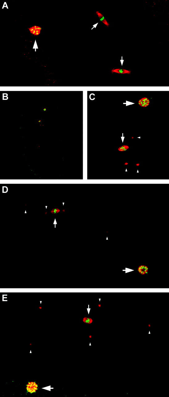
Figure 3.—

Phenotype of embryos from klp3A females. In A–E, embryos are stained with antihistones (green) to visualize chromatin and antitubulin (red) to visualize microtubules. (A) Embryo from a klp3Amei-352/FM7w female in mitotic cycle 2. Two zygotic nuclei are present on mitotic spindles with centrosomes at each pole (small arrows). The polar body chromatin mass is visible at left (large arrow). (B) Low-magnification image of an embryo from a klp3Amei-352 homozygous female showing that only two masses of chromatin are present: a large mass of polar body chromatin and a smaller nucleus on a spindle. (C) Higher-magnification image of the embryo from B. The mass of polar body chromatin is associated with a circular concentration of microtubules (large arrow) and the small nucleus sits on a spindle (small arrow). However, the spindle appears to lack asters associated with the spindle poles but several free-lying centrosomes are present (arrowheads). (D) Embryo from a klp3Amei-352/klp3A521 female. The only antihistone signals are the polar body chromatin mass (large arrow) and a small nucleus residing on a spindle (small arrow). Centrosomes (arrowheads) appear to have dissociated from the spindle and additional centrosomes are visible in the embryo. (E) Embryo from a klp3Amei-352/klp3A1124 female. Five independent centrosomes are visible (arrowheads), but none appear associated with the single spindle (small arrow) or polar body chromatin mass (large arrow).
