
Figure 4.—
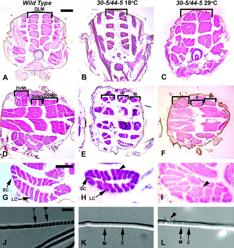
Histological analyses of wild-type and 30-5/44-5 mutants raised at the permissive and restrictive temperatures. Wild-type animals are 96-hr APF, and mutant animals raised at 18° and 29° were of equivalent stages. (A–C) Transverse thoracic sections to visualize DLMs (bracketed), dorsal to top. (D–F) Horizontal thoracic sections to visualize the DVM I-III fibers (bracketed), anterior to the left. (G–I) Horizontal view of TDT muscle, anterior to left. (J–L), Dissected myofibrils. Sections in A–I were stained with hematoxylin and eosin. (A) Wild type. (B) 30-5/44-5 mutant raised at 18°, where DLM fiber number and arrangement were similar to wild type. (C) 30-5/44-5 mutant raised at 29°, note the reduced number of DLM fibers. (D) Wild type. (E) 30-5/44-5 mutant raised at 18°. DVM fiber number remained similar to wild type but some patterning defects were seen. (F) 30-5/44-5 mutant raised at 29°, fiber number was not notably different from permissive temperature. (G) Wild-type TDT, indicating the small cells (SC) and the large cells (LC) of the muscle. (H) TDT of a 30-5/44-5 mutant raised at 18°, showing a nearly normal structure, but with a single abnormally shaped large cell (arrowhead). (I) TDT of a 30-5/44-5 mutant raised at 29°: the central lumen characteristic of this muscle was missing and many of the fibers were shaped oddly. (J) Myofibril isolated from the DLM of wild-type animal. Note the regular structure of the sarcomere and prominent Z-lines and M-lines (arrows). (K) Myofibril isolated from a DLMof 30-5/44-5 animal raised at 18° was apparently normal. (L) Myofibril isolated from a DLM of 30-5/44-5 animal raised at 29°; note the frayed appearance of the fiber at left (arrowhead). Bar for A–F, 100 μm; for G–I, 50 μm; for J–L, 5 μm.