

Abstract
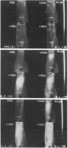
Digital thermographic imaging and laser-Doppler velocimetry techniques were used to study the sequence of microcirculatory changes in typical Type IV (DHS-type) skin reactions to intradermal injection of PPD into seven healthy tuberculin-positive subjects. Thermography has shown that there is very little change in thermal status at 6 hr, that there is an area of raised temperature roughly corresponding to that of erythema around the site of antigen injection at 24 hr, and that the area of 'hot' skin starts to exceed that of the clinically observed reaction at 48 hr and remains extensive even at 96 hr. The area of the increased RBC flux detected by the laser-Doppler technique corresponded generally to that of erythema for the first 48 hr, but thereafter became smaller than the clinically observable reaction. Cuff occlusion experiments showed that the phenomenon of reactive hyperaemia is abolished at 24 and 48 hr in conditions of maximal hyperaemia, but that this response is recovering by 96 hr. Vasomotion was significantly exaggerated between 48 hr and 96 hr. These studies indicate that there is maximal arteriolar vasodilatation during the first 2 days of the reaction with rapid blood flow in the congested capillaries of the dermal papillae, and that engorgement of deeper venous plexus of the dermis is the predominant vascular change in the resolving response at 72-96 hr.
Full text
PDF


Images in this article
Selected References
These references are in PubMed. This may not be the complete list of references from this article.
- Gibbs J. H., Ferguson J., Brown R. A., Kenicer K. J., Potts R. C., Coghill G., Swanson Beck J. Histometric study of the localisation of lymphocyte subsets and accessory cells in human Mantoux reactions. J Clin Pathol. 1984 Nov;37(11):1227–1234. doi: 10.1136/jcp.37.11.1227. [DOI] [PMC free article] [PubMed] [Google Scholar]
- Holloway G. A., Jr Cutaneous blood flow responses to injection trauma measured by laser Doppler velocimetry. J Invest Dermatol. 1980 Jan;74(1):1–4. doi: 10.1111/1523-1747.ep12514556. [DOI] [PubMed] [Google Scholar]
- NICOLL P. A., WEBB R. L. Vascular patterns and active vasomotion as determiners of flow through minute vessels. Angiology. 1955 Aug;6(4):291–308. doi: 10.1177/000331975500600403. [DOI] [PubMed] [Google Scholar]
- Nilsson G. E., Tenland T., Oberg P. A. Evaluation of a laser Doppler flowmeter for measurement of tissue blood flow. IEEE Trans Biomed Eng. 1980 Oct;27(10):597–604. doi: 10.1109/TBME.1980.326582. [DOI] [PubMed] [Google Scholar]
- Nilsson G. E., Tenland T., Obert P. A. A new instrument for continuous measurement of tissue blood flow by light beating spectroscopy. IEEE Trans Biomed Eng. 1980 Jan;27(1):12–19. doi: 10.1109/TBME.1980.326686. [DOI] [PubMed] [Google Scholar]
- Rodgers G. P., Schechter A. N., Noguchi C. T., Klein H. G., Nienhuis A. W., Bonner R. F. Periodic microcirculatory flow in patients with sickle-cell disease. N Engl J Med. 1984 Dec 13;311(24):1534–1538. doi: 10.1056/NEJM198412133112403. [DOI] [PubMed] [Google Scholar]
- Salerud E. G., Tenland T., Nilsson G. E., Oberg P. A. Rhythmical variations in human skin blood flow. Int J Microcirc Clin Exp. 1983;2(2):91–102. [PubMed] [Google Scholar]
- Sokal J. E. Editorial: Measurement of delayed skin-test responses. N Engl J Med. 1975 Sep 4;293(10):501–502. doi: 10.1056/NEJM197509042931013. [DOI] [PubMed] [Google Scholar]
- Spence V. A., Walker W. F. The relationship between temperature isotherms and skin blood flow in the ischemic limb. J Surg Res. 1984 Mar;36(3):278–281. doi: 10.1016/0022-4804(84)90099-4. [DOI] [PubMed] [Google Scholar]