Abstract
A vesicular stomatitis virus (VSV) pseudotype bearing hantavirus envelope glycoproteins was produced and used in a neutralization test as a substitute for native hantavirus. The recombinant VSV, in which the enveloped protein gene (G) was replaced by the green fluorescent protein gene and complemented with G protein expressed in trans (VSVΔG*G), was kindly provided by M. A. Whitt. 293T cells were transfected with plasmids for the expression of envelope glycoproteins (G1 and G2) of HTNV or SEOV and were then infected with VSVΔG*G. Pseudotype VSV with the Hantaan (VSVΔG*-HTN) or Seoul (VSVΔG*-SEO) envelope glycoproteins were harvested from the culture supernatant. The number of infectious units (IU) of the pseudotype VSVs ranged from 105 to 106/ml. The infectivity of VSVΔG*-HTN and VSVΔG*-SEO was neutralized with monoclonal antibodies, immune rabbit sera, and sera from patients with hemorrhagic fever with renal syndrome, and the neutralizing titers were similar to those obtained with native hantaviruses. These results show that VSVΔG*-HTN and -SEO can be used as a rapid, specific, and safe neutralization test for detecting hantavirus-neutralizing antibodies as an effective substitute for the use of native hantaviruses. Furthermore, the IU of VSVΔG*-HTN and -SEO did not decrease by more than 10-fold when stored at 4°C for up to 30 days. The stability of the pseudotype viruses allows distribution of the material to remote areas by using conventional cooling boxes for use as a diagnostic reagent.
Hemorrhagic fever with renal syndrome (HFRS) and hantavirus pulmonary syndrome (HPS) are rodent-borne viral zoonoses that occur worldwide. They are caused by viruses in the genus Hantavirus, family Bunyaviridae. Currently, at least 22 species have been identified in the genus (6). Each hantavirus is carried predominantly by its own natural host, usually a rodent or insectivore (18). At least six of the viruses are known to be transmitted to humans via the inhalation of infected rodent excreta. In Asia, Hantaan virus (HTNV) and Seoul virus (SEOV) are the causative agents of HFRS, which affects 100,000 patients per year in China. In Europe, Puumala virus, Dobrava virus (DOBV), and Saaremaa virus cause HFRS (12). Sin Nombre virus and related viruses cause HPS in the Americas (19).
Hantaviruses possess a single-stranded, negative-sense RNA genome that consists of three segments: the large (L), medium (M), and small (S) segments. The S segment encodes the nucleocapsid (N) protein, and the L segment is believed to encode RNA-dependent RNA polymerase. The N protein, RNA polymerase, and the genome for the viral ribonucleocapsid are packaged in the virion. The M segment encodes a glycoprotein precursor that is cleaved into envelope glycoproteins G1 and G2 cotranslationally. G1 and G2 form projections on the virion surface and are the targets of neutralizing antibodies (2, 5, 7).
Like many other viruses, Hantavirus serotypes are determined from using the focus or plaque reduction neutralization test (FRNT) to compare the neutralizing antibody titers of reference immune sera to those of homologous titers. In addition, the neutralizing antibody response of patient serum is widely thought to be an important factor in protective immunity. Although it has these benefits, the FRNT for hantaviruses takes 1 to 2 weeks to detect a plaque or focus, requires containment facilities for virus manipulation, and remains a laborious and time-consuming assay.
Vesicular stomatitis virus (VSV), the prototype rhabdovirus, has been used as a model system for studying the replication and assembly of enveloped RNA viruses due to its ability to grow to high titers in a variety of cell lines. Furthermore, the reverse genetic system of VSV allows the generation of recombinant viruses that express foreign proteins (8, 23). VSVΔG* is a recombinant VSV in which the G gene has been replaced by the green fluorescent protein (GFP) gene (21). Consequently, VSVΔG* is not infectious unless the envelope protein responsible for receptor binding and membrane fusion is provided in trans. In the present study, we generated pseudotype VSVΔG* possessing HTNV or SEOV envelope glycoproteins and developed a simple, safe, and rapid neutralization test for the use of native viruses.
MATERIALS AND METHODS
Cells.
The 293T cell line is derived from human embryonic kidney cell line 293 and contains the simian virus 40 large T antigen. 293T cells were maintained in Dulbecco modified Eagle medium (Nissui, Tokyo, Japan) supplemented with 0.45% glucose and 10% heat-inactivated fetal calf serum (FCS). The E6 clone of Vero cells (ATCC C1008; Cell Repository line 1586) was grown in Eagle minimal essential medium (Nissui) supplemented with 2 mM glutamine and 5% heat-inactivated FCS.
Plasmids.
cDNA clones containing the coding region of the M segment of HTNV strain 76-118 and SEOV strain SR-11 were kindly provided by C. S. Schmaljohn. The cDNA of the M segment of HTNV was excised by using BglII and subcloned into the BglII site of expression vector pCAGGS/MCS (15). The cDNA of the M segment of SEOV was excised by using SmaI and XhoI and subcloned into the SmaI and XhoI sites of pCAGGS/MCS. The plasmids containing the M segment cDNA of HTNV and SEOV were designated pCHTNM and pCSEOM, respectively. pCVSVG is the expression plasmid made by cloning the cDNA encoding the VSV G protein into the expression vector pCAGGS/MCS. pCVSVG was kindly provided by Michael A. Whitt.
Viruses.
HTNV strain 76-118 and SEOV strain SR-11 were used as representative strains of the HTNV and SEOV serotypes, respectively. These strains were propagated in the E6 clone of Vero cells. VSVΔG*G is the recombinant VSV derived from a full-length cDNA clone of the VSV genome (Indiana serotype) in which the coding region of the G protein was replaced by the coding region of the GFP gene and the G protein was expressed in trans (21). VSVΔG*G was kindly provided by Michael A. Whitt.
Expression of G1 and G2 of HTNV and SEOV.
The expression of G1 and G2 was analyzed by using an indirect immunofluorescent antibody (IFA) test and flow cytometry. 293T cell monolayers in six-well plates (80% confluent) were transfected with 2 μg of pCHTNM and pCSEOM by using Mirus TransIT LT1 transfection reagents (Panvera, Madison, Wis.), as recommended by the manufacturer. After 48 h, cells were examined for protein expression. Cells from six-well plastic plate (Coster) were detached by using a pipette and then suspended in culture medium. They were then centrifuged, and the pellet was resuspended with 1 ml of phosphate-buffered saline (PBS). For intracellular staining, the IFA test was carried out. Briefly, after centrifugation, the pelleted cells were resuspended again with an appropriate volume of PBS. The resuspended cells were spotted onto a 24-well glass slide, air dried, and fixed with acetone for 10 min. Acetone-fixed smears of 293T cells were treated with the culture supernatant from hybridoma clones, producing the monoclonal antibodies (MAbs) 8B6 against G1 and HCO2 against G2, for 1 h at 37°C. After being washed with PBS, fluorescein isothiocyanate (FITC)-conjugated rabbit polyclonal antibodies against anti-mouse immunoglobulin G (IgG; Zymed) at 1:400 were added to the well. After 1 h of incubation at 37°C, cells were washed and examined with a fluorescence microscope. For cell surface staining, flow cytometry analysis was done. The transfected 293T cells were detached with a pipette, washed with PBS, and fixed with 3% paraformaldehyde in PBS for 5 min at room temperature. After being washed with 0.5% bovine serum albumin in Dulbecco PBS (fluorescence-activated cell sorting [FACS] solution), the cells were treated with purified MAb (clone HCO2) at 10 μg/100 μl for 30 min on ice. After being washed with FACS solution, the cells were treated with FITC-conjugated anti-mouse IgG (1:100) for 30 min on ice. After another wash with FACS solution, the cells were suspended in 500 μl of FACS solution and analyzed by using a FACSCalibur (Becton Dickinson). The data were evaluated by using Becton Dickinson FACScan research software (CellQuest, v3.0.1.f).
Preparation of pseudotype viruses.
At 36 h after transfection of 293T cells with pCHTNM, pCSEOM, pCVSVG, or pCAGGS/MCS, the cells were infected with VSVΔG*G at a multiplicity of infection of 1 for 1 h at 37°C. The 293T cell monolayer was then washed with 1% heat-inactivated FCS-PBS three times, and culture medium was added. After 24 h of incubation at 37°C in a CO2 incubator, the culture supernatant was clarified by low-speed centrifugation and stored at −80°C.
Titration of pseudotype viruses.
For pseudotype virus titration in Vero E6 cells, Vero E6 cells grown on 96-well plates were infected with 50 μl of serially diluted virus stock. After a 1-h adsorption period, the inoculum was removed, fresh culture medium was added, and the cells were incubated at 37°C in a CO2 incubator. At 16 h postinfection (hpi), the cells were fixed with 1% paraformaldehyde in PBS for 10 min at room temperature, washed with distilled water, and air dried. GFP-expressing cells were counted under a fluorescence microscope. Since pseudotype VSVs are unable to produce infectious progeny virus, the numbers of GFP-positive cells were regarded as infectious units (IU).
Detection of viral proteins incorporated into pseudotype VSV.
The incorporation of the G1 and G2 envelope glycoproteins of HTNV and SEOV into the pseudotype viruses was examined by Western blotting. Culture supernatants of 293T cells were partially purified by ultracentrifugation at 100,000 rpm for 1 h through 20% sucrose in TNE buffer (10 mM Tris, 135 mM NaCl, 2 mM EDTA). The pellets were dissolved in sodium dodecyl sulfate-polyacrylamide gel electrophoresis (SDS-PAGE) sample buffer (2% SDS, 10% glycerol, 5% 2-ME, 0.002% bromophenol blue, 0.0625 M Tris-Cl; pH 6.8) for VSV M protein detection or in radioimmunoprecipitation assay buffer (10 mM Tris-HCl [pH 7.8], 2% Triton X-100, 150 mM NaCl, 600 mM KCl, 5 mM EDTA, 1% aprotinin, 1 mM phenylmethylsulfonyl fluoride) for G1 and G2 glycoprotein detection. The virus samples dissolved in SDS-PAGE sample buffer were separated by SDS-PAGE in 12% gels under reducing conditions. The virus samples dissolved in radioimmunoprecipitation assay buffer were separated by PAGE in 8% gels under nonreducing conditions. After electrophoresis, the proteins were blotted onto polyvinylidene difluoride membrane (ATTO, Tokyo, Japan). The membrane was soaked in 3% skim milk in PBS at room temperature to saturate unsaturated areas. For VSV M staining, mouse MAb to M protein (working dilution of 1:1,000; a gift from M. A. Whitt) was used (10). For hantavirus G1 and G2 staining, the undiluted culture supernatants of hybridoma clones of 8B6 against G1 of HTNV and 8E10 against G2 of HTNV were used (2). The membrane was incubated with MAb for 1 h at room temperature. The membrane was then washed three times with 0.05% Tween 20 in PBS for 5 min each and incubated with biotinylated anti-mouse IgG (Zymed) at a 1:5,000 dilution for 1 h. After incubation for 1 h, the membrane was washed according to the same procedure used in the second step and was incubated with horseradish peroxidase-conjugated avidin (1:400; Cappel). The band was detected by using the ECL Plus Western blotting detection system (Amersham Pharmacia Biotech, Ltd.). Briefly, the membrane were treated with a mixture of A solution and B solution (supplied by the manufacturer at a 40:1 volume), incubated for 5 min at room temperature, drained off from the membrane, exposed to Hyperfilm ECL for 15 s, developed with Rendol, and fixed with Renfix.
MAbs, rabbit immune sera, and HFRS patient sera.
Clones producing MAbs directed against the HTNV envelope glycoproteins were prepared as previously described (2). Rabbit immune sera against HTNV and SEOV were obtained from rabbits infected with live viruses, as previously described (11). A total of 22 serum specimens from HFRS patients previously diagnosed as being infected by HTNV and SEOV were kindly provided by H. Wang of the Institute of Virology, Chinese Academy of Preventive Medicine, Beijing, China, and 5 specimens were provided by Y. Nishimune of the Research Institute for Microbial Diseases, Osaka University. Ten serum specimens from the United Arab Emirates obtained from patients with high fever of unknown etiology were provided by M. K. Ijaz and T. Al Karmi of the Department of Medical Microbiology, Faculty of Medicine and Health Science, United Arab Emirates, through H.-K. Ooi of the Department of Parasitology, Faculty of Veterinary Medicine, Hokkaido University. These sera were confirmed to be negative for hantavirus infection, as previously reported (24).
FRNT.
The endpoint titers of the neutralizing antibodies were determined by FRNT as described earlier (1). Briefly, sera were serially diluted and mixed with an equal volume containing 30 to 70 focus-forming units of virus per 50 μl. The mixtures were incubated for 1 h at 37°C and then inoculated onto Vero E6 cell monolayers in 96-well tissue culture plates (Iwaki 3860-096; Asahi Technoglass Co., Tokyo, Japan). After adsorption for 1 h at 37°C, the wells were overlaid with medium containing 1.5% carboxymethyl cellulose. After incubation for 7 days in a CO2 incubator, the monolayers were fixed with acetone-methanol (1:1) and dried. The focus of virus-infected cells was detected by staining the cells with polyclonal serum from a rabbit immunized with truncated N protein (amino acids 1 to 244) from HTNV, followed by the addition of horseradish peroxidase-labeled goat antibodies and substrate. The FRNT titer was expressed as the reciprocal of the highest serum dilution resulting in a reduction in a number of infected cell foci greater than 80%.
Neutralization of VSV pseudotypes.
A total of 30 μl of medium containing 200 IU of VSV pseudotypes was incubated with an equal volume of serially diluted sample serum for 1 h at 37°C. Then, 50 μl of the mixture was inoculated onto Vero E6 cell monolayers in 96-well tissue culture plates. After adsorption for 1 h, the mixture was replaced with Eagle minimal essential medium. After a 16-h incubation period, the cells were fixed with 2% paraformaldehyde for 10 min, washed with distilled water, air dried, and stored at −80°C until assay. Cells infected with VSV pseudotypes were examined and counted based on GFP expression under a fluorescence microscope.
RESULTS
Expression of G1 and G2 of HTNV and SEOV.
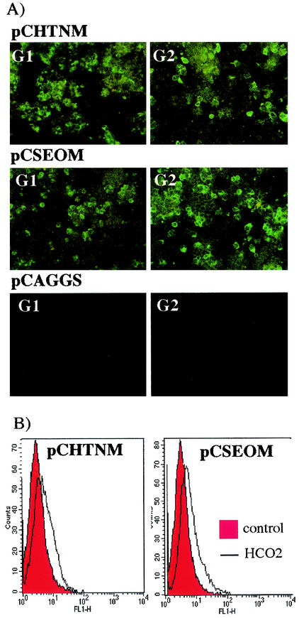
The cDNA containing the coding region of G1 and G2 of HTNV strain 76-118 and SEOV strain SR-11 was subcloned into the mammalian expression vector, pCAGGS/MCS, which contains a chicken β-actin promoter. We investigated the antigenicity of these recombinant proteins and their cell surface localization. Figure 1A shows representative staining patterns of acetone-fixed 293T cells with MAbs to both G1 and G2 of HTNV and SEOV, which show strong expression and high transfection efficiency of both the G1 and G2 proteins. The recombinant G1 and G2 proteins derived from HTNV and SEOV retained the same reactivity to MAbs as Vero E6 cells infected with the native virus. It has been reported that for the efficient generation of pseudotype VSV by using this system, the transport of envelope glycoproteins to the cell surface is critical (15). Therefore, we also tested the cell surface expression of G1 and G2 by flow cytometry. The expressed envelope glycoproteins were efficiently transported to the cell surface, as indicated in Fig. 1B. The cell surface IFA test also confirmed the surface localization of G1 and G2 (data not shown).
FIG. 1.
Intracellular and cell surface expression of G1 and G2 glycoproteins in 293T cells. (A) Intracellular staining. Transfected 293T cells were resuspended in PBS, spotted onto a 24-well glass slide, and air dried. After being fixed with acetone, the cells were treated with the culture supernatant from hybridoma clones producing the MAb 8B6 for G1 and MAb HCO2 for G2. (B) Cell surface staining analyzed by flow cytometry. Transfected cells were stained with purified HCO2 (10 μg/100 μl), and then FITC-labeled anti-mouse IgG was added.
Generation of recombinant VSV pseudotyped with HTNV and SEOV glycoproteins G1 and G2.
We inoculated 293T cells expressing hantavirus G1 and G2 with the recombinant VSV complemented with G protein (VSVΔG*G), as previously described (18), to examine the incorporation of G1 and G2 proteins into VSV particles. At 30 h after the transfection of pCHTNM, pCSEOM, or pCVSVG, 293T cells were inoculated with VSVΔG*G at a multiplicity of infection of 1. After 24 h, the culture supernatant was harvested and pseudotype viruses were purified by centrifugation through a sucrose cushion (20 and 60%) and analyzed by Western blotting.
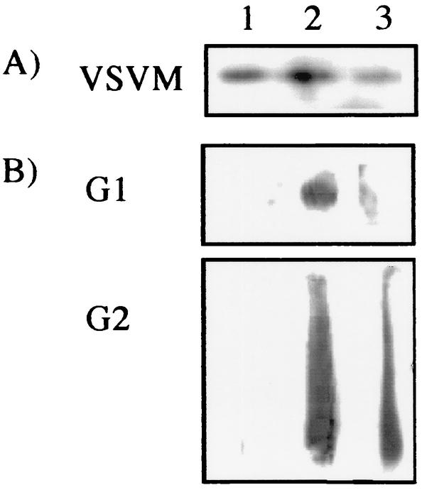
M proteins were detected in all of the recombinant VSVs complemented with G protein (Fig. 2, lane 1), the G1 and G2 proteins of HTNV (lane 2), or SEOV (lane 3) by Western blotting analysis after SDS-PAGE separation (Fig. 2A). None of the antibodies that we have tested thus far detects G1 or G2 protein after reducing SDS-PAGE separation, probably because of the highly conformational structure of hantavirus envelope glycoproteins. Therefore, we used a different procedure (native PAGE) to detect G1 and G2 incorporated into each recombinant VSV particle. The smeared, blurred shape of the G1 or G2 bands may be due to the lower performance of native PAGE in separating the proteins compared to SDS-PAGE (Fig. 2B, lanes 2 and 3). In addition, glycosylation of a protein also affects its charge. To dissociate G1 and G2, high concentrations of salt and the nonionic detergent Triton X-100 (2% Triton X-100, 150 mM NaCl, 600 mM KCl) were needed. As a result, surplus chloride ions that reduced the performance of electrophoresis remained in the electrophoresis buffer. These results indicated that G1 and G2 proteins expressed in trans were incorporated into the recombinant VSV particles. The SEOV G1 band was weaker reaction of SEOV G1 compared to HTNV G1. This experiment was performed twice, and both gave a smudged G1 band of the same intensity and pattern. Therefore, the weaker signal of SEOV G1 was due to the heterogeneous reaction of MAb 8B6 produced against HTNV.
FIG. 2.
Incorporation of G1 and G2 glycoproteins into pseudotype VSVΔG*. Pseudotype viruses were partially purified by centrifugation at 100,000 rpm through 20% sucrose on a 60% sucrose cushion. (A) Detection of VSV M protein by MAb in pseudotype VSVΔG* after separation by SDS-12% PAGE. (B) Detection of hantaviral G1 and G2 proteins by MAbs in pseudotype VSVΔG* after native 8% PAGE separation. Lanes: 1, VSV ΔG*G; 2, VSVΔ G*-HTN; 3, VSVΔG*-SEO.
Infection of Vero E6 cells with pseudotype VSV bearing HTNV and SEOV glycoproteins.
To test whether hantavirus G1 and G2 rescued the infectivity of recombinant VSV lacking G protein, we examined the infectivity of VSVΔG*-HTN and -SEO in Vero E6 cells. GFP-expressing cells were first detected 8 hpi. With time, the number of positive cells increased, as did the fluorescence intensity. The number of positive cells plateaued at ca. 16 hpi and then declined. At ca. 23 hpi, the infected cells detached from the dishes. The same growth kinetics were observed with both pseudotype VSVs. The titers of VSVΔG*-HTN and -SEO were ca. 105 to 106 IU/ml and consistently 10-fold higher than those of native HTNV and SEOV produced in Vero E6 cells in three independent experiments performed in our laboratory.
Application of the pseudotype VSVs in a neutralization test with Vero E6 cells.
Based on the growth kinetics of the pseudotype VSVs described above, we set the fixation time for the neutralization test at 16 h after inoculation. To confirm the applicability of VSVΔG*-HTN and VSVΔG*-SEO as substitutes for the native virus in the neutralization test, we compared the neutralizing activities of 12 MAbs characterized previously and immune rabbit sera. As shown in Table 1, the MAbs recognizing antigenic sites G1-b, G2-a, and G2-c (3) neutralized the VSVΔG*-HTN infection at the same concentrations as native HTNV. HCO2, which also neutralizes SEOV infection, neutralized VSVΔG*-SEO at the same concentration. A cross-neutralization test with rabbit HTNV and SEOV immune sera showed that VSVΔG*-HTN and VSVΔG*-SEO gave 8- to 32-fold-higher neutralizing titers in response to the homologous antiserum than in response to the heterologous one. This finding is comparable to the results obtained with native viruses (Table 1).
TABLE 1.
Comparison of neutralizing activities at an 80% reduction of infectivity of MAbs and polyclonal antibodies among pseudotype viruses and native hantaviruses
| Antibody | Neutralizing activityb
|
||||
|---|---|---|---|---|---|
| HTNV | SEOV | VSVΔG∗HTN | VSVΔG∗SEO | VSVΔG∗G | |
| MAbsa | |||||
| G1-aa 6D4/10F11 | - | - | - | - | - |
| G1-b 2D5c/3D5/16D2c | 64 | - | 64 | - | - |
| G2-a | |||||
| HCO2 | 16 | 16 | 16 | 16 | - |
| 16E6 | 64 | - | 64 | - | - |
| G2-c 11E10-2-2 | 2 | - | 2 | - | - |
| G2-d 5B7 | - | - | - | - | - |
| G2-e 20D3 | - | - | - | - | - |
| G2-f 8E10/1C6/7G6/23G10-1 | - | - | - | - | - |
| Rabbit polyclonal antibodies | |||||
| Anti-HTNV | 320 | <40 | 320 | 40 | <40 |
| Anti-SEOV | <40 | 1,280 | 40 | 1,280 | <40 |
Antigenic sites were characterized by Arikawa et al. (2).
-, No neutralizing activity was detected at maximum concentration of MAbs (250 μg/ml). For the MAbs, values refer to the reciprocal dilution of purified MAbs (250 μg/ml) at an 80% reduction of infectivity. For the rabbit polyclonal antibodies, values indicate the reciprocal titers of rabbit immune serum at an 80% reduction of infectivity.
Not tested.
Neutralization test with pseudotype VSVs with HFRS patient sera.
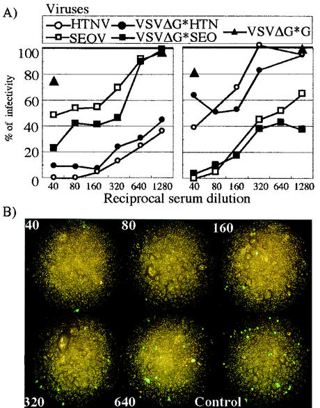
To examine the applicability of the neutralization test for measuring the neutralizing value of human sera and serotyping, we compared the neutralizing titers of the pseudotype VSVs and native viruses by using representative patient serum specimens of each serotype (Fig. 3A). These sera neutralized the native and pseudotype viruses in a dilution factor-dependent manner. The titers based on an 80% reduction of infectivity (neutralizing titer) were almost as high for native viruses as for the pseudotype viruses. The neutralizing titers were roughly two to three times higher for homologous reactions than for heterologous reactions. There was no neutralizing activity with VSVΔG*G. We also took pictures to count the number of GFP-expressing cells in the neutralization test. These showed strong GFP fluorescence, and the number of GFP-positive cells decreased in a dilution factor-dependent manner (Fig. 3B).
FIG. 3.
Neutralization activities of HFRS patient sera against native and pseudotype viruses. (A) Serum from an HFRS patient infected with HTNV (left panel). Serum from an HFRS patient infected with SEOV(right panel). (B) GFP-expressing Vero E6 cells. Cells were inoculated with a mixture of VSVΔG*-HTN and serum from an HFRS patient infected with HTNV at the indicated dilutions.
Sera from 22 patients previously diagnosed by using the FRNT or IFA test were subjected to the neutralization test with VSVΔG*-HTN, VSVΔG*-SEO, and VSVΔG*G. The neutralizing titers of each virus are shown in Table 2. It was clearly possible to distinguish between HTNV and SEOV infection in human sera with VSVΔG*-HTN and -SEO. All of the sera from healthy individuals and patients suffering from unknown febrile illnesses, which were previously determined to be seronegative for hantavirus infection by the IFA test or the FRNT, were also negative for the pseudotypes VSVΔG*-HTN and -SEO.
TABLE 2.
Neutralization titers of sera from HFRS patients and control individuals against pseudotype and native hantaviruses
| Serum type and designation | Neutralization titer
|
||||
|---|---|---|---|---|---|
| HTNV | SEOV | VSVΔG∗HTN | VSVΔG∗SEO | VSVΔG∗G | |
| HFRS patient with HTNV infection | |||||
| A1 | 160 | 40 | 160 | 40 | <10 |
| A2 | 320 | 10 | 160 | 40 | <10 |
| A3 | 320 | 10 | 160 | <10 | <10 |
| A4 | 160 | 10 | 40 | <10 | <10 |
| I1 | 320 | 10 | 80 | <10 | <10 |
| I2 | 640 | 10 | 80 | <10 | <10 |
| I3 | 320 | 20 | 160 | 80 | <10 |
| CS#2 | 320 | <10 | 80 | <10 | <10 |
| CS#7 | 10 | <10 | 40 | <10 | <10 |
| CS#8 | 40 | 10 | 40 | <10 | <10 |
| CS#17 | 80 | <10 | 160 | <10 | <10 |
| HFRS patient with SEOV infection | |||||
| B1 | 10 | 80 | <10 | 80 | <10 |
| B2 | 10 | 40 | <10 | 160 | <10 |
| B3 | 10 | 80 | <10 | 160 | <10 |
| B4 | 10 | 160 | <10 | 160 | <10 |
| D2 | 20 | 640 | 20 | 160 | <10 |
| D3 | 20 | 80 | 10 | 80 | <10 |
| D4 | 10 | 160 | 20 | 160 | <10 |
| H | 10 | 80 | 40 | 160 | <10 |
| IH | 80 | 640 | 40 | 1280 | <10 |
| M | <10 | 20 | <10 | 40 | <10 |
| Y | <10 | 20 | <10 | 80 | <10 |
| Patient with febrile illness, negative for hantavirus infection | |||||
| 44 | NTa | <16 | <10 | <10 | <10 |
| 45 | NT | <16 | <10 | <10 | <10 |
| 46 | NT | <16 | <10 | <10 | <10 |
| 47 | NT | <16 | <10 | <10 | <10 |
| 48 | NT | <16 | <10 | <10 | <10 |
| 49 | NT | <16 | <10 | <10 | <10 |
| 50 | NT | <16 | <10 | <10 | <10 |
| 51 | NT | <16 | <10 | <10 | <10 |
| 52 | NT | <16 | <10 | <10 | <10 |
| 53 | NT | <16 | <10 | <10 | <10 |
| Healthy individual, negative for hantavirus infection | |||||
| NHS1 | NT | NT | <10 | <10 | <10 |
| YU | NT | NT | <10 | <10 | <10 |
| IW | NT | NT | <10 | <10 | <10 |
| NHS19 | NT | NT | <10 | <10 | <10 |
| HUTI | NT | NT | <10 | <10 | <10 |
NT, not tested.
Combined with these results, the recombinant VSVs complemented with hantavirus envelope glycoproteins should be useful for measuring neutralizing antibodies and serotyping.
Stability of the pseudotype VSVs at 4°C and room temperature.
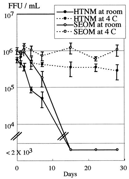
We also tested the stability of the pseudotype VSVs subjected to repeated freezing-thawing, stored at 4°C, and stored at room temperature (25°C). Repeated freezing-thawing (stored at −80°C for 1 year, thawed once, and frozen again) did not affect the infectivity of pseudotype VSVs (data not shown). As shown in Fig. 4, the infectivity titers of the pseudotype VSVs were retained at >105 IU/ml when stored for up to 28 days at 4°C. On the other hand, after 14 days of incubation at room temperature, the infectivity of the pseudotype VSVs decreased to undetectable levels.
FIG. 4.
Stability of the pseudotype viruses at 4°C and room temperature. VSVΔ G*-HTN and VSVΔG*-SEO were kept at 4°C or room temperature for the indicated number of days. Then, the IU value was determined as described in Materials and Methods.
DISCUSSION
To find an alternative to the conventional laborious and time-consuming neutralization test for hantaviruses, we established a neutralization test with pseudotype VSV containing HTNV or SEOV envelope glycoproteins (VSVΔG*-HTN and -SEO). The infection of VSVΔG*-HTN and -SEO was neutralized with MAbs, immune sera, and patient sera with similar kinetics and titers, indicating that the pseudotype VSVs were effective alternatives to native hantaviruses in the neutralization test. Moreover, this neutralization system does not require a high-level containment laboratory, since the genome of the pseudotypes contains the GFP gene instead of the VSV G protein gene, and is consequently unable to produce infectious progeny viruses unless the envelope proteins mediating cellular entry are provided in trans. Furthermore, due to the powerful expression of GFP in the VSV replication system, infected cells can be counted 16 h after infection by using a simple fluorescence assay. The ordinary neutralization test requires immunofluorescence procedures, immunocytochemical staining, and plaque detection to detect infected foci. The time required for the virus to replicate to detectable levels for the immunofluorescence of infected cell foci on Vero E6 monolayers is 2 to 5 days, depending on the hantavirus species. Immunocytochemical staining takes even longer: from 5 to 10 days depending on the hantavirus species. The VSVΔG*-HTN and -SEO substitutes for native viruses make the neutralization test faster and simpler.
This recombinant VSVΔG* system efficiently incorporates foreign viral envelope proteins, as previously reported for Ebola virus, measles virus, human T-cell leukemia virus type 1, hepatitis C virus, and Borna disease virus (14, 16, 17, 21, 22). VSVΔG*-HTN and -SEO were generated efficiently at titers in the range of 105 to 106 IU/ml, which are higher titers than those of native HTNV and SEOV (104 to 105 focus-forming units/ml). Another pseudotype system with murine leukemia virus has been previously reported to produce a hantavirus pseudotype; however, the titers in that system were much lower, i.e., in the range of 103 IU/ml (13). It was thought that these results reflect low expression levels of Hantaan envelope glycoproteins or inefficient virus assembly.
The SEOV titers with virus and pseudotype are generally similar, whereas the HTNV titers are often higher (one- or twofold dilution) when authentic virus is used, although this tendency was not observed for rabbit immune serum (Table 2). This observation may be unique in humans infected with HTNV and may not occur in an envelope glycoprotein-dependent manner. Serum from a patient infected with HTNV, but not SEOV, might contain an inhibitor or antibodies induced against other viral components. This might hinder the steps of replication after attachment or adsorption, which would be caused in an envelope glycoprotein-dependent manner. Another interpretation of Table 2 is that, although there were small differences in the antigenicity of HTNV and VSVG*HTN, immune rabbit serum could not distinguish the difference in the cross-neutralization test. VSV incorporates a variety of foreign membrane proteins. The efficiency and conformation, which might affect the function of foreign proteins incorporated into VSV particles, depend on the proteins (20). The amino acid homology of HTNV and SEOV envelope glycoproteins is approximately 80%. In fact, little effort has been made to compare the precise conformation of the envelope glycoproteins between serotypes; only genetic and serological approaches have been used. Therefore, the antigenicity of HTN envelope glycoproteins incorporated into the recombinant VSV particle might differ slightly from that of HTNV.
In the manual for HFRS and HPS diagnosis published by the World Health Organization (9), the cross-neutralization test is recommended as the standard serotyping method for a newly isolated hantavirus. According to the definition in Virus Taxonomy, the Hantavirus species show at least a fourfold difference in two-way cross-neutralization tests in serum from convalescent patients. Hantavirus species occupy a unique ecological niche, i.e., they occur in different primary rodent reservoir species or subspecies (6). Therefore, measuring neutralizing antibodies provides the basic information needed to characterize newly discovered species of hantavirus and to prevent disease. The greater stability of pseudotype VSVs at 4°C allows the transport of the viruses to remote areas by using conventional iceboxes. Since pseudotype VSV-based neutralization is simple, rapid, and safe, this system can replace that using the native virus. Recently, we developed an enzyme-linked immunosorbent assay using truncated recombinant nucleocapsid proteins to differentiate HTNV, SEOV, and DOBV infections (1). This test could complement the neutralization assays when multiple samples are being screened. Another type-specific diagnostic method has been reported: reverse transcriptase PCR (RT-PCR) with specific primer pairs. The detection and characterization of amplicons after the RT-PCR test provide definite genetic information for genotyping. Thus, a combination of different diagnostic procedures should provide useful information for understanding the etiology of hantavirus and preventing infection.
There have been several reports of serological cross-reactions and absent reactions involving hantaviruses. Therefore, a set of pseudotypes that covers all of the hantaviruses causing HFRS or HPS will be more valuable and necessary for the purpose of a global epidemiological survey. We are now trying to prepare pseudotypes that possess the envelope glycoproteins of other HFRS-causing hantaviruses: DOBV and Puumala virus.
In addition, to define newly discovered species of hantaviruses, the neutralization assay provides information that evaluates the protective immune status. The neutralizing antibody titer in patient serum likely affects the patient's prognosis in hantavirus pulmonary syndrome. Bharadwaj et al. (4) reported that, on admission, patients with severe disease had lower neutralizing antibody titers than did patients with mild disease. These authors suggested that a strong neutralizing antibody response might be a predictor of effective clearance of and recovery from Sin Nombre virus infection. Experimentally, the passive transfer of neutralizing MAbs protects test animals from hantavirus infection (3). Therefore, the titer of neutralizing antibodies may be an essential determinant of the protective immune status of individuals.
In summary, we have established a novel neutralization test that is simple, safe, and rapid by using pseudotype viruses. In addition to providing a convenient diagnostic method, the pseudotypes also offer a unique tool for analyzing cellular entry and the mechanism of neutralization of hantaviruses.
Acknowledgments
We thank M. A. Whitt for allowing us to use the VSVΔG* system. We aknowledge H. Wang, Y. Nishimune, M. K. Ijaz, T. Al Karmi, and H.-K. Ooi for patient sera. We thank Textcheck (English consultants) for grammatical revision of the final draft of the manuscript.
This work was supported in part by Grants-in-Aid for Scientific Research and the Development of Scientific Research from the Japanese Ministry of Education, Science, Sports, and Culture and by grants from the Swedish Medical Research Council (projects 12177 and 12642). M.O. and H.E. were Research Fellows of the Japan Society for the Promotion of Science (JSPS) and were supported by JSPS Research Fellowships for Young Scientists.
REFERENCES
- 1.Araki, K., K. Yoshimatsu, M. Ogino, H. Ebihara, A. Lundkvist, H. Kariwa, I. Takashima, and J. Arikawa. 2001. Truncated hantavirus nucleocapsid proteins for serotyping hantaan, seoul, and dobrava hantavirus infections. J. Clin. Microbiol. 39:2397-2404. [DOI] [PMC free article] [PubMed] [Google Scholar]
- 2.Arikawa, J., A. L. Schmaljohn, J. M. Dalrymple, and C. S. Schmaljohn. 1989. Characterization of Hantaan virus envelope glycoprotein antigenic determinants defined by monoclonal antibodies. J. Gen. Virol. 70:615-624. [DOI] [PubMed] [Google Scholar]
- 3.Arikawa, J., J. S. Yao, K. Yoshimatsu, I. Takashima, and N. Hashimoto. 1992. Protective role of antigenic sites on the envelope protein of Hantaan virus defined by monoclonal antibodies. Arch. Virol. 126:271-281. [DOI] [PMC free article] [PubMed] [Google Scholar]
- 4.Bharadwaj, M., R. Nofchissey, D. Goade, F. Koster, and B. Hjelle. 2000. Humoral immune responses in the hantavirus cardiopulmonary syndrome. J. Infect. Dis. 182:43-48. [DOI] [PubMed] [Google Scholar]
- 5.Dantas, J. R., Jr., Y. Okuno, H. Asada, M. Tamura, M. Takahashi, O. Tanishita, Y. Takahashi, T. Kurata, and K. Yamanishi. 1986. Characterization of glycoproteins of viruses causing hemorrhagic fever with renal syndrome (HFRS) using monoclonal antibodies. Virology 151:379-384. [DOI] [PubMed] [Google Scholar]
- 6.Elliot, R., M. Bouloy, C. H. Calisher, R. Goldbach, J. T. Moyer, S. T. Nichol, R. Pettersson, A. Plyusnin, and C. S. and Schmaljohn. 2001. Family Bunyaviridae, p. 599-621. In V. Regenmortel (ed.), Virus taxonomy: seventh report of the International Commitee on Taxonomy of Viruses. Academic Press, Inc., New York, N.Y.
- 7.Hooper, J. W., D. M. Custer, E. Thompson, and C. S. Schmaljohn. 2001. DNA vaccination with the Hantaan virus M gene protects Hamsters against three of four HFRS hantaviruses and elicits a high-titer neutralizing antibody response in rhesus monkeys. J. Virol. 75:8469-8477. [DOI] [PMC free article] [PubMed] [Google Scholar]
- 8.Lawson, N. D., E. A. Stillman, M. A. Whitt, and J. K. Rose. 1995. Recombinant vesicular stomatitis viruses from DNA. Proc. Natl. Acad. Sci. USA 92:4477-4481. [DOI] [PMC free article] [PubMed] [Google Scholar]
- 9.Lee, H. W., C. Calisher, and C. Schmaljohn (ed.). 1998. Manual of hemorrhagic fever with renal syndrome and hantavirus pulmonary syndrome. W.H.O. Collaborating Center for Virus Reference and Research (Hantaviruses), Asan Institute for Life Sciences, Seoul, Korea.
- 10.Lefrancois, L., and D. S. Lyles. 1982. The interaction of antibody with the major surface glycoprotein of vesicular stomatitis virus. I. Analysis of neutralizing epitopes with monoclonal antibodies. Virology 121:157-167. [PubMed] [Google Scholar]
- 11.Lundkvist, A., M. Hukic, J. Horling, M. Gilljam, S. Nichol, and B. Niklasson. 1997. Puumala and Dobrava viruses cause hemorrhagic fever with renal syndrome in Bosnia-Herzegovina: evidence of highly cross-neutralizing antibody responses in early patient sera. J. Med. Virol. 53:51-59. [PubMed] [Google Scholar]
- 12.Lundkvist, Å., and A. Plyusnin. Molecular epidemiology of hantavirus infections. In T. Leitner (ed.), Molecular epidemiology of human viruses, in press. Kluwer Academic Publishers, Dordrecht, The Netherlands.
- 13.Ma, M., D. B. Kersten, K. I. Kamrud, R. J. Wool-Lewis, C. Schmaljohn, and F. Gonzalez-Scarano. 1999. Murine leukemia virus pseudotypes of La Crosse and Hantaan bunyaviruses: a system for analysis of cell tropism. Virus Res. 64:23-32. [DOI] [PubMed] [Google Scholar]
- 14.Matsuura, Y., H. Tani, K. Suzuki, T. Kimura-Someya, R. Suzuki, H. Aizaki, K. Ishii, K. Moriishi, C. S. Robison, M. A. Whitt, and T. Miyamura. 2001. Characterization of pseudotype VSV possessing HCV envelope proteins. Virology 286:263-275. [DOI] [PubMed] [Google Scholar]
- 15.Niwa, H., K. Yamamura, and J. Miyazaki. 1991. Efficient selection for high-expression transfectants with a novel eukaryotic vector. Gene 108:193-199. [DOI] [PubMed] [Google Scholar]
- 16.Okuma, K., Y. Matsuura, H. Tatsuo, Y. Inagaki, M. Nakamura, N. Yamamoto, and Y. Yanagi. 2001. Analysis of the molecules involved in human T-cell leukaemia virus type 1 entry by a vesicular stomatitis virus pseudotype bearing its envelope glycoproteins. J. Gen. Virol. 82:821-830. [DOI] [PubMed] [Google Scholar]
- 17.Perez, M., M. Watanabe, M. A. Whitt, and J. C. de la Torre. 2001. N-terminal domain of Borna disease virus G (p56) protein is sufficient for virus receptor recognition and cell entry. J. Virol. 75:7078-7085. [DOI] [PMC free article] [PubMed] [Google Scholar]
- 18.Plyusnin, A., O. Vapalahti, and A. Vaheri. 1996. Hantaviruses: genome structure, expression and evolution. J. Gen. Virol. 77:2677-2687. [DOI] [PubMed] [Google Scholar]
- 19.Schmaljohn, C., and B. Hjelle. 1997. Hantaviruses: a global disease problem. Emerg. Infect. Dis. 3:95-104. [DOI] [PMC free article] [PubMed] [Google Scholar]
- 20.Schnell, M., J. Buonocore, L. Kretzschmar, E. Johnson, and J. K. Rose. 1996. Foreign glycoproteins expressed from recombinant vesicular stomatitis viruses are incorporated efficiently into virus particles. Proc. Natl. Acad. Sci. USA 93:11359-11365. [DOI] [PMC free article] [PubMed] [Google Scholar]
- 21.Takada, A., C. Robison, H. Goto, A. Sanchez, K. G. Murti, M. A. Whitt, and Y. Kawaoka. 1997. A system for functional analysis of Ebola virus glycoprotein. Proc. Natl. Acad. Sci. USA 94:14764-14769. [DOI] [PMC free article] [PubMed] [Google Scholar]
- 22.Tatsuo, H., K. Okuma, K. Tanaka, N. Ono, H. Minagawa, A. Takade, Y. Matsuura, and Y. Yanagi. 2000. Virus entry is a major determinant of cell tropism of Edmonston and wild-type strains of measles virus as revealed by vesicular stomatitis virus pseudotypes bearing their envelope proteins. J. Virol. 74:4139-4145. [DOI] [PMC free article] [PubMed] [Google Scholar]
- 23.Whelan, S. P., L. A. Ball, J. N. Barr, and G. T. Wertz. 1995. Efficient recovery of infectious vesicular stomatitis virus entirely from cDNA clones. Proc. Natl. Acad. Sci. USA 92:8388-8392. [DOI] [PMC free article] [PubMed] [Google Scholar]
- 24.Yoshimatsu, K., J. Arikawa, H. Li, H. Kariwa, and N. Hashimoto. 1996. Western blotting using recombinant Hantaan virus nucleocapsid protein expressed in silkworm as a serological confirmation of hantavirus infection in human sera. J. Vet. Med. Sci. 58:71-74. [DOI] [PubMed] [Google Scholar]


