

Abstract
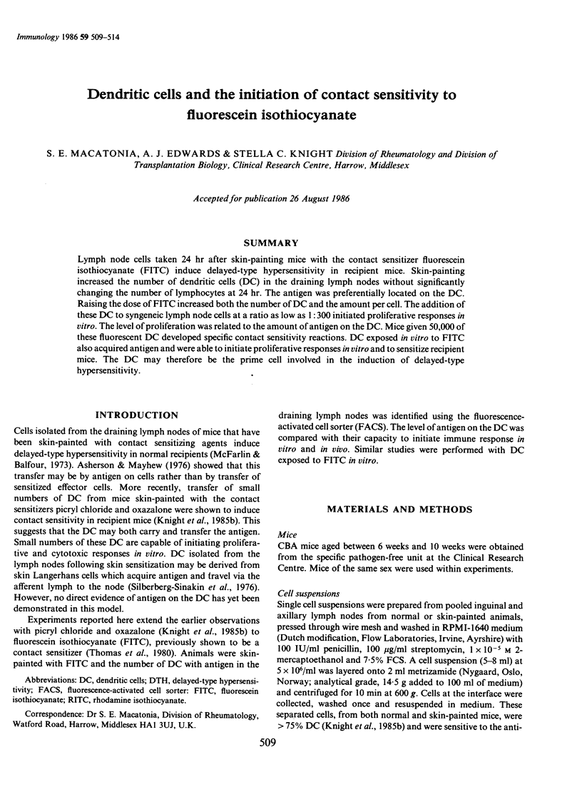
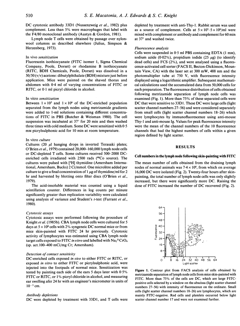
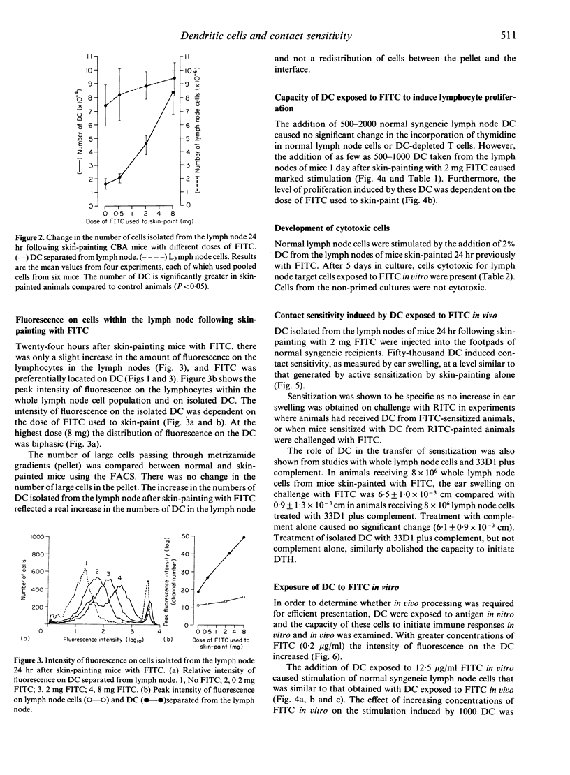
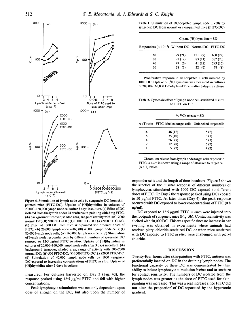
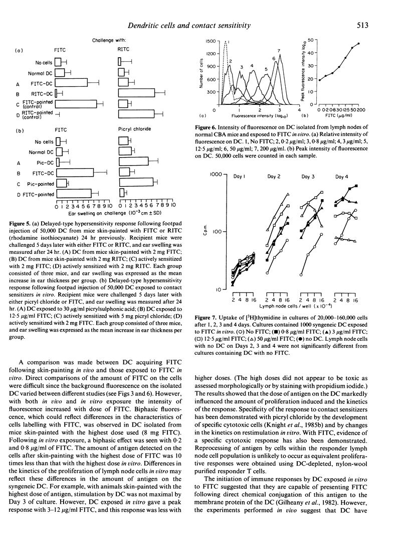
Lymph node cells taken 24 hr after skin-painting mice with the contact sensitizer fluorescein isothiocyanate (FITC) induce delayed-type hypersensitivity in recipient mice. Skin-painting increased the number of dendritic cells (DC) in the draining lymph nodes without significantly changing the number of lymphocytes at 24 hr. The antigen was preferentially located on the DC. Raising the dose of FITC increased both the number of DC and the amount per cell. The addition of these DC to syngeneic lymph node cells at a ratio as low as 1:300 initiated proliferative responses in vitro. The level of proliferation was related to the amount of antigen on the DC. Mice given 50,000 of these fluorescent DC developed specific contact sensitivity reactions. DC exposed in vitro to FITC also acquired antigen and were able to initiate proliferative responses in vitro and to sensitize recipient mice. The DC may therefore be the prime cell involved in the induction of delayed-type hypersensitivity.
Full text
PDF
Selected References
These references are in PubMed. This may not be the complete list of references from this article.
- Asherson G. L., Mayhew B. Induction of cell-mediated immunity in the mouse: circumstantial evidence for highly immunogenic antigen in the regional lymph nodes following skin painting with contact sensitizing agents. Isr J Med Sci. 1976 Apr-May;12(4-5):454–467. [PubMed] [Google Scholar]
- Austyn J. M., Gordon S. F4/80, a monoclonal antibody directed specifically against the mouse macrophage. Eur J Immunol. 1981 Oct;11(10):805–815. doi: 10.1002/eji.1830111013. [DOI] [PubMed] [Google Scholar]
- Butcher E. C., Weissman I. L. Direct fluorescent labeling of cells with fluorescein or rhodamine isothiocyanate. I. Technical aspects. J Immunol Methods. 1980;37(2):97–108. doi: 10.1016/0022-1759(80)90195-7. [DOI] [PubMed] [Google Scholar]
- Julius M. H., Simpson E., Herzenberg L. A. A rapid method for the isolation of functional thymus-derived murine lymphocytes. Eur J Immunol. 1973 Oct;3(10):645–649. doi: 10.1002/eji.1830031011. [DOI] [PubMed] [Google Scholar]
- Knight S. C., Bedford P., Hunt R. The role of dendritic cells in the initiation of immune responses to contact sensitizers. II. Studies in nude mice. Cell Immunol. 1985 Sep;94(2):435–439. doi: 10.1016/0008-8749(85)90267-9. [DOI] [PubMed] [Google Scholar]
- Knight S. C., Krejci J., Malkovsky M., Colizzi V., Gautam A., Asherson G. L. The role of dendritic cells in the initiation of immune responses to contact sensitizers. I. In vivo exposure to antigen. Cell Immunol. 1985 Sep;94(2):427–434. doi: 10.1016/0008-8749(85)90266-7. [DOI] [PubMed] [Google Scholar]
- Knight S. C. Veiled cells--"dendritic cells" of the peripheral lymph. Immunobiology. 1984 Dec;168(3-5):349–361. doi: 10.1016/S0171-2985(84)80122-9. [DOI] [PubMed] [Google Scholar]
- Nussenzweig M. C., Steinman R. M., Witmer M. D., Gutchinov B. A monoclonal antibody specific for mouse dendritic cells. Proc Natl Acad Sci U S A. 1982 Jan;79(1):161–165. doi: 10.1073/pnas.79.1.161. [DOI] [PMC free article] [PubMed] [Google Scholar]
- O'Brien J., Knight S., Quick N. A., Moore E. H., Platt A. S. A simple technique for harvesting lymphocytes cultured in Terasaki plates. J Immunol Methods. 1979;27(3):219–223. doi: 10.1016/0022-1759(79)90219-9. [DOI] [PubMed] [Google Scholar]
- Silberberg-Sinakin I., Thorbecke G. J., Baer R. L., Rosenthal S. A., Berezowsky V. Antigen-bearing langerhans cells in skin, dermal lymphatics and in lymph nodes. Cell Immunol. 1976 Aug;25(2):137–151. doi: 10.1016/0008-8749(76)90105-2. [DOI] [PubMed] [Google Scholar]
- Stohlman S. A., Matsushima G. K., Casteel N., Frelinger J. A. The defect in delayed-type hypersensitivity of young adult SJL mice is due to a lack of functional antigen-presenting cells. Eur J Immunol. 1985 Sep;15(9):913–916. doi: 10.1002/eji.1830150909. [DOI] [PubMed] [Google Scholar]
- Thomas W. R., Edwards A. J., Watkins M. C., Asherson G. L. Distribution of immunogenic cells after painting with the contact sensitizers fluorescein isothiocyanate and oxazolone. Different sensitizers form immunogenic complexes with different cell populations. Immunology. 1980 Jan;39(1):21–27. [PMC free article] [PubMed] [Google Scholar]
- Van Loveren H., Kato K., Meade R., Green D. R., Horowitz M., Ptak W., Askenase P. W. Characterization of two different Ly-1+ T cell populations that mediate delayed-type hypersensitivity. J Immunol. 1984 Nov;133(5):2402–2411. [PubMed] [Google Scholar]