Abstract
SsrA, or tmRNA, is a small RNA that interacts with selected translating ribosomes to target the nascent polypeptides for degradation. Here we report that SsrA activity is required for normal timing of the G1-to-S transition in Caulobacter crescentus. A deletion of the ssrA gene, or of the gene encoding SmpB, a protein required for SsrA activity, results in a specific delay in the cell cycle during the G1-to-S transition. The ssrA deletion phenotype is not due to accumulation of stalled ribosomes, because the deletion is not complemented by a mutated version of SsrA that releases ribosomes but does not target proteins for degradation. Degradation of the CtrA response regulator normally coincides with initiation of DNA replication, but in strains lacking SsrA activity there is a 40-min delay between the degradation of CtrA and replication initiation. This uncoupling of initiation of replication from CtrA degradation indicates that there is an SsrA-dependent pathway required for correct timing of DNA replication.
Regulated proteolysis is a fundamental mechanism of cell cycle control, both in eukaryotes and in the bacterium Caulobacter crescentus (6). We report here that a small RNA that targets proteins for degradation in bacteria is required for normal timing of the G1-to-S phase transition in Caulobacter. This RNA, known as SsrA (also referred to as tmRNA and 10Sa RNA), is a small, highly structured RNA that intervenes in selected translation reactions to release ribosomes from the mRNAs and target the nascent polypeptides for proteolysis. SsrA RNAs are ubiquitous in bacteria and are found in some chloroplasts and mitochondria of eukaryotes (10, 21, 37).
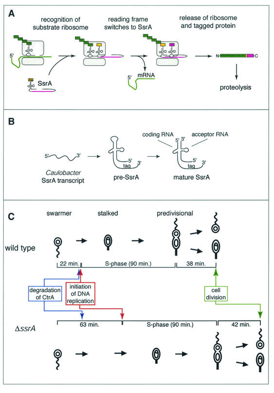
SsrA RNA has been called tmRNA because it has properties of both a tRNA and an mRNA. The 5′ and 3′ ends of the RNA are folded into a tRNA-like structure, and the RNA can be charged with alanine by alanyl-tRNA synthetase (23, 35). Another portion of the RNA contains a specialized open reading frame that encodes a peptide containing determinants for multiple proteases (11, 14, 17, 22, 34). In the model proposed for SsrA activity (22) (Fig. 1A), SsrA RNA charged with alanine enters the A site of selected ribosomes and acts like a tRNA analog. The nascent polypeptide is transferred to alanyl-SsrA by transpeptidation, and the reading frame of the translating ribosome switches from the original mRNA to the mRNA-like portion of SsrA RNA. Translation of the SsrA open reading frame results in the addition of a peptide tag to the nascent polypeptide, and termination at the SsrA-encoded stop codon releases the ribosome. The SsrA-encoded peptide tag targets the nascent polypeptide for rapid degradation by a number of intracellular proteases (14, 17, 22).
FIG. 1.
SsrA peptide-tagging activity, processing, and the Caulobacter cell cycle. (A) Model for SsrA activity (22). A ribosome translating an mRNA becomes a substrate for SsrA RNA with the nascent polypeptide and tRNA still engaged. SsrA RNA charged with alanine on its tRNA-like 3′ end enters the ribosomal A site. Transpeptidation occurs to the alanine on SsrA, the mRNA is removed from the ribosome, and the translational reading frame switches to the mRNA-like portion of SsrA RNA. Translation of the SsrA reading frame adds a peptide to the incomplete protein and targets the protein for rapid proteolysis by several intracellular proteases. (B) Model for pre-SsrA transcription and processing into matureSsrA (21). A single transcript made from the ssrA gene folds into a structure similar to those of one-piece SsrA's, except that the 5′ and 3′ ends are in different parts of the molecule. Processing by nucleases produces the tRNA-like 5′ and 3′ ends, resulting in mature SsrA composed of a coding RNA and an acceptor RNA. (C) Comparison of the timing of major cell cycle events in wild-type Caulobacter and the ΔssrA strain. Cartoon diagrams of the Caulobacter cell cycle show the swarmer cell with a polar flagellum (curved line) and a single, nonreplicating chromosome (open circle), the stalked cell with a replicating chromosome (theta structure), and the predivisional cell with two completely replicated chromosomes (open circles).
SsrA RNA activity requires at least three RNA-binding proteins, SmpB (20), EF-Tu (4, 32), and ribosomal protein S1 (40), in addition to general translation factors. SmpB and EF-Tu bind simultaneously to the tRNA-like domain of SsrA RNA and are required for efficient aminoacylation by alanyl-tRNA synthetase and for interaction with target ribosomes (3, 15, 20). The ribosomal protein S1 binds to the mRNA-like domain of SsrA RNA and is proposed to play a role in entry of the SsrA peptide reading frame into the ribosome (40). Both EF-Tu and S1 are required for other canonical translation reactions, but SmpB has no known role other than in SsrA-mediated peptide tagging. A deletion of smpB has the same phenotype as a deletion of ssrA in Escherichia coli (20) and Bacillus subtilis (36), consistent with a dedicated and essential role in SsrA RNA activity.
The targets that have been identified for SsrA tagging activity in E. coli suggest that stalling of the ribosome is an important determinant for substrate selectivity. SsrA activity has been demonstrated with ribosomes that are stalled during translation by four different mechanisms: ribosomes stalled at the 3′ end of an mRNA that does not have a stop codon (22), ribosomes stalled due to limiting amounts of cognate tRNA for the next codon (31), ribosomes delayed in termination because of a C-terminal proline residue (16), and ribosomes stalled on an mRNA that is incomplete due to a block of transcriptional elongation during coupled transcription-translation (1). Because SsrA resolves stalled translational complexes by ridding the cell of the incompletely translated proteins and freeing the ribosomes, it has been proposed to perform a quality control function for translation (19, 22, 39). In this study we present evidence that SsrA RNA has a regulatory role in the G1-to-S transition of the Caulobacter cell cycle.
In Caulobacter and related α-proteobacteria, the ssrA gene is unusual in that it contains a circular permutation that results in a mature SsrA composed of two RNA molecules (Fig. 1B) (21). Despite this two-piece composition, the Caulobacter SsrA RNA is predicted to have a structure very similar to those of the one-piece SsrA RNAs from other bacteria. As in E. coli, SsrA RNA in Caulobacter tags proteins made from mRNA with no stop codon, and the Caulobacter SsrA tag targets proteins for rapid degradation in vivo (21).
Caulobacter is ideal for cell cycle studies because it is easy to isolate a population of cells in G1 phase, and this population will pass synchronously through the cell cycle. In addition, the cell cycle of Caulobacter is tied to a developmental program (Fig. 1C) (18). The G1 phase of the Caulobacter cell cycle coincides with the swarmer cell stage, in which cells are motile and have a single polar flagellum. Swarmer cells cannot initiate DNA replication or undergo cell division until they differentiate into stalked cells. This differentiation includes loss of the flagellum and of the chemotaxis apparatus, growth of a new appendage called a stalk, and initiation of DNA replication. Thus, the swarmer-to-stalked cell transition is coincident with the G1-to-S phase transition. After differentiation, the stalked cell continues DNA replication, elongates, and eventually forms a division plane, becoming a predivisional cell. The predivisional cell divides asymmetrically to produce a swarmer cell and regenerate the stalked cell.
MATERIALS AND METHODS
Strains and plasmids.
The wild-type Caulobacter strain used in this work is CB15N (9). Caulobacter strains were grown in M2G or PYE at 28°C (8), and growth was monitored by the increase in optical density at 660 nm. The pssrA plasmid was constructed by amplifying a 916-bp DNA fragment containing the ssrA gene and promoter sequences from Caulobacter genomic DNA by PCR and cloning this fragment into the broad-host-range vector pJS14 (21). The pssrA-DD plasmid was derived from pssrA by using PCR mutagenesis to alter the last two codons of the reading frame to Asp-Asp (21). The ΔssrA and ΔsmpB strains were constructed by the two-step recombination method with sacB counterselection (12) as follows. A copy of the chromosomal region surrounding ssrA or smpB, but with a deletion in the relevant gene, was made by cloning 1 kb of DNA flanking each end of the gene, with a spectinomycin resistance marker inserted in place of the gene, into pNPTS138, an integration plasmid carrying the sacB gene and a kanamycin resistance marker (M. R. K. Alley, unpublished data). The plasmid was transformed into wild-type Caulobacter, and integrants were selected by growth on kanamycin. Subsequent growth on PYE-agarose plates containing 3% sucrose was used to select for recombination, and recombinants containing the disrupted copy were screened by growth on spectinomycin and verified by Southern blotting. For the ssrA deletion, only wild-type recombinants were recovered due to slow growth of cells with the ssrA deletion under selective conditions. In this case, the pNPTS138-based integrants were transformed with the pssrA plasmid prior to growth on sucrose, and the ssrA deletion was transduced from the spectinomycin-resistant recombinants into wild-type Caulobacter by using bacteriophage φCr30 (8). Because a disruption of the chromosomal ssrA gene was observed only when a copy of ssrA was supplied on a plasmid, it was initially reported that ssrA is essential in Caulobacter (21). All plasmids were verified by DNA sequencing and were mobilized into Caulobacter by electroporation or conjugation from the E. coli strain S17 (8).
Cell cycle studies.
Synchronized cultures of Caulobacter were obtained by centrifugation in a Ludox density gradient followed by isolation of swarmer cells (9). Aliquots were removed from synchronized cultures every 15 min for analysis by flow cytometry or Western blotting, and the timing of loss of motility and cell division in these cultures was estimated by visual inspection using light microscopy. Analyses of the levels of CtrA and McpA were performed by Western blotting followed by chemiluminescence detection and quantification using ImageQuant software (Molecular Dynamics). Flow cytometry assays for DNA content and initiation of replication were performed as previously described (38). Replication initiation assays employed either rifampin (15 μg/ml) or chloramphenicol (25 μg/ml) as an inhibitor of initiation, with no observable differences between inhibitors. The percentage of cells that had initiated DNA replication when the inhibitor was added was determined by fitting the data to two normal distributions, corresponding to DNA contents of one or two chromosomes, derived from control experiments.
RNA analysis.
Total RNA was isolated using the hot phenol method (33). Northern blotting was performed after separation of equal quantities of total RNA on polyacrylamide-urea gels. 32P-labeled DNA probes were generated from PCR products by using the QuickPrime protocol (Amersham Biosciences), visualized by use of a PhosphorImager (Molecular Dynamics), and quantified with ImageQuant. To compare the relative amounts of SsrA RNA in different strains, Northern blots were corrected for differences in loading and transfer by normalizing the SsrA RNA signal to the amount of 5S rRNA. Northern blots were stripped and reprobed for 5S rRNA, and the amount of 5S rRNA was detected and quantified as above.
RESULTS
Absence of SsrA RNA causes a stall in the G1-to-S transition.
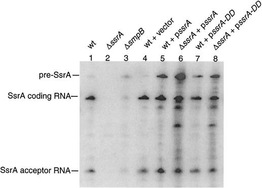
To investigate the role of SsrA RNA in Caulobacter, a strain lacking SsrA RNA was generated by replacing the ssrA gene with an omega cassette containing a spectinomycin resistance marker. Deletion strains were verified by Southern blotting to detect the omega cassette insertion (data not shown) and by Northern blot analysis for SsrA RNA (Fig. 2, lanes 1 and 2). This strain had no detectable SsrA RNA, indicating that ssrA is not essential for viability. However, the ΔssrA strain grew more slowly than the wild type and had an aberrant cell cycle (Fig. 1C). In minimal medium (M2G), the doubling time of the ΔssrA strain during log phase growth, as measured by optical density, was 26 min longer than that of the wild type (179 versus 153 min [Table 1]). To determine if this altered growth rate was the result of slower growth throughout the cell cycle or if it was due to a disruption in one stage of the cell cycle, swarmer cells from the ΔssrA and wild-type strains were isolated and observed by light microscopy as they passed synchronously through the cell cycle. Under these conditions, the time to complete a cell cycle was 45 min longer for the ΔssrA strain than for the wild type (195 versus 150 min [Table 1]). The increase in the length of the cell cycle in the ΔssrA strain is greater than the increase in the doubling time (45 versus 26 min), suggesting that a lack of SsrA affects cell cycle timing more than it affects accumulation of cell mass. In fact, cells lacking SsrA were larger than wild-type cells (data not shown). The swarmer stage, as measured by loss of motility, was approximately 45 min longer in the ΔssrA strain (75 versus 30 min), but the lengths of the stalked and predivisional stages in the two strains were the same. Thus, the swarmer stage is more than twice as long in the ΔssrA strain, and this difference accounts for the slower cell cycle.
FIG. 2.
Levels of SsrA RNA in wild-type (wt) Caulobacter and mutant strains, detected by Northern blotting. Equal quantities of total RNA were loaded in each lane and probed for SsrA RNA. Bands corresponding to pre-SsrA RNA, the coding RNA, and the acceptor RNA are indicated.
TABLE 1.
Growth and cell cycle parameters of Caulobacter strains
| Straina | Time (min) of the indicated process in:
|
|||||||||
|---|---|---|---|---|---|---|---|---|---|---|
| Minimal medium (M2G)
|
PYE
|
|||||||||
| Doublingb | Cell divisionc | Loss of motilityc | Initiation of replicationd | Length of S phasee | Doublingb | Cell divisionc | Loss of motilityc | Initiation of replicationd | Length of S phasee | |
| wt | 153 ± 11 | 135-150 | 15-30 | 22 ± 3 | 90 | 102 ± 4 | 75-90 | 0-15 | 5 ± 3 | 75 |
| ΔssrA | 179 ± 9 | 180-195 | 60-75 | 63 ± 3 | 90 | 127 ± 5 | 120-135 | 45-60 | 49 ± 8 | 75 |
| ΔsmpB | 182 ± 10 | 180-195 | 60-75 | 62 ± 2 | 90 | 128 ± 6 | 120-135 | 45-60 | 54 ± 9 | 75 |
| wt vector alone | 163 ± 8 | 135-150 | 15-30 | 31 ± 3 | 90 | 110 ± 2 | ||||
| wt pssrA | 166 ± 10 | 135-150 | 15-30 | 33 ± 2 | 90 | 115 ± 4 | ||||
| wt pssrA-DD | 186 ± 7 | 150-165 | 30-45 | 45 ± 3 | 90 | 126 ± 6 | ||||
| ΔssrA pssrA | 170 ± 10 | 135-150 | 15-30 | 35 ± 3 | 90 | 117 ± 5 | ||||
| ΔssrA pssrA-DDf | 235 ± 28 | 128 ± 16 | ||||||||
wt, wild type.
During exponential growth. The standard deviation is given.
Estimated from light microscopy studies of synchronized cultures as the interval during which more than 80% of the cells lost motility or divided.
Time at which 50% of cells in the culture have initiated DNA replication, as determined in Fig. 3.
Time required after initiation for the average DNA content to reach the level corresponding to two chromosomes.
The ΔssrA pssrA-DD strain is not synchronizable.
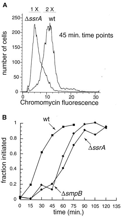
As described above, the G1-to-S transition normally coincides with swarmer-to-stalked cell differentiation in Caulobacter. To determine if a lack of SsrA causes a delay in the initiation of DNA replication as well as in the swarmer-to-stalked cell transition, the timing of initiation of DNA replication was determined by using a flow cytometry-based assay (Table 1; Fig. 3). In this assay, the percentage of cells that have initiated replication at a given time is determined by adding an inhibitor of replication initiation to samples of a synchronous culture, incubating the samples to allow chromosomes that have already initiated replication to complete the process, and measuring the total DNA content in each cell by flow cytometry (38). Cells that had initiated replication when the inhibitor was added will have a DNA content equivalent to two chromosomes, whereas cells that had not initiated replication will have a DNA content equivalent to one chromosome. In M2G, the ΔssrA strain initiated replication 41 min later than the wild type (63 ± 3 min versus 22 ± 3 min). This timing correlates with the ∼45-min delay in the swarmer-to-stalked cell transition. To estimate the length of S phase, flow cytometry was used to monitor the increase in DNA content in a synchronized culture through the cell cycle in the absence of inhibitors. Once replication initiated in the ΔssrA strain, the time required to complete S phase was indistinguishable from that for the wild type (Table 1). Thus, the timing of initiation of DNA replication is 41 min later in the ΔssrA strain, but the S phase is the same length.
FIG. 3.
Replication initiation is delayed in SsrA-deficient strains. (A) Sample histogram showing the numbers of cells that had initiated replication for the wild-type (wt) and the ΔssrA strain at 45 min after synchronization. The regions of the histogram corresponding to one chromosome (1 X) and two chromosomes (2 X) are indicated at the top. (B) Histograms such as the one in panel A were used to determine the fraction of cells that had initiated replication at each time point in synchronized cultures of wild-type, ΔssrA, and ΔsmpB strains.
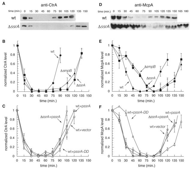
A likely cause of delay in the G1-to-S transition is a change in the timing of proteolysis of the CtrA response regulator. CtrA binds to five sites in the origin of DNA replication and represses initiation of replication in the swarmer cell (26), and CtrA is normally cleared from the cell immediately before initiation of replication (6, 27). To test whether the absence of SsrA RNA affects the expression of CtrA, cell cycle regulation of CtrA levels in synchronized cultures of the ΔssrA strain and the wild type was assayed by Western blotting (Fig. 4A and B). In both strains the amount of CtrA decreased 15 min after synchronization, and CtrA was no longer detected by 30 min. For the wild type, this decrease occurred during the swarmer-to-stalked cell transition, correlating with the timing of initiation of replication at 22 min, and this timing is in accord with previous observations (6, 27). In the ΔssrA strain, CtrA was eliminated at the same time with respect to synchronization as in the wild type, but this timing was more than 40 min before replication was initiated at the swarmer-to-stalked cell transition. These results demonstrate that SsrA RNA is not required for degradation of CtrA. In addition, they show that the degradation of CtrA is not sufficient for initiation of replication and that there must be an SsrA-dependent process that is required for proper timing of replication initiation.
FIG. 4.
Expression of cell cycle-regulated proteins in mutant strains. Western blots of lysates from synchronized cultures of the wild type (wt) and ΔssrA strains were probed with antibodies for CtrA (A) and McpA (D). Blots such as these for wild-type, ΔssrA, and ΔsmpB strains (B and E) and plasmid-bearing strains (C and F) were quantified, normalized to the initial time point, and plotted to determine how levels change with respect to the cell cycle. Each curve represents average results of at least three synchronization experiments, and each error bar represents 1 standard deviation.
Deletion of ssrA does not alter the relative timing of events after replication initiation.
We assayed the timing of events after the initiation of replication to determine if the absence of SsrA affects other portions of the cell cycle. In wild-type cells, CtrA is synthesized in the stalked cell after replication initiation, thereby preventing inappropriate reinitiation events (27). CtrA was synthesized at the same point in S phase in both the ΔssrA strain and the wild type, even though this point was delayed by 45 min in the ΔssrA strain relative to its timing in the wild type (Fig. 4A and B). Therefore, in strains lacking SsrA RNA, there is no disruption in the timing of synthesis of CtrA with respect to the cell cycle.
Another event in the swarmer-to-stalked cell transition is the removal of proteins involved in chemotaxis (2). The effect of SsrA RNA on this process was investigated by assaying expression of McpA, a methyl-accepting chemotaxis protein. Like CtrA, McpA is normally degraded during the swarmer-to-stalked cell transition and accumulates in the predivisional stage due to temporally controlled transcription. In the ΔssrA strain, McpA degradation was delayed by 30 min relative to its timing in the wild type, consistent with the extended swarmer cell stage (Fig. 4D and E). Accumulation in the predivisional cell was delayed by 45 min in the ΔssrA strain, occurring at the same time as in the wild type with respect to S phase. The delay in McpA degradation indicates that a lack of SsrA RNA does not have a general effect on the turnover of proteins in the swarmer-to-stalked cell transition: CtrA is degraded at the same time as in the wild type, but McpA is not. Therefore, the absence of SsrA RNA causes a delay in the cell cycle at a time after the proteolysis of CtrA but before the initiation of DNA replication and the proteolysis of McpA.
Complementation of the ssrA deletion phenotype.
To ensure that the phenotypes observed for the ΔssrA strain are due to the lack of SsrA RNA and not to a secondary mutation, we determined if the phenotype could be complemented by a multicopy plasmid bearing the ssrA gene under the control of its own promoter (pssrA). There is a small cost to the cell for maintaining a plasmid and growing under antibiotic selection, so the ΔssrA strain with pssrA was compared to the wild type with pssrA and to the wild type with vector alone. The ΔssrA strain bearing pssrA expressed mature SsrA RNA at a twofold-higher steady-state level than the wild type with vector alone, but at a lower level than the wild type with pssrA (Fig. 2, lanes 4 to 6). There were no significant differences among these three strains in the growth rate, length of the swarmer stage, timing of initiation of DNA replication (Table 1), or degradation and expression of CtrA and McpA (Fig. 4C and F). Therefore, the ΔssrA phenotype is complemented by the ssrA gene in trans, and the ΔssrA phenotype is due to the absence of SsrA and not to a secondary mutation.
In E. coli, SsrA mutants have growth defects under conditions of nutrient deprivation (25). To ensure that the cell cycle defects of the ΔssrA strain observed in minimal medium are not the result of limiting nutrients, all the assays described above were repeated in a rich medium (PYE). In PYE, the ΔssrA strain showed slower growth than the wild type (doubling time, 127 ± 5 min versus 102 ± 4 min) and a 45-min delay in the swarmer-to-stalked cell transition relative to the timing of the wild type (60 versus 15 min [Table 1]). There was also a 44-min delay in the initiation of DNA replication (49 ± 8 min versus 5 ± 3 min [Table 1]). These results indicate that SsrA RNA is required for correct timing of the swarmer-to-stalked cell transition in rich medium as well as minimal medium; thus, the requirement for SsrA RNA is not due to nutrient deprivation.
A deletion in smpB causes the SsrA− phenotype.
SmpB is an SsrA RNA-binding protein known to be required for SsrA activity in E. coli (20) and B. subtilis (36). The Caulobacter homologue of SmpB was identified by similarity to the E. coli protein; Caulobacter SmpB is 52% identical and 67% similar to E. coli SmpB. In E. coli, smpB is found immediately upstream of ssrA, but the Caulobacter smpB gene is more than 956 kb from ssrA. To determine if SmpB, like SsrA RNA, is required for normal timing of the G1-to-S transition in Caulobacter, a deletion in the smpB gene was constructed and the ΔsmpB strain was analyzed. The ΔsmpB strain had a delay in the swarmer-to-stalked cell transition which was identical to that of the ΔssrA strain. The growth rate in log-phase and synchronous cultures (Table 1), the delay in initiation of DNA replication (Fig. 3B), and the times of degradation and expression of CtrA and McpA (Fig. 4B and E) were indistinguishable from those of the ΔssrA strain, demonstrating that SmpB is also required for control of the G1-to-S transition in Caulobacter. In addition, since the SsrA− phenotype resulted from a mutation in the smpB gene, which is distant on the chromosome from ssrA, these results confirm that the phenotype observed for the ΔssrA strain was due to a lack of SsrA activity and not to a linked suppressor mutation.
The smpB deletion also caused a 10-fold decrease in the steady-state level of mature SsrA RNA (Fig. 2, lanes 1 and 3). This depletion of mature SsrA RNA indicates that SmpB is required for the transcription, processing, or stability of SsrA RNA. It is not yet clear if SmpB acts only on the steady-state level of SsrA RNA in Caulobacter or if it is also required for other steps in SsrA activity such as charging with alanine or association with ribosomes.
A proteolysis-resistant SsrA tag induces the SsrA− phenotype.
Why is SsrA activity required for control of the G1-to-S transition? One possibility is that SsrA-mediated proteolysis is required to rapidly eliminate certain proteins that are being translated in G1 phase, thereby enabling the G1-to-S transition. To test whether proteolysis of SsrA-tagged substrates is important for the G1-to-S transition, we used a variant of SsrA that can release stalled ribosomes and tag the nascent proteins but does not target these tagged proteins for degradation. In this variant, SsrA-DD, the last two codons of the tag reading frame have been changed from Ala-Ala to Asp-Asp. SsrA-DD tags proteins in Caulobacter (21), but the half-life of these tagged proteins is >120 min, compared to <3 min for proteins tagged by wild-type SsrA (data not shown). If proteolysis of SsrA-tagged substrates is essential for control of the cell cycle by SsrA, then SsrA-DD should not complement the ΔssrA phenotype. Conversely, if proteolysis is not essential and SsrA relies on a different aspect of its activity, such as release of stalled ribosomes, then SsrA-DD should complement the SsrA− phenotype. Accordingly, ΔssrA cells were transformed with a multicopy plasmid (pssrA-DD) bearing the SsrA-DD variant under the control of the ssrA promoter. The ΔssrA strain bearing pssrA-DD had a phenotype that was more severe than either that of the ΔssrA strain with no plasmid or that of wild-type cells bearing pssrA-DD. The growth of the ΔssrA strain bearing pssrA-DD was significantly slower than that of any of the other strains described here (Table 1). Although swarmer cells from the ΔssrA pssrA-DD strain could be isolated, they remained as swarmer cells so long that they did not pass synchronously through the cell cycle. These defects were not due to a lack of SsrA RNA, because the ΔssrA pssrA-DD strain had the same steady-state levels of mature SsrA RNA as the wild type (Fig. 2, lanes 1 and 8). Thus, SsrA-DD does not complement the SsrA− phenotype, indicating that tagging with the wild-type SsrA peptide is required for control of the G1-to-S transition.
In wild-type Caulobacter, pssrA-DD induces a phenotype similar to, but weaker than, that induced by a deletion of ssrA. The wild-type strain bearing pssrA-DD grew more slowly than isogenic strains bearing the vector with no insert or pssrA (Table 1), and this slower growth was due to a delay in the swarmer-to-stalked cell transition. The cell cycle was 15 min longer in the wild type with pssrA-DD than in the wild type with vector alone, and this difference was due entirely to a longer swarmer cell stage (Table 1). The wild-type strain with pssrA-DD also had a disruption of the cell cycle. In synchronized cultures of the wild type with pssrA-DD, initiation of DNA replication occurred 14 min later than for the wild type with vector alone (Table 1). Although this delay was not as long as that for the ΔssrA strain, the timing of CtrA degradation was similarly uncoupled from the initiation of DNA replication. In the wild type bearing pssrA-DD, CtrA was degraded 14 min before the initiation of replication (Fig. 4C). As in the ΔssrA strain, in the wild type with pssrA-DD the synthesis of CtrA, the degradation of McpA, and the synthesis of McpA occurred at the same time with respect to S phase, even though this timing was delayed by 14 min relative to the timing in the wild type with vector alone (Fig. 4C and F). The SsrA-DD phenotype was not due to a failure to express SsrA RNA, because the wild-type strain with pssrA-DD had the same steady-state level of SsrA RNA as the wild type with no plasmid or with vector alone (Fig. 2, lanes 1, 4, 7). Thus, the presence of SsrA-DD caused a delay in the G1-to-S transition similar to that caused by a deletion of ssrA, although the delay was not as long. These defects were observed even though there was a wild-type copy of SsrA on the chromosome. This dominant-negative phenotype indicates that inhibiting the degradation of SsrA-tagged substrates has an effect similar to that of a complete loss of SsrA activity.
DISCUSSION
The data presented here show that SsrA activity is required for control of the G1-to-S transition in Caulobacter. Lack of SsrA RNA or SmpB protein causes a specific delay in the G1-to-S transition, between the degradation of CtrA and the initiation of replication. Absence of SsrA activity does not cause a general growth defect, since there is little change in the relative timing of cell cycle-regulated events after replication initiation. The uncoupling of the timing of CtrA proteolysis from initiation of replication in strains lacking SsrA activity demonstrates that removal of CtrA is not sufficient to trigger replication initiation. Inactivation of CtrA, by either degradation or dephosphorylation, is necessary for initiation of replication, and normally replication initiates immediately after CtrA is removed (6, 26, 27). However, the ∼40-min delay between CtrA degradation and replication initiation in the ΔssrA and ΔsmpB strains indicates that replication does not necessarily initiate as soon as CtrA is removed. In addition to the inactivation of CtrA, there must be an SsrA-dependent pathway for regulation of the G1-to-S transition.
How does SsrA activity affect the timing of the G1-to-S transition? SsrA activity could be acting positively to promote replication initiation, or SsrA activity could be needed to pass a checkpoint required for the G1-to-S transition. If SsrA RNA is acting directly on DNA replication, it could affect an unidentified initiation factor or any of the events known to be required for initiation of replication in Caulobacter other than the inactivation of CtrA, including synthesis of the DnaA activator protein (13); transcription from Cori Ps, a strong promoter within the origin of replication; and assembly and activity of DNA polymerase (24). It is also possible that SsrA does not act directly on an initiation factor, but that absence of SsrA activity results in stress that stalls the cell cycle specifically at the G1-to-S transition. Since the SsrA-DD variant does not complement the ssrA deletion phenotype, this stress cannot be due to the accumulation of stalled ribosomes. However, because SsrA RNA targets incomplete proteins made from truncated mRNAs for proteolysis, the absence of SsrA activity might result in an accumulation of these incomplete proteins and stress the cell. This stress would have to affect only the G1-to-S transition, because the absence of SsrA activity does not affect growth throughout the remainder of the cell cycle. If the requirement for SsrA activity is due to such an indirect effect, there must be an unknown checkpoint to sense this stress and delay initiation of replication until the stress has been resolved.
There is increasing evidence that SsrA plays a regulatory role in the cell. Proteomic studies have shown that many proteins, including several transcription factors, are preferentially tagged by SsrA (1, 28, 30). The phenotypes of ssrA mutations in a number of species are consistent with a regulatory role: SsrA RNA is required for invasion of macrophages by Salmonella enterica serovar Typhimurium (5), for symbiotic growth in root nodules by Bradyrhizobium japonicum (7), and for development of some variants of phages λ and Mu (20, 29). Whether SsrA is acting directly on initiation of replication in Caulobacter or whether there is a checkpoint that requires SsrA activity, there is clearly an unknown pathway for regulating the G1-to-S transition that will be clarified by further analysis of DNA replication in SsrA-deficient strains and identification of substrates for SsrA activity.
Acknowledgments
We thank Sarah Ades for critical reading of the manuscript.
This work was supported by National Institutes of Health grants GM32506/5120 M2 and GM51426. K.C.K. was supported in part by a DOE-Energy Biosciences Research Fellowship of the Life Sciences Research Foundation.
REFERENCES
- 1.Abo, T., T. Inada, K. Ogawa, and H. Aiba. 2000. SsrA-mediated tagging and proteolysis of LacI and its role in the regulation of lac operon. EMBO J. 19:3762-3769. [DOI] [PMC free article] [PubMed] [Google Scholar]
- 2.Alley, M. R. K., J. R. Maddock, and L. Shapiro. 1993. Requirement of the carboxyl terminus of the bacterial chemoreceptor for its targeted proteolysis. Science 259:1754-1757. [DOI] [PubMed] [Google Scholar]
- 3.Barends, S., A. W. Karzai, R. T. Sauer, J. Wower, and B. Kraal. 2001. Simultaneous and functional binding of SmpB and EF-Tu-TP to the alanyl acceptor arm of tmRNA. J. Mol. Biol. 314:9-21. [DOI] [PubMed] [Google Scholar]
- 4.Barends, S., J. Wower, and B. Kraal. 2000. Kinetic parameters for tmRNA binding to alanyl-tRNA synthetase and elongation factor Tu from Escherichia coli. Biochemistry 39:2652-2658. [DOI] [PubMed] [Google Scholar]
- 5.Baumler, A. J., J. G. Kusters, I. Stojiljkovic, and F. Heffron. 1994. Salmonella typhimurium loci involved in survival within macrophages. Infect. Immun. 62:1623-1630. [DOI] [PMC free article] [PubMed] [Google Scholar]
- 6.Domian, I. J., K. C. Quon, and L. Shapiro. 1997. Cell type-specific phosphorylation and proteolysis of a transcriptional regulator controls the G1-to-S transition in a bacterial cell cycle. Cell 90:415-424. [DOI] [PubMed] [Google Scholar]
- 7.Ebeling, S., C. Kundig, and H. Hennecke. 1991. Discovery of a rhizobial RNA that is essential for symbiotic root nodule development. J. Bacteriol. 173:6373-6382. [DOI] [PMC free article] [PubMed] [Google Scholar]
- 8.Ely, B. 1991. Genetics of Caulobacter crescentus. Methods Enzymol. 204:372-384. [DOI] [PubMed] [Google Scholar]
- 9.Evinger, M., and N. Agabian. 1977. Envelope-associated nucleoid from Caulobacter crescentus stalked and swarmer cells. J. Bacteriol. 132:294-301. [DOI] [PMC free article] [PubMed] [Google Scholar]
- 10.Felden, B., R. F. Gesteland, and J. F. Atkins. 1999. Eubacterial tmRNAs: everywhere except the alpha-proteobacteria? Biochim. Biophys. Acta 1446:145-148. [DOI] [PubMed] [Google Scholar]
- 11.Flynn, J. M., I. Levchenko, M. Seidel, S. H. Wickner, R. T. Sauer, and T. A. Baker. 2001. Overlapping recognition determinants within the ssrA degradation tag allow modulation of proteolysis. Proc. Natl. Acad. Sci. USA 98:10584-10589. [DOI] [PMC free article] [PubMed] [Google Scholar]
- 12.Gay, P., D. Le Coq, M. Steinmetz, T. Berkelman, and C. I. Kado. 1985. Positive selection procedure for entrapment of insertion sequence elements in gram-negative bacteria. J. Bacteriol. 164:918-921. [DOI] [PMC free article] [PubMed] [Google Scholar]
- 13.Gorbatyuk, B., and G. T. Marczynski. 2001. Physiological consequences of blocked Caulobacter crescentus dnaA expression, an essential DNA replication gene. Mol. Microbiol. 40:485-497. [DOI] [PubMed] [Google Scholar]
- 14.Gottesman, S., E. Roche, Y. Zhou, and R. T. Sauer. 1998. The ClpXP and ClpAP proteases degrade proteins with carboxy-terminal peptide tails added by the SsrA-tagging system. Genes Dev. 12:1338-1347. [DOI] [PMC free article] [PubMed] [Google Scholar]
- 15.Hanawa-Suetsugu, K., M. Takagi, H. Inokuchi, H. Himeno, and A. Muto. 2002. SmpB functions in various steps of trans-translation. Nucleic Acids Res. 30:1620-1629. [DOI] [PMC free article] [PubMed] [Google Scholar]
- 16.Hayes, C. S., B. Bose, and R. T. Sauer. 2002. Proline residues at the C-terminus of nascent chains induce SsrA-tagging during translation termination. J. Biol. Chem. 277:33825-33832. [DOI] [PubMed] [Google Scholar]
- 17.Herman, C., D. Thevenet, P. Bouloc, G. C. Walker, and R. D'Ari. 1998. Degradation of carboxy-terminal-tagged cytoplasmic proteins by the Escherichia coli protease HflB (FtsH). Genes Dev. 12:1348-1355. [DOI] [PMC free article] [PubMed] [Google Scholar]
- 18.Hung, D., H. McAdams, and L. Shapiro. 1999. Regulation of the Caulobacter cell cycle, p. 361-378. In Y. V. Brun and L. J. Shimkets (ed.), Prokaryotic development. ASM Press, Washington, D.C.
- 19.Karzai, A. W., E. D. Roche, and R. T. Sauer. 2000. The SsrA-SmpB system for protein tagging, directed degradation, and ribosome rescue. Nat. Struct. Biol. 7:449-455. [DOI] [PubMed] [Google Scholar]
- 20.Karzai, A. W., M. M. Susskind, and R. T. Sauer. 1999. SmpB, a unique RNA-binding protein essential for the peptide-tagging activity of SsrA (tmRNA). EMBO J. 18:3793-3799. [DOI] [PMC free article] [PubMed] [Google Scholar]
- 21.Keiler, K. C., L. Shapiro, and K. P. Williams. 2000. tmRNAs that encode proteolysis-inducing tags are found in all known bacterial genomes: a two-piece tmRNA functions in Caulobacter. Proc. Natl. Acad. Sci. USA 97:7778-7783. [DOI] [PMC free article] [PubMed] [Google Scholar]
- 22.Keiler, K. C., P. R. Waller, and R. T. Sauer. 1996. Role of a peptide tagging system in degradation of proteins synthesized from damaged messenger RNA. Science 271:990-993. [DOI] [PubMed] [Google Scholar]
- 23.Komine, Y., M. Kitabatake, T. Yokogawa, K. Nishikawa, and H. Inokuchi. 1994. A tRNA-like structure is present in 10Sa RNA, a small stable RNA from Escherichia coli. Proc. Natl. Acad. Sci. USA 91:9223-9227. [DOI] [PMC free article] [PubMed] [Google Scholar]
- 24.Marczynski, G. T., K. Lentine, and L. Shapiro. 1995. A developmentally regulated chromosomal origin of replication uses essential transcription elements. Genes Dev. 9:1543-1557. [DOI] [PubMed] [Google Scholar]
- 25.Oh, B. K., and D. Apirion. 1991. 10Sa RNA, a small stable RNA of Escherichia coli, is functional. Mol. Gen. Genet. 229:52-56. [DOI] [PubMed] [Google Scholar]
- 26.Quon, K. C., G. T. Marczynski, and L. Shapiro. 1996. Cell cycle control by an essential bacterial two-component signal transduction protein. Cell 84:83-93. [DOI] [PubMed] [Google Scholar]
- 27.Quon, K. C., B. Yang, I. J. Domian, L. Shapiro, and G. T. Marczynski. 1998. Negative control of bacterial DNA replication by a cell cycle regulatory protein that binds at the chromosome origin. Proc. Natl. Acad. Sci. USA 95:120-125. [DOI] [PMC free article] [PubMed] [Google Scholar]
- 28.Ranquet, C., J. Geiselmann, and A. Toussaint. 2001. The tRNA function of SsrA contributes to controlling repression of bacteriophage Mu prophage. Proc. Natl. Acad. Sci. USA 98:10220-10225. [DOI] [PMC free article] [PubMed] [Google Scholar]
- 29.Retallack, D. M., L. L. Johnson, and D. I. Friedman. 1994. Role for 10Sa RNA in the growth of lambda-P22 hybrid phage. J. Bacteriol. 176:2082-2089. [DOI] [PMC free article] [PubMed] [Google Scholar]
- 30.Roche, E. D., and R. T. Sauer. 2001. Identification of endogenous SsrA-tagged proteins reveals tagging at positions corresponding to stop codons. J. Biol. Chem. 276:28509-28515. [DOI] [PubMed] [Google Scholar]
- 31.Roche, E. D., and R. T. Sauer. 1999. SsrA-mediated peptide tagging caused by rare codons and tRNA scarcity. EMBO J. 18:4579-4589. [DOI] [PMC free article] [PubMed] [Google Scholar]
- 32.Rudinger-Thirion, J., R. Giege, and B. Felden. 1999. Aminoacylated tmRNA from Escherichia coli interacts with prokaryotic elongation factor Tu. RNA 5:989-992. [DOI] [PMC free article] [PubMed] [Google Scholar]
- 33.Sambrook, J., E. F. Fritsch, and T. Maniatis. 1989. Molecular cloning: a laboratory manual, 2nd ed. Cold Spring Harbor Laboratory Press, Cold Spring Harbor, N.Y.
- 34.Tu, G. F., G. E. Reid, J. G. Zhang, R. L. Moritz, and R. J. Simpson. 1995. C-terminal extension of truncated recombinant proteins in Escherichia coli with a 10Sa RNA decapeptide. J. Biol. Chem. 270:9322-9326. [DOI] [PubMed] [Google Scholar]
- 35.Ushida, C., H. Himeno, T. Watanabe, and A. Muto. 1994. tRNA-like structures in 10Sa RNAs of Mycoplasma capricolum and Bacillus subtilis. Nucleic Acids Res. 22:3392-3396. [DOI] [PMC free article] [PubMed] [Google Scholar]
- 36.Wiegert, T., and W. Schumann. 2001. SsrA-mediated tagging in Bacillus subtilis. J. Bacteriol. 183:3885-3889. [DOI] [PMC free article] [PubMed] [Google Scholar]
- 37.Williams, K. P. 2002. The tmRNA Website: invasion by an intron. Nucleic Acids Res. 30:179-182. [DOI] [PMC free article] [PubMed] [Google Scholar]
- 38.Winzeler, E., and L. Shapiro. 1995. Use of flow cytometry to identify a Caulobacter 4.5S RNA temperature-sensitive mutant defective in the cell cycle. J. Mol. Biol. 251:346-365. [DOI] [PubMed] [Google Scholar]
- 39.Withey, J., and D. Friedman. 1999. Analysis of the role of trans-translation in the requirement of tmRNA forλimmP22 growth in Escherichia coli. J. Bacteriol. 181:2148-2157. [DOI] [PMC free article] [PubMed] [Google Scholar]
- 40.Wower, I. K., C. W. Zwieb, S. A. Guven, and J. Wower. 2000. Binding and cross-linking of tmRNA to ribosomal protein S1, on and off the Escherichia coli ribosome. EMBO J. 19:6612-6621. [DOI] [PMC free article] [PubMed] [Google Scholar]


