Abstract
A negative control mutant of the nitrogen assimilation control protein, NAC, has been isolated. Mutants with the leucine at position 111 changed to a nonhydrophobic residue activate transcription from hut and ure promoters, but fail to repress gdhA expression. This failure does not result from failure to bind to either of the two sites required for gdhA repression, but the binding at those sites is altered in the mutant. It appears that the NAC negative control mutants fail to form the complex structures (probably tetramers) formed by wild-type NAC at the gdhA promoter.
The nitrogen assimilation control protein (NAC) of Klebsiella aerogenes and Escherichia coli is a transcriptional regulator that serves as both an activator and repressor and belongs to the LysR family of regulators (19). A member of the nitrogen regulatory (Ntr) regulon, NAC serves to couple the regulation of σ70-dependent genes and operons to the σ54-dependent Ntr system (1). NAC activates operons that catabolize histidine (hut), urea (ure), proline (put), alanine (dad), and cytosine (cod) (11, 13, 16) and represses operons involved in the assimilation of ammonia into glutamate (gdhA and gltBD) (13). In addition, NAC negatively regulates its own expression (7).
The mechanisms by which NAC can activate or repress transcription are also diverse. NAC can activate transcription from a variety of positions upstream of RNA polymerase (RNAP) (17), perhaps via distinct contacts with the various subunits of RNAP, as has been shown with other activator proteins, including the LysR family members CatR, TrpI, and OxyR (6, 10, 21). The consensus sequence of the sites where NAC activates transcription is ATA-N6-TNGTAT, and the site can be divided into two functional half-sites—a promoter-distal site involved in the binding of NAC to the DNA and a promoter-proximal site. The promoter-proximal site is important for binding, but also contains information necessary for NAC to be proficient at activating transcription, presumably by inducing a conformational change in the protein (18).
The consensus sequence of the sites at which NAC represses transcription is slightly different, ATAA-N8-GAT. When one of these sites was substituted for an activation site, DNA-binding activity was retained, but NAC bound at this site failed to activate transcription (18). Repression by NAC falls into distinct categories. Repression at nac requires the binding of NAC in a single region upstream from the start of transcription and is thought to act by interfering with the necessary interactions between σ54-RNAP and its enhancer-bound activator, NtrC (7). In contrast, repression at gdhA requires two well-separated NAC-binding sites, one upstream and the other downstream from the start of transcription (8). This arrangement of binding sites suggests a mechanism of repression by which NAC molecules bound at the two distinct sites interact.
Positive control (PC) mutants of transcriptional regulators have been isolated for a variety of activators, most notably the catabolite activator protein (4). These mutants retain the ability to bind to DNA, yet fail to activate transcription, and the mutations typically map to portions of the protein that normally make contacts with RNAP (3, 9). A similar class of mutant could exist, negative control (NC) mutants, which would retain all normal functions of the protein but lose the ability to repress transcription. In the case of NAC, two classes of NC mutants could be predicted: those that fail to recognize the slightly different binding site found at operons where transcription is repressed, and those that still bind to these sites normally yet still fail to repress transcription. For the first class, the ability of NAC to activate transcription should be unaffected. For the second class, this ability should only be unaffected if the mechanism for repression can be separated from the mechanism of activation. Here we describe the isolation and characterization of an NC mutant of NAC (NACNC) that belongs to the second class.
NC mutants of NAC are defined here as those mutants that retained the ability to activate transcription at ure and hut, but failed to repress transcription at gdhA. The isolation of such NC mutants involved first the generation of a collection of mutants of NAC that failed to repress gdhA and the subsequent screening of these mutants for the subset that retained the ability to activate transcription. In strains lacking glutamate synthase (GOGAT) activity (gltB or gltD), net synthesis of glutamate requires a high level of expression of glutamate dehydrogenase (GDH) (2). Introduction of plasmid pCB1051, which expresses nac constitutively from the lacZ promoter, into strain K. aerogenes KC4728 (nac-204 gltD702) resulted in severe repression of GDH and a consequent inability to grow on minimal medium unless glutamate was added. In addition to this growth phenotype, this strain exhibited NAC-activated levels of histidase and urease and repressed levels of GDH, whereas KC4728 containing only vector plasmid did not (data not shown, but similar to the wild-type or no-NAC values shown in Table 1).
TABLE 1.
Characterization of amino acid substitutions at position L111
| Substitutiona | Sp act (nmol of product formed/min/mg of protein)b
|
||
|---|---|---|---|
| Urease | Histidase | GDH | |
| No NAC | 17 | 45 | 875 |
| Wild type | 857 | 326 | 28 |
| L111P | 597 | 209 | 335 |
| L111K | 1,080 | 239 | 431 |
| L111R | 959 | 219 | 330 |
| L111Q | 1,326 | 265 | 365 |
| L111T | 1,052 | 211 | 253 |
| L111D | 748 | 233 | 282 |
| L111E | 810 | 253 | 262 |
| L111N | 825 | 233 | 215 |
| L111G | 778 | 237 | 170 |
| L111S | 745 | 233 | 163 |
| L111A | 806 | 222 | 73 |
| L111H | 756 | 250 | 90 |
| L111C | 708 | 305 | 31 |
| L111I | 735 | 409 | 28 |
| L111M | 762 | 294 | 31 |
| L111V | 722 | 302 | 31 |
| L111F | 856 | 343 | 18 |
| L111W | 563 | 268 | 37 |
| L111Y | 17 | 60 | 695 |
All mutants were carried as cloned fragments in pCB1041. No NAC, pCB1041; Wild type, pCB1051. The background strain was KC4598 (hutC515 nac-204::λplac Mu53 srl-7012::Tn5-131).
Cells were grown in W4 minimal salts (18) supplemented with glucose (0.4%), ammonium sulfate (0.2%), glutamine (0.2%), 2 μM nickel sulfate, and 100 μg of ampicillin per ml. Assay values are reported as specific activities and are the mean of at least three independent experiments in which the standard error was <15% of the mean.
Plasmid pCB1051 was mutagenized by passage through the mutator strain XL-1 Red (Stratagene) and introduced into strain KC4728 (14). Transformants were selected and purified on plates containing glucose, ammonia, and ampicillin (GNamp), but no glutamate. Liquid cultures of each colony were grown in GNamp medium supplemented with 2 μM Ni2SO4 and assayed for urease activity. A total of 271 of the 1,052 colonies tested contained high levels of urease and were characterized further. There is a hierarchy of sensitivities among the targets for NAC based on the amount of NAC present: hut requires the greatest amount of NAC to elicit a response, gdhA requires less NAC, and ure requires the smallest amount of NAC to be fully activated by NAC (20). Thus, levels of histidase were measured (13) to distinguish mutants that had reduced expression or stability from those with a defect specific to gdhA. Plasmid DNA from potential NC mutants (those that grew on minimal medium and retained high levels of urease and histidase) was isolated and reintroduced into strain KC4728. Urease, histidase, and GDH levels were determined on these secondary transformants as described above. The plasmid DNA was also isolated by Qiagen miniprep kits, and the DNA sequence was determined at the University of Michigan DNA Sequencing Core Facility.
Three classes of mutants were isolated from this procedure. The majority of the mutants tested were either null mutants of nac (these failed to repress gdhA and failed to activate ure) or “reduced expression” mutants of nac (those that showed a reduced ability to repress gdhA and activate ure, and failed to activate hut). A few mutants fit the criteria for NC mutants of NAC. They were impaired in their ability to repress gdhA, yet maintained the ability to activate both ure and hut. These were all isolated from the same pool of mutagenized DNA, so they were not necessarily independent. Each of the five nac mutants contained more than one mutation, but each of them contained a T-to-C change at position 331 of the nac coding sequence, which resulted in a leucine-to-proline change in codon 111 (L111P).
By using GeneEditor site-directed mutagenesis (Promega), we constructed the 19 substitutions at codon 111, and the effects of these mutations on the expression of ure, hut, and gdhA were measured (Table 1). With one exception (L111Y was a null mutant), the collection fell into two categories: wild type (GDH activity <150 nmol of product formed per min per mg of protein) and NC (GDH activity >100 nmol of product formed per min per mg of protein). The change of leucine to another hydrophobic residue or to cysteine or histidine resulted in a wild-type NAC phenotype. The change of leucine to a charged or polar residue or to proline or glycine resulted in a NACNC phenotype.
Of all the substitutions at position 111, NACL111K had the clearest NC phenotype, with strong activation of both urease and histidase and only the two- to threefold repression of GDH that is discussed below. Therefore, NACL111K was used to characterize the NC phenotype further. Plasmids pCB1289 and pCB1305 carry NACWT and NACL111K, respectively, expression of which is under the control of the native nac promoter. Thus, formation of NACWT or NACL111K is controlled by the Ntr system in response to the quality of the nitrogen source in the medium. The two plasmids were introduced into strain KC4727 (nac-204), and the specific activities of several NAC-regulated targets were analyzed under nitrogen-excess and nitrogen-limiting conditions (Table 2). Under conditions of nitrogen limitation, where nac is expressed, NACL111K activated urease and histidase expression, but repressed GDH only threefold.
TABLE 2.
NACWT and NACNC activities at various NAC-dependent targets
| Plasmida | Sp act (nmol of product formed/min/mg of protein)b
|
|||||||||
|---|---|---|---|---|---|---|---|---|---|---|
| Urease
|
Histidase
|
GDH
|
GOGAT
|
β-Galacto- sidase
|
||||||
| +N | −N | +N | −N | +N | −N | +N | −N | +N | −N | |
| None | 23 | 167 | 46 | 45 | 916 | 980 | 112 | 207 | 7 | 9,890 |
| pCB1289 | 19 | 1,070 | 33 | 533 | 863 | 42 | 102 | 36 | 8 | 2,160 |
| pCB1305 | 35 | 1,680 | 35 | 370 | 942 | 309 | 147 | 108 | 6 | 2,740 |
The strain background was KC4727 (hutC515, Δ[bla-2], nac-204::λplacMu53). pCB1289 contains wild-type nac (expression from the native nac promoter), and pCB1305 is identical, except that it contains the NACNC mutant NACL111K.
Each value is the average of at least three separate experiments with a standard error of ≤15% of the mean. The media were W4 minimal salts (18) supplemented with 0.4% glucose, 0.2% ammonium sulfate, and 0.2% glutamine (+N) or glucose and 0.04% glutamine (−N). (In addition, ampicillin was present at 100 μg/ml when a plasmid was present in the strain.) The β-galactosidase values reflect the expression of the chromosomal nac promoter.
NAC also repressed its own promoter, as demonstrated by monitoring the expression of β-galactosidase from the nac-204::λplacMu53 allele (in which the nac promoter is fused to lacZ). NACWT repressed the nac promoter 4.6-fold, and NACL111K repressed it nearly as well, 3.6-fold. Thus NACL111K appeared to retain the ability to repress transcription at the nac promoter.
The effect at GOGAT was intermediate. Comparing nitrogen-rich and nitrogen-limiting conditions, NACWT repressed GOGAT formation only 3-fold, so it is unclear whether the reduced ability of NACL111K to repress gltBD transcription (1.4-fold) more resembles the slight loss of activation seen at hut or the more severe loss of repression seen at gdhA. Comparing the mutant to the wild type under nitrogen-limited conditions only, a greater effect can be seen: NACWT repressed GOGAT formation nearly sixfold when compared to the null mutant. In contrast, NACL111K repressed GOGAT formation only 1.9-fold under these conditions, suggesting that the expression of gltBD may also be impaired in a manner similar to that of gdhA. Two NAC-binding site motifs are located in the promoter of gltBD (T. J. Goss and R. A. Bender, unpublished observation).
The threefold repression of gdhA by NACL111K is significant. The repression of gdhA expression by NAC requires two NAC-binding sites (8). If the upstream binding site is deleted, all NAC-mediated repression is abolished. If the downstream site is deleted, strong repression is lost, but a weak (approximately threefold) NAC-dependent repression remains. This repression appears to result from NAC's ability to compete with a lysine-sensitive positive effector needed for full gdhA expression (8). To test whether the residual repression of gdhA by NACL111K might be attributed to its interaction with the upstream binding site, we introduced plasmid-borne NACWT and NACL111K into strain KC4989 (nac-1). In this strain, a gdhA promoter that contains only the upstream NAC-binding site is fused to lacZ on a λ prophage. NACWT repressed β-galactosidase expression 1.9-fold, and NACL111K repressed expression to 1.7-fold (data not shown). Thus it would appear that NACL111K can bind to the upstream NAC-binding site in a manner analogous to that of the wild type.
We had no in vivo test for DNA binding at the downstream site at gdhA, so it remained a possibility that NACL111K failed to repress transcription because it failed to bind to the downstream site. Therefore, we purified the wild-type and mutant proteins and tested their abilities to bind DNA in vitro by using gel mobility shift assays. Both NACWT and NACL111K were cloned with six-histidine codons at the carboxy terminus and purified by nickel-affinity chromatography (Qiagen). DNA fragments end labeled with [32P]dATP were incubated with purified NACWT or NACL111K. When increasing concentrations of NACWT-his or NACL111K-his were incubated with DNA fragments containing either the hut promoter or the ure promoter, only one shifted band was seen, which was representative of one NAC dimer bound to the DNA (data not shown).
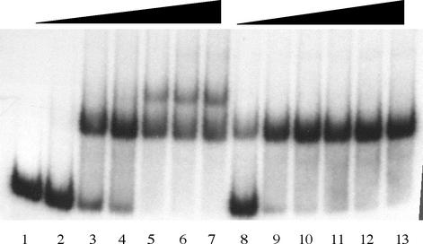
Figure 1 shows the interaction of NACL111K-his with DNA carrying the gdhA promoter region and the upstream NAC-binding site, but lacking the downstream NAC-binding site. With increasing concentrations of protein, NACWT-his was able to shift the DNA to two slower-migrating complexes (Fig. 1, lanes 2 to 7). NACL111K-his also bound to the upstream binding site (Fig. 1, lanes 8 to 13); however, gel shifts with NACL111K-his resulted in only one discrete band when comparably active protein concentrations were used.
FIG. 1.
Gel mobility shift assay of NACWT and NACNC at the upstream NAC-binding site from gdhA. Increasing amounts of purified NACWT-his (lanes 2 to 7) or NACL111K-his (lanes 8 to 13) were incubated with 128 nM DNA fragment containing the upstream NAC-binding site from gdhA. The NACWT-his and NACL111K-his preparations were found to be 24 and 4.2% active, respectively, in terms of DNA binding (specific activity). Concentrations are reported as the amount of active protein used in the assay. Lanes 1 to 7 contained 0, 25, 50, 75, 100, 125, and 150 nM active NACWT-his, respectively. Lanes 8 to 13 contained 25, 50, 75, 100, 125, and 150 nM NACL111K-his, respectively.
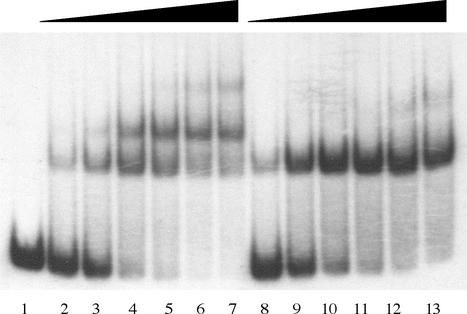
NACWT-his also bound to the DNA fragment carrying the downstream NAC-binding site, but lacking the upstream NAC-binding site, yielding multiple shifted bands. Two bands, resembling those seen with the upstream site alone, were formed with increasing concentrations of protein, and a third diffuse band was seen at very high concentrations of NAC (Fig. 2, lanes 2 to 7). NAC has a weak affinity for many sites in DNA, and we have detected four such sites in the cloning vector pUC19 that have no physiological significance (Goss and Bender, unpublished). NACL111K-his also bound to the downstream NAC-binding site and again formed only one distinctly shifted band with increasing concentrations of NACL111K-his (Fig. 2, lanes 8 to 13). The diffuse band that appeared at very high concentrations of NACL111K-his (Fig. 2, lane 13) did not align with any of the other slower-migrating bands formed by NACWT-his. Clearly NACL111K-his is able to bind to both the upstream and downstream binding sites, but fails to form the second shifted species seen with NACWT.
FIG. 2.
Gel mobility shift assay of NACWT and NACNC at the downstream NAC-binding site from gdhA. Increasing amounts of purified NACWT-his (lanes 2 to 7) or NACL111K-his (lanes 8 to 13) were incubated with 128 nM DNA fragment containing the downstream NAC-binding site from gdhA. Lanes 1 to 7 contained 0, 25, 50, 75, 100, 125, and 150 nM active NACWT-his, respectively. Lanes 8 to 13 contained 25, 50, 75, 100, 125, and 150 nM NACL111K-his, respectively.
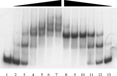
Figure 3 shows the gel mobility shift pattern when the DNA target contained the entire gdhA promoter containing both the upstream and downstream NAC-binding sites. NACWT-his exhibited a complex shifting pattern; at least five discretely shifted species were seen (Fig. 3, lanes 2 to 7). NACL111K-his gave quite a different shifting pattern, with only two discretely shifted bands (Fig. 3, lanes 8 to 13). These two shifted species most likely represent the target bound at one binding site (the initial shift) and then target bound at both sites (the second shift). A third, unidentified shift appeared at higher protein concentrations of NACL111K-his (Fig. 3, lane 8). It is unclear what structures the multiple bands represent when NACWT-his is bound to the gdhA promoter; however, it is clear that NACL111K does not form them.
FIG. 3.
Gel mobility shift assays of NACWT or NACNC with the full-length gdhA promoter. Increasing amounts of purified NACWT-his (lanes 2 to 7) or NACL111K-his (lanes 8 to 13) were incubated with 128 nM DNA fragment containing both NAC-binding sites. Lanes 1 to 7 contained 0, 25, 50, 75, 100, 125, and 150 nM active NACWT-his, respectively. Lanes 8 to 13 contained 150, 125, 100, 75, 50, and 25 nM NACL111K-his, respectively.
The isolation of the NACNC mutant, NACL111K, clearly demonstrates that the functions of activation at hut and ure and repression at gdhA are separable. We have reported previously that both NAC-binding sites in gdhA are required for repression and that an interaction between the two sites must occur for this repression (8). NACL111K further supports that protein interactions must occur for repression. We believe that the DNA binding region (a helix-turn-helix domain) and the dimerization domain are located at the N terminus of NAC, because NAC100, containing only the N-terminal 100 amino acids of NAC, exists as a dimer in solution and is able to activate transcription at hut (15). Therefore, it is not clear what the nature of the defect is that makes NACL111K unable to repress gdhA. We suspect that NACL111K is unable to form a tetramer. Although we show no direct evidence for the formation of NAC tetramers in this study, the gel shift patterns seen with isolated NAC binding sites from gdhA, and especially the complex gel shift patterns observed for wild-type NAC binding to the gdhA promoter, suggest that more NAC-DNA species form than simply the DNA bound with one or two NAC dimers (as is the case for NACL111K). Many proteins from the LysR family oligomerize into tetramers in order to perform their various functions. It is possible that NAC contains the information necessary to form tetramers. Studies with mutants of OxyR have shown domains within the carboxy terminus of OxyR that are involved in multimerization (5, 12). Structural studies with the carboxy-terminal end of OxyR show that I110 and L124 participate in bidentate hydrophobic interactions with A233 of another subunit of OxyR in the unmodified form (5). Mutation of A233 to a valine resulted in only dimeric forms of OxyR (12). L111 in NAC is homologous to I110 in OxyR. Thus, one would expect that a polar residue here might also prevent multimerization. This of course assumes a basic similarity in the structures of these two LysR-type proteins. The three-dimensional structure of NAC has not been determined, but the structures of the two LysR-type proteins that have been determined (OxyR and CysB) are quite similar in this region, and we have no reason to expect that NAC will be different (NAC is 40% identical to OxyR) (5, 15, 22). Thus, whatever the mechanism of the interaction between the two sites required for gdhA repression (DNA looping, cooperative filling of the space between them, phased DNA bends, etc.), we expect that the formation of NAC tetramers or higher oligomers will be fundamental to that process.
Acknowledgments
This work was supported by Public Health Service grant GM 47156 from the National Institutes of Health to R.A.B.
REFERENCES
- 1.Bender, R. A. 1991. The role of the NAC protein in the nitrogen regulation of Klebsiella aerogenes. Mol. Microbiol. 5:2575-2580. [DOI] [PubMed] [Google Scholar]
- 2.Brenchley, J. E., M. J. Prival, and B. Magasanik. 1973. Regulation of the synthesis of enzymes responsible for glutamate formation in Klebsiella aerogenes. J. Biol. Chem. 248:6122-6128. [PubMed] [Google Scholar]
- 3.Busby, S., and R. H. Ebright. 1994. Promoter structure, promoter recognition, and transcription activation in prokaryotes. Cell 79:743-746. [DOI] [PubMed] [Google Scholar]
- 4.Busby, S., and R. H. Ebright. 1999. Transcription activation by catabolite activator protein (CAP). J. Mol. Biol. 293:199-213. [DOI] [PubMed] [Google Scholar]
- 5.Choi, H.-J., S.-J. Kim, P. Mukhopadhyay, S. Cho, J.-R. Woo, G. Storz, and S.-E. Ryu. 2001. Structural basis of the redox switch in the OxyR transcription factor. Cell 105:103-113. [DOI] [PubMed] [Google Scholar]
- 6.Chugani, S. A., M. R. Parsek, C. D. Hershberger, K. Murakami, A. Ishihama, and A. M. Chakrabarty. 1997. Activation of the catBCA promoter: probing the interaction of CatR and RNA polymerase through in vitro transcription. J. Bacteriol. 179:2221-2227. [DOI] [PMC free article] [PubMed] [Google Scholar]
- 7.Feng, J., T. J. Goss, R. A. Bender, and A. J. Ninfa. 1995. Repression of the Klebsiella aerogenes nac promoter. J. Bacteriol. 177:5535-5538. [DOI] [PMC free article] [PubMed] [Google Scholar]
- 8.Goss, T. J., B. K. Janes, and R. A. Bender. 2002. Repression of glutamate dehydrogenase formation in Klebsiella aerogenes requires two binding sites for the nitrogen assimilation control protein, NAC. J. Bacteriol. 184:6966-6975. [DOI] [PMC free article] [PubMed] [Google Scholar]
- 9.Ishihama, A. 1992. Role of the RNA polymerase alpha subunit in transcription activation. Mol. Microbiol. 6:3283-3288. [DOI] [PubMed] [Google Scholar]
- 10.Ishihama, A. 1993. Protein-protein communication within the transcription apparatus. J. Bacteriol. 175:2483-2489. [DOI] [PMC free article] [PubMed] [Google Scholar]
- 11.Janes, B. K., and R. A. Bender. 1998. Alanine catabolism in Klebsiella aerogenes: molecular characterization of the dadAB operon and its regulation by the nitrogen assimilation control protein. J. Bacteriol. 180:563-570. [DOI] [PMC free article] [PubMed] [Google Scholar]
- 12.Kullik, I., J. Stevens, M. B. Toledano, and G. Storz. 1995. Mutational analysis of the redox-sensitive transcriptional regulator OxyR: regions important for DNA binding and multimerization. J. Bacteriol. 177:1285-1291. [DOI] [PMC free article] [PubMed] [Google Scholar]
- 13.Macaluso, A., E. A. Best, and R. A. Bender. 1990. Role of the nac gene product in the nitrogen regulation of some NTR-regulated operons of Klebsiella aerogenes. J. Bacteriol. 172:7249-7255. [DOI] [PMC free article] [PubMed] [Google Scholar]
- 14.Maniatis, T., E. F. Fritsch, and J. Sambrook. 1982. Molecular cloning: a laboratory manual. Cold Spring Harbor Laboratory Press, Cold Spring Harbor, N.Y.
- 15.Muse, W. B., and R. A. Bender. 1999. The amino-terminal 100 residues of the nitrogen assimilation control protein (NAC) encode all known properties of NAC from Klebsiella aerogenes and Escherichia coli. J. Bacteriol. 181:934-940. [DOI] [PMC free article] [PubMed] [Google Scholar]
- 16.Muse, W. B., and R. A. Bender. 1998. The nac (nitrogen assimilation control) gene from Escherichia coli. J. Bacteriol. 180:1166-1173. [DOI] [PMC free article] [PubMed] [Google Scholar]
- 17.Pomposiello, P. J., and R. A. Bender. 1995. Activation of the Escherichia coli lacZ promoter by the Klebsiella aerogenes nitrogen assimilation control protein (NAC), a LysR family transcription factor. J. Bacteriol. 177:4820-4824. [DOI] [PMC free article] [PubMed] [Google Scholar]
- 18.Pomposiello, P. J., B. K. Janes, and R. A. Bender. 1998. Two roles for the DNA recognition site of the Klebsiella aerogenes nitrogen assimilation control protein. J. Bacteriol. 180:578-585. [DOI] [PMC free article] [PubMed] [Google Scholar]
- 19.Schell, M. A. 1993. Molecular biology of the LysR family of transcriptional regulators. Annu. Rev. Microbiol. 47:597-626. [DOI] [PubMed] [Google Scholar]
- 20.Schwacha, A., and R. A. Bender. 1993. The product of the Klebsiella aerogenes nac (nitrogen assimilation control) gene is sufficient for activation of the hut operons and repression of the gdh operon. J. Bacteriol. 175:2116-2124. [DOI] [PMC free article] [PubMed] [Google Scholar]
- 21.Tao, K., C. Zou, N. Fujita, and A. Ishihama. 1995. Mapping of the OxyR protein contact site in the C-terminal region of RNA polymerase α subunit. J. Bacteriol. 177:6740-6744. [DOI] [PMC free article] [PubMed] [Google Scholar]
- 22.Tyrrell, R., H. H. G. Verschieren, E. J. Dodson, G. N. Murshudov, C. Addy, and A. J. Wilkinson. 1997. The structure of the cofactor-binding fragment of the LysR family member, CysB: a familiar fold with a surprising subunit arrangement. Structure 5:1017-1032. [DOI] [PubMed] [Google Scholar]


