Abstract
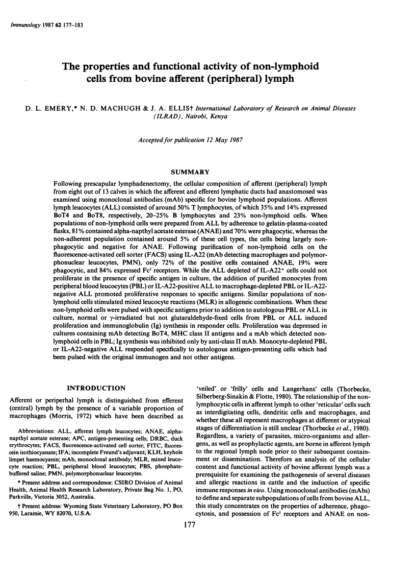
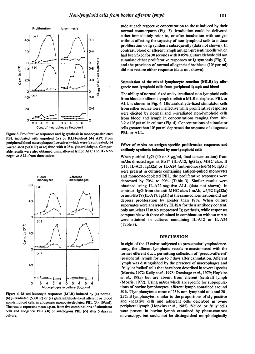
Following prescapular lymphadenectomy, the cellular composition of afferent (peripheral) lymph from eight out of 13 calves in which the afferent and efferent lymphatic ducts had anastomosed was examined using monoclonal antibodies (mAb) specific for bovine lymphoid populations. Afferent lymph leucocytes (ALL) consisted of around 50% T lymphocytes, of which 35% and 14% expressed BoT4 and BoT8, respectively, 20-25% B lymphocytes and 23% non-lymphoid cells. When populations of non-lymphoid cells were prepared from ALL by adherence to gelatin-plasma-coated flasks, 81% contained alpha-napthyl acetate esterase (ANAE) and 70% were phagocytic, whereas the non-adherent population contained around 5% of these cell types, the cells being largely non-phagocytic and negative for ANAE. Following purification of non-lymphoid cells on the fluorescence-activated cell sorter (FACS) using IL-A22 (mAb detecting macrophages and polymorphonuclear leucocytes, PMN), only 72% of the positive cells contained ANAE, 19% were phagocytic, and 84% expressed Fc1 receptors. While the ALL depleted of IL-A22+ cells could not proliferate in the presence of specific antigen in culture, the addition of purified monocytes from peripheral blood leucocytes (PBL) or IL-A22-positive ALL to macrophage-depleted PBL or IL-A22-negative ALL promoted proliferative responses to specific antigens. Similar populations of non-lymphoid cells stimulated mixed leucocyte reactions (MLR) in allogeneic combinations. When these non-lymphoid cells were pulsed with specific antigens prior to addition to autologous PBL or ALL in culture, normal or gamma-irradiated but not glutaraldehyde-fixed cells from PBL or ALL induced proliferation and immunoglobulin (Ig) synthesis in responder cells. Proliferation was depressed in cultures containing mAb detecting BoT4, MHC class II antigens and a mAb which detected non-lymphoid cells in PBL; Ig synthesis was inhibited only by anti-class II mAb. Monocyte-depleted PBL or IL-A22-negative ALL responded specifically to autologous antigen-presenting cells which had been pulsed with the original immunogen and not other antigens.
Full text
PDF


Selected References
These references are in PubMed. This may not be the complete list of references from this article.
- Baldwin C. L., Teale A. J., Naessens J. G., Goddeeris B. M., MacHugh N. D., Morrison W. I. Characterization of a subset of bovine T lymphocytes that express BoT4 by monoclonal antibodies and function: similarity to lymphocytes defined by human T4 and murine L3T4. J Immunol. 1986 Jun 15;136(12):4385–4391. [PubMed] [Google Scholar]
- Drexhage H. A., Mullink H., de Groot J., Clarke J., Balfour B. M. A study of cells present in peripheral lymph of pigs with special reference to a type of cell resembling the Langerhans cell. Cell Tissue Res. 1979 Nov;202(3):407–430. doi: 10.1007/BF00220434. [DOI] [PubMed] [Google Scholar]
- Ellis J. A., Baldwin C. L., MacHugh N. D., Bensaid A., Teale A. J., Goddeeris B. M., Morrison W. I. Characterization by a monoclonal antibody and functional analysis of a subset of bovine T lymphocytes that express BoT8, a molecule analogous to human CD8. Immunology. 1986 Jul;58(3):351–358. [PMC free article] [PubMed] [Google Scholar]
- Emery D. L., Morrison W. I., Jack R. M. Induction of immunity against infection with Theileria parva (East Coast fever) in cattle using plasma membranes from parasitized lymphoblasts. Vet Parasitol. 1986 Feb;19(3-4):321–327. doi: 10.1016/0304-4017(86)90079-8. [DOI] [PubMed] [Google Scholar]
- Fithian E., Kung P., Goldstein G., Rubenfeld M., Fenoglio C., Edelson R. Reactivity of Langerhans cells with hybridoma antibody. Proc Natl Acad Sci U S A. 1981 Apr;78(4):2541–2544. doi: 10.1073/pnas.78.4.2541. [DOI] [PMC free article] [PubMed] [Google Scholar]
- Goddeeris B. M., Baldwin C. L., ole-MoiYoi O., Morrison W. I. Improved methods for purification and depletion of monocytes from bovine peripheral blood mononuclear cells. Functional evaluation of monocytes in responses to lectins. J Immunol Methods. 1986 May 22;89(2):165–173. doi: 10.1016/0022-1759(86)90354-6. [DOI] [PubMed] [Google Scholar]
- Hall J. G., Robertson D. Phagocytosis, in vivo, of immune complexes by dendritic cells in the lymph of sheep. Int Arch Allergy Appl Immunol. 1984;73(2):155–161. doi: 10.1159/000233457. [DOI] [PubMed] [Google Scholar]
- Kishimoto T. Factors affecting B-cell growth and differentiation. Annu Rev Immunol. 1985;3:133–157. doi: 10.1146/annurev.iy.03.040185.001025. [DOI] [PubMed] [Google Scholar]
- Pinder M., Musoke A. J., Morrison W. I., Roelants G. E. The bovine lymphoid system. III. A monoclonal antibody specific for bovine cell surface and serum IgM. Immunology. 1980 Jul;40(3):359–365. [PMC free article] [PubMed] [Google Scholar]
- Roska A. K., Lipsky P. E. Dissection of the functions of antigen-presenting cells in the induction of T cell activation. J Immunol. 1985 Nov;135(5):2953–2961. [PubMed] [Google Scholar]
- Shimonkevitz R., Kappler J., Marrack P., Grey H. Antigen recognition by H-2-restricted T cells. I. Cell-free antigen processing. J Exp Med. 1983 Aug 1;158(2):303–316. doi: 10.1084/jem.158.2.303. [DOI] [PMC free article] [PubMed] [Google Scholar]
- Splitter G. A., Everlith K. M. Collaboration of bovine T lymphocytes and macrophages in T-lymphocyte response to Brucella abortus. Infect Immun. 1986 Mar;51(3):776–783. doi: 10.1128/iai.51.3.776-783.1986. [DOI] [PMC free article] [PubMed] [Google Scholar]
- Thorbecke G. J., Silberberg-Sinakin I., Flotte T. J. Langerhans cells as macrophages in skin and lymphoid organs. J Invest Dermatol. 1980 Jul;75(1):32–43. doi: 10.1111/1523-1747.ep12521083. [DOI] [PubMed] [Google Scholar]


