

Abstract
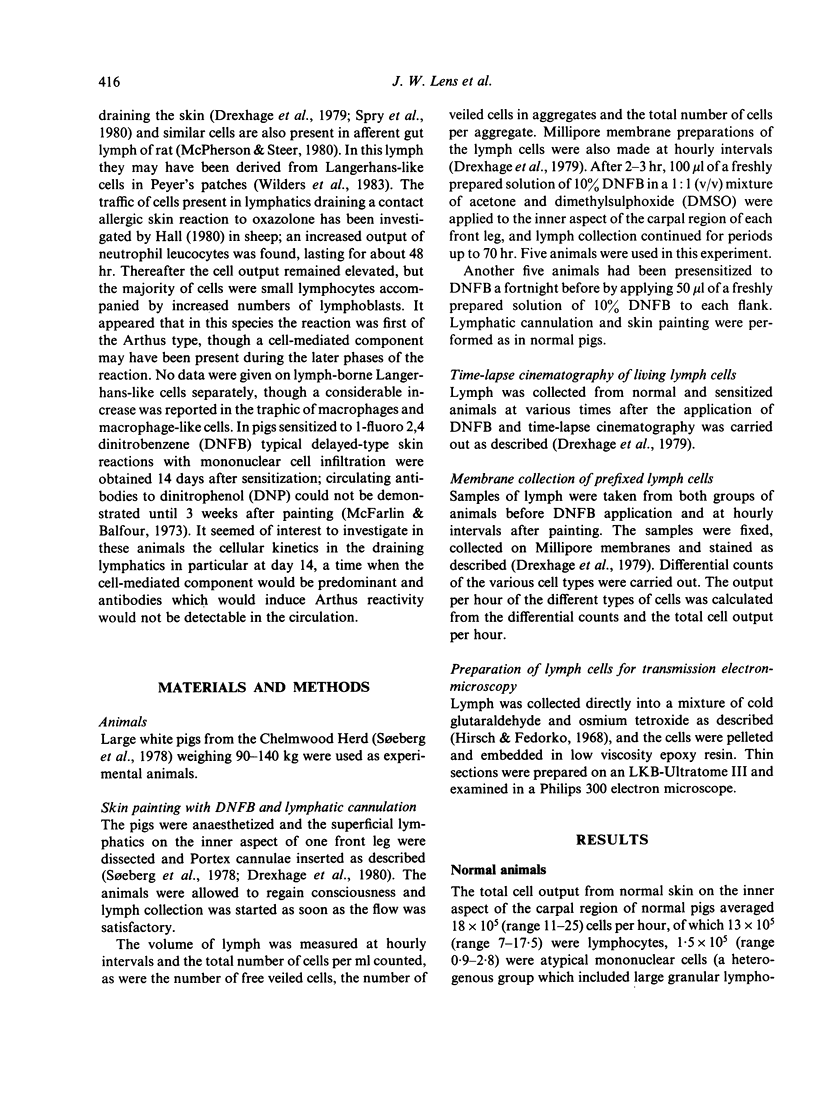
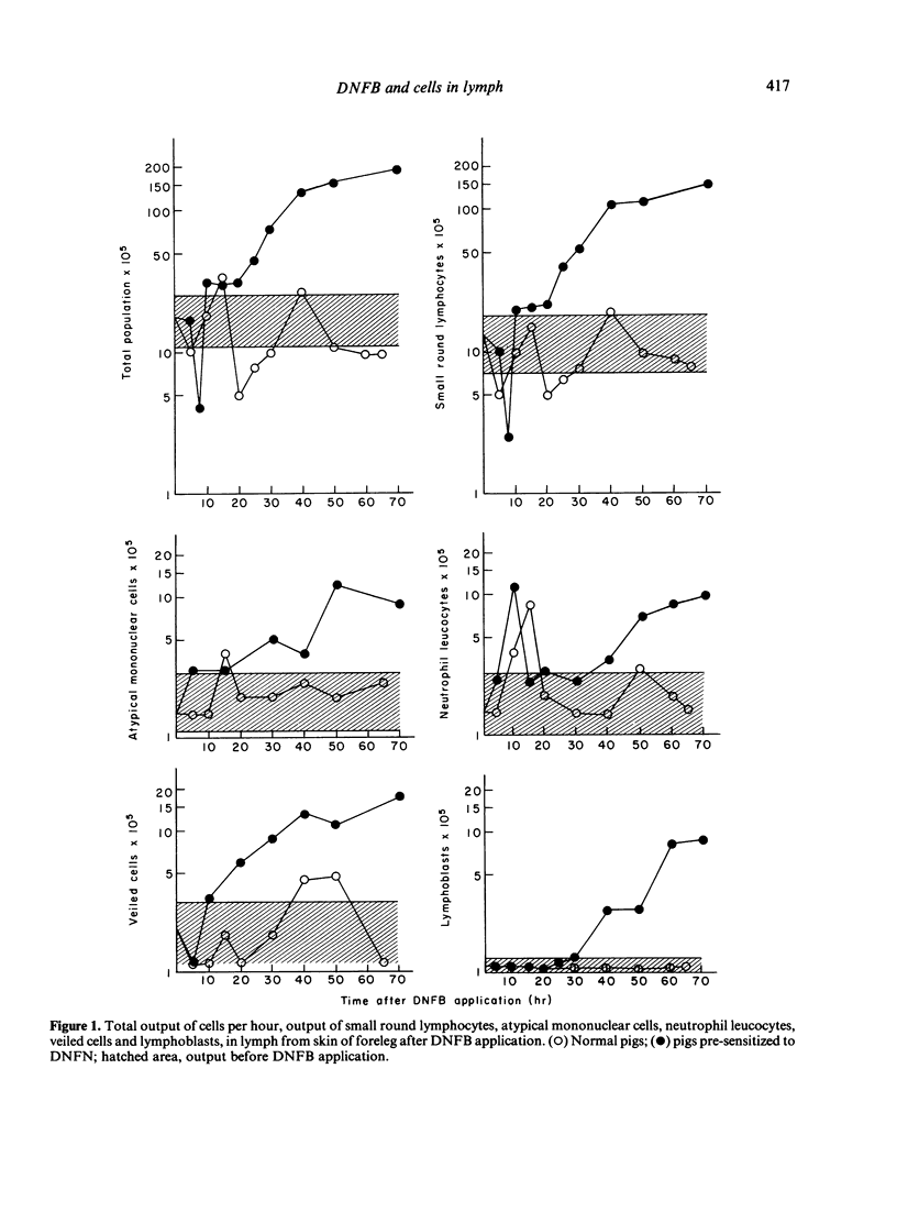
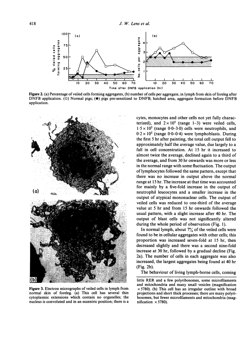
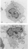
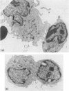
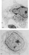
Pigs were skin-painted with the contact sensitizing agent 1-fluoro 2,4 dinitrobenzene (DNFB) and lymph cells coming from the site were collected. Half the animals were sensitive to DNFB and half were normal controls. Special attention was paid to cells belonging to the veiled cell series. At 20 hr after DNFB application, some blast cells--on morphological grounds belonging to the veiled cell series--could be observed in the lymph of presensitized animals. But the most predominant finding in these pigs was that, approximately 30 hr after painting, the total cell output began to increase, reaching 10 times the normal level at 70 hr. The increase in the output of lymphoblasts was the most marked, suggesting peripheral sensitization. Such changes were not observed in normal animals. Here the most striking alterations were seen in the veiled cells. Some of these cells were moving ponderously about, apparently in pursuit of other lymph-borne cells, and this activity resulted in the formation of large cellular aggregates. Since it is known that veiled cells are involved in antigen handling, this behaviour probably facilitates the induction of the T-cell immune response. By contrast, the veiled cells from presensitized animals behaved in a more normal manner and the proportion found in aggregates was only briefly increased. In these animals lymphocytes were seen to interact with veiled cells in a manner reminiscent of peripolesis, apparently recognizing antigenic signals on the surface of the veiled cells. It is discussed that this might result in deletion of the antigen-presenting veiled cells, thus controlling the magnitude of the immune response.
Full text
PDF
Images in this article
Selected References
These references are in PubMed. This may not be the complete list of references from this article.
- Drexhage H. A., Mullink H., de Groot J., Clarke J., Balfour B. M. A study of cells present in peripheral lymph of pigs with special reference to a type of cell resembling the Langerhans cell. Cell Tissue Res. 1979 Nov;202(3):407–430. doi: 10.1007/BF00220434. [DOI] [PubMed] [Google Scholar]
- Hirsch J. G., Fedorko M. E. Ultrastructure of human leukocytes after simultaneous fixation with glutaraldehyde and osmium tetroxide and "postfixation" in uranyl acetate. J Cell Biol. 1968 Sep;38(3):615–627. doi: 10.1083/jcb.38.3.615. [DOI] [PMC free article] [PubMed] [Google Scholar]
- Klareskog L., Tjernlund U., Forsum U., Peterson P. A. Epidermal Langerhans cells express Ia antigens. Nature. 1977 Jul 21;268(5617):248–250. doi: 10.1038/268248a0. [DOI] [PubMed] [Google Scholar]
- McFarlin D. E., Balfour B. Contact sensitivity in the pig. Immunology. 1973 Dec;25(6):995–1009. [PMC free article] [PubMed] [Google Scholar]
- Morris B. Migration intratissulaire des lymphocytes du mouton. Nouv Rev Fr Hematol. 1968 Sep-Oct;8(5):525–534. [PubMed] [Google Scholar]
- Rowden G., Lewis M. G., Sullivan A. K. Ia antigen expression on human epidermal Langerhans cells. Nature. 1977 Jul 21;268(5617):247–248. doi: 10.1038/268247a0. [DOI] [PubMed] [Google Scholar]
- Silberberg I., Baer R. L., Rosenthal S. A. The role of langerhans cells in contact allergy. I. An ultrastructural study in actively induced contact dermatitis in guinea pigs. Acta Derm Venereol. 1974;54(5):321–331. [PubMed] [Google Scholar]
- Silberberg I., Baer R. L., Rosenthal S. A., Thorbecke G. J., Berezowsky V. Dermal and intravascular Langerhans cells at sites of passively induced allergic contact sensitivity. Cell Immunol. 1975 Aug;18(2):435–453. doi: 10.1016/0008-8749(75)90071-4. [DOI] [PubMed] [Google Scholar]
- Spry C. J., Pflug A. J., Janossy G., Humphrey J. H. Large mononuclear (veiled) cells like 'Ia-like' membrane antigens in human afferent lympn. Clin Exp Immunol. 1980 Mar;39(3):750–755. [PMC free article] [PubMed] [Google Scholar]
- Søeberg B., Sumerska T., Binns R. M., Balfour B. M. Contact sensitivity in the pig. II. Induction by intralymphatic infusion of DNP conjugated cells. Int Arch Allergy Appl Immunol. 1978;57(2):114–125. [PubMed] [Google Scholar]
- Wilders M. M., Drexhage H. A., Weltevreden E. F., Mullink H., Duijvestijn A., Meuwissen S. G. Large mononuclear Ia-positive veiled cells in Peyer's patches. I. Isolation and characterization in rat, guinea-pig and pig. Immunology. 1983 Mar;48(3):453–460. [PMC free article] [PubMed] [Google Scholar]