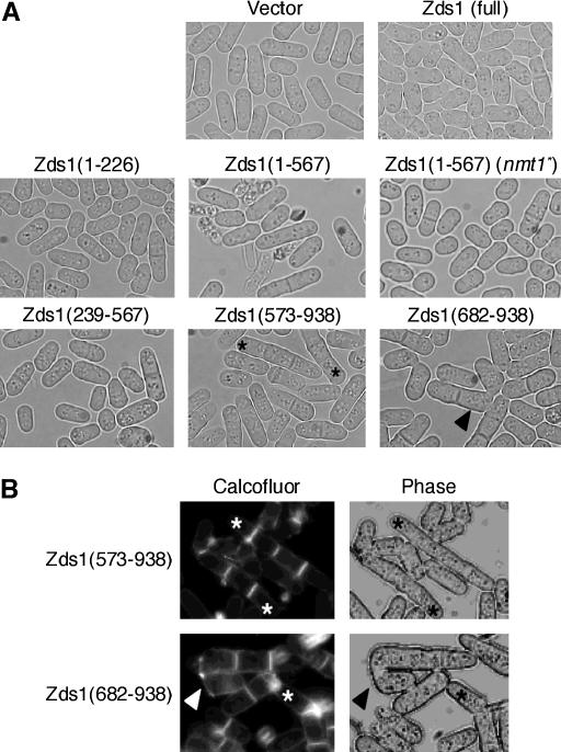
Figure 6.
The morphology of cells expressing part of Zds1. (A) Wild-type SP66 was transformed with various plasmids that contain various lengths of the zds1 gene. Each transformant was cultured in PMA medium at 30° for 24 hr. The strains tested were SP66 harboring pSLF172L GFPS65A, pSLF172L Zds1–GFP, pSLF172L Zds1(1–226)–GFP, pSLF172L Zds1(1–567)–GFP, pSLF272L Zds1(1–567)–GFP, pSLF172L Zds1(239–567)–GFP, pSLF172L Zds1(573–938)–GFP, and pSLF172L Zds1(682–938)–GFP. (B) MY6010 cells expressing Zds1(573–938) and Zds1(682–938) were stained with calcofluor white. Asterisks show multi-septated cells and arrowheads show the abnormal zygotes.

