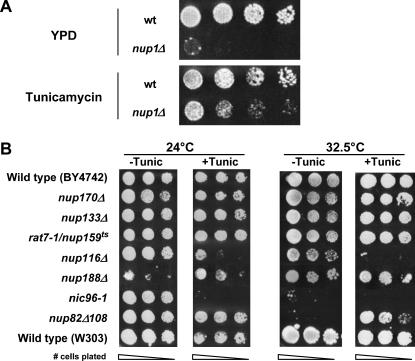
Figure 4.
Inhibition of glycosylation alters the growth rate of nup1, nup116, nup82, and nic96 cells. (A) Wild-type (W303) and nup1Δ (KBY423) cells were grown to log phase in YPD at 25° and then serially diluted and plated on YPD and YPD containing 0.5 μg/ml tunicamycin. Plates were incubated at 30° for 72 hr. (B) Overnight cultures of wild-type (W303 and BY4742), nup170Δ (KBY795), nup133Δ (KBY644), rat7-1/nup159ts (LGY101), nup188Δ (KBY1293), nup116Δ (SWY29), nic96-1 (Y-728), and nup82Δ108 (NUP82-Δ108) cells were grown to log phase, resuspended at identical concentrations, serially diluted, and plated on YPD (−Tunic) or on YPD containing 1.0 μg/ml tunicamycin (+Tunic). Plates were incubated at 24° and 32.5° for 72 hr. (C) Wild-type (BY4742) and nup1Δ (LDY461) cells were transformed with pGAL∷SSA1 (2μ LEU2 GAL∷SSA1) or pRS315 (CEN LEU2). Cells were grown to log phase and serial dilutions were spotted onto YPD media containing glucose or galactose. Plates were incubated at 32.5° for 72 hr.


