

Abstract
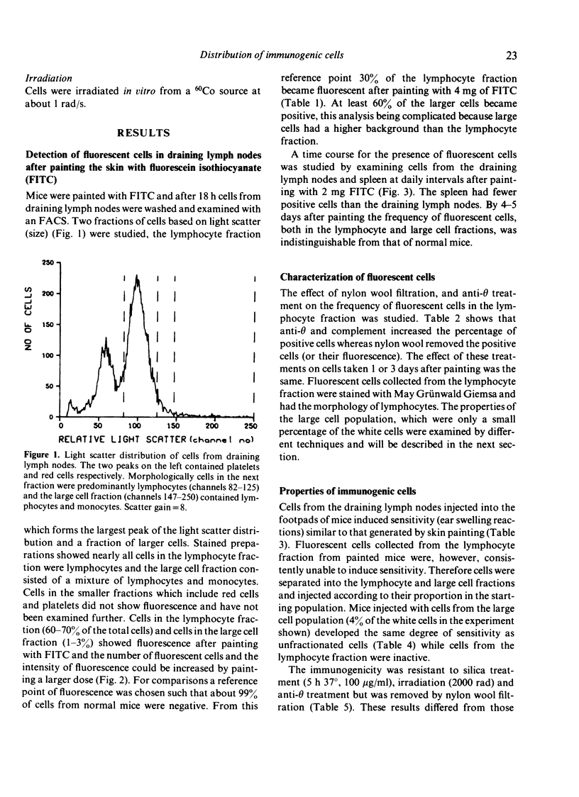
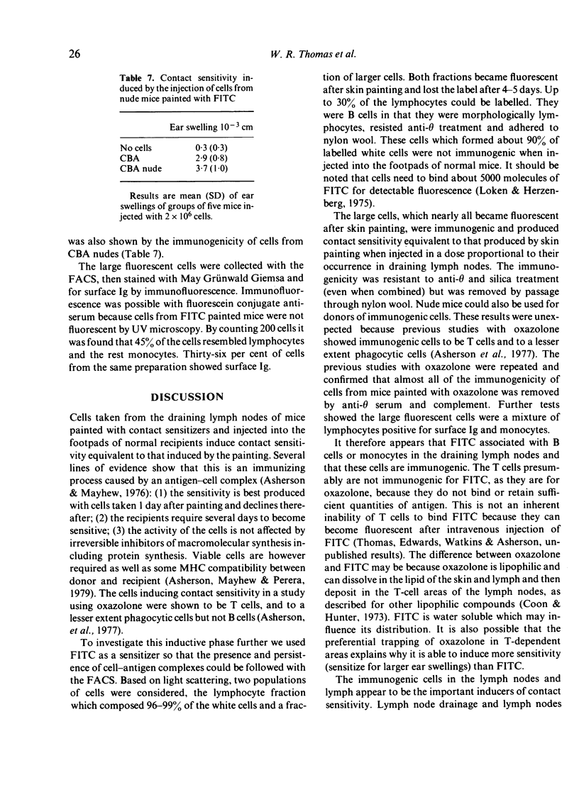
The distribution of fluorescent cells in the draining lymph nodes of mice painted with the contact sensitizing agent fluorescein isothiocyanate (FITC) was investigated using a fluorescence-activated cell sorter. Up to 30% of the cells were fluorescent after 18 h and this decreased thereafter becoming undetectable after 4-5 days. Most of the fluorescent cells were morphologically lymphocytes, theta - ve and adherent to nylon wool. Immunogenicity of these cells was tested by injecting them into the footpads of normal mice and measuring contact sensitivity after 6 days. This was restricted to large cells which represented less than 5% of the white cell population and nearly all of which became fluorescent after skin painting. The large fluorescent cells were a mixture of monocytes and lymphocytes. Most of the lymphocytes had surface immunoglobulin. The immunogenicity was reduced by nylon filtration but was not affected by silica and anti-theta. These results showed that the immunogenicity is not associated with T cells. In contrast, similar immunogenic activity in the draining lymph nodes of mice painted with oxazolone is associated with T cells. The results therefore showed that different sensitizers form immunogenic complexes with different cell populations, perhaps in this case becuase of the different water solubilities of FITC and oxazolone. They also suggested that this may cause important differences in antigen presentation, for example in their association with different MHC products.
Full text
PDF


Selected References
These references are in PubMed. This may not be the complete list of references from this article.
- Asherson G. L., Allwood G. G., Mayhew B. Contact sensitivity in the mouse. XI. Movement of T blasts in the draining lymph nodes to sites of inflammation. Immunology. 1973 Sep;25(3):485–494. [PMC free article] [PubMed] [Google Scholar]
- Asherson G. L., Mayhew B. Induction of cell-mediated immunity in the mouse: circumstantial evidence for highly immunogenic antigen in the regional lymph nodes following skin painting with contact sensitizing agents. Isr J Med Sci. 1976 Apr-May;12(4-5):454–467. [PubMed] [Google Scholar]
- Asherson G. L., Mayhew B., Perera M. A. The production of contact sensitivity by the injection into the footpads of recipients of the lymph node cells from mice 1 day after painting the skin with contact sensitizing agent: requirement for matching at the major histocompatibility complex between donor and recipient mice. Immunology. 1979 May;37(1):241–245. [PMC free article] [PubMed] [Google Scholar]
- Asherson G. L., Zembala M., Mayhew B. Passive transfer of contact sensitivity by bone marrow cells and evidence for their origin from immunized lymph nodes. Int Arch Allergy Appl Immunol. 1974;46(2):256–260. doi: 10.1159/000231128. [DOI] [PubMed] [Google Scholar]
- Cantor H., Simpson E. Regulation of the immune response by subclasses of T lymphocytes. I. Interactions between pre-killer T cells and regulatory T cells obtained from peripheral lymphoid tissues of mice. Eur J Immunol. 1975 May;5(5):330–336. doi: 10.1002/eji.1830050508. [DOI] [PubMed] [Google Scholar]
- Coon J., Hunter R. Selective induction of delayed hypersensitivity by a lipid conjugated protein antigen which is localized in thymus dependent lymphoid tissue. J Immunol. 1973 Jan;110(1):183–190. [PubMed] [Google Scholar]
- EISEN H. N., KERN M., NEWTON W. T., HELMREICH E. A study of the distribution of 2,4-dinitrobenzene sensitizers between isolated lymph node cells and extracellular medium in relation to induction of contact skin sensitivity. J Exp Med. 1959 Aug 1;110(2):187–206. doi: 10.1084/jem.110.2.187. [DOI] [PMC free article] [PubMed] [Google Scholar]
- EISEN H. N., ORRIS L., BELMAN S. Elicitation of delayed allergic skin reactions with haptens; the dependence of elicitation on hapten combination with protein. J Exp Med. 1952 May 1;95(5):473–487. doi: 10.1084/jem.95.5.473. [DOI] [PMC free article] [PubMed] [Google Scholar]
- FREY J. R., WENK P. Experimental studies on the pathogenesis of contact eczema in the guinea-pig. Int Arch Allergy Appl Immunol. 1957;11(1-2):81–100. doi: 10.1159/000228405. [DOI] [PubMed] [Google Scholar]
- GELL P. G., BENACERRAF B. Studies on hypersensitivity. IV. The relationship between contact and delayed sensitivity: a study of the specificity of cellular immune reactions. J Exp Med. 1961 Mar 1;113:571–585. doi: 10.1084/jem.113.3.571. [DOI] [PMC free article] [PubMed] [Google Scholar]
- McFarlin D. E., Balfour B. Contact sensitivity in the pig. Immunology. 1973 Dec;25(6):995–1009. [PMC free article] [PubMed] [Google Scholar]
- Nakagawa S., Amos H. E., Gotoh M., Tanioku K. The distribution of 2,4-dinitrophenyl groups in lymphoid tissue of guinea-pigs following skin painting with 2,4-dinitrochlorobenzene. Immunology. 1979 Apr;36(4):851–857. [PMC free article] [PubMed] [Google Scholar]
- Parker D., Aoki T., Turk J. L. Studies on the ability of the soluble proteins from skin, painted in vivo with DNFB, to cause contact sensitivity in the guinea pig. Int Arch Allergy Appl Immunol. 1970;38(1):42–56. doi: 10.1159/000230258. [DOI] [PubMed] [Google Scholar]
- Søeberg B., Sumerska T., Binns R. M., Balfour B. M. Contact sensitivity in the pig. II. Induction by intralymphatic infusion of DNP conjugated cells. Int Arch Allergy Appl Immunol. 1978;57(2):114–125. [PubMed] [Google Scholar]
- Thomas W. R., Asherson G. L., Perera M. A. Induction of contact sensitivity. Selective induction of delayed hypersensitivity by the injection of cells from draining lymph nodes into the footpads of normal recipients. Immunology. 1978 Apr;34(4):725–731. [PMC free article] [PubMed] [Google Scholar]