Abstract
The effects of γ-aminobutyric acid (GABA) on the electrophysiological properties of intracardiac neurones were investigated in the intracardiac ganglion plexus in situ and in dissociated neurones from neonatal, juvenile and adult rat hearts. Focal application of GABA evoked a depolarizing, excitatory response in both intact and dissociated intracardiac ganglion neurones. Under voltage clamp, both GABA and muscimol elicited inward currents at −60 mV in a concentration-dependent manner. The fast, desensitizing currents were mimicked by the GABAA receptor agonists muscimol and taurine, and inhibited by the GABAA receptor antagonists, bicuculline and picrotoxin. The GABAA0 antagonist (1,2,5,6-tetrahydropyridin-4-yl)methyl phosphonic acid (TPMPA), had no effect on GABA-induced currents, suggesting that GABAA receptor-channels mediate the response. The GABA-evoked current amplitude recorded from dissociated neurones was age dependent whereby the peak current density measured at −100 mV was ∼ 20 times higher for intracardiac neurones obtained from neonatal rats (P2–5) compared with adult rats (P45–49). The decrease in GABA sensitivity occurred during the first two postnatal weeks and coincides with maturation of the sympathetic innervation of the rat heart. Immunohistochemical staining using antibodies against GABA demonstrate the presence of GABA in the intracardiac ganglion plexus of the neonatal rat heart. Taken together, these results suggest that GABA and taurine may act as modulators of neurotransmission and cardiac function in the developing mammalian intrinsic cardiac nervous system.
Neural control of the heart is under the influence of the sympathetic and the parasympathetic divisions of the autonomic nervous system. Activation of the parasympathetic division, arising from neurones in the medulla of the brain stem, has negative chronotropic, dromotropic and inotropic actions (Adams & Cuevas, 2004). The intracardiac ganglia (ICG) of the rat are organized in four major ganglion clusters (Sampaio et al. 2003) that form the final common pathway for the cardiac autonomic nervous system. Parasympathetic intracardiac neurones receive excitatory synaptic input from extrinsic and/or intrinsic nerves (Seabrook et al. 1990; Selyanko & Skok, 1992; Selyanko & Skok, 1992; Edwards et al. 1995). The ICG plexus has been demonstrated to be capable of maintaining local circuit reflexes (see Armour, 1999).
GABA is the major inhibitory neurotransmitter in the central nervous system (Sieghart et al. 1999) and the physiological actions of GABA are mediated via ionotropic GABAA and GABAA0 (also known as GABAC) receptors and via G-protein coupled metabotropic GABAB receptors. GABAA receptors are pentameric Cl−-permeable channels that are opened by GABA and modulated by a variety of clinically relevant drugs, such as benzodiazepines, barbiturates, steroids, anaesthetics and convulsants (Macdonald & Olsen, 1994; Sieghart et al. 1999). GABA has also been shown to be stored and released in peripheral ganglia and GABAA receptors have been reported on guinea-pig myenteric plexus neurones (Zhou & Galligan, 2000; Reis et al. 2002), rat superior cervical ganglion neurones (Brown et al. 1979), rat major pelvic ganglion neurones (Akasu et al. 1999) and a subpopulation of parasympathetic neurones in cat pancreatic ganglia (Sha et al. 2001).
Intravenous administration of GABA lowers arterial blood pressure and induces bradycardia in several mammalian species (Loscher, 1982). The prolonged cardiovascular depression in response to GABA was attenuated by subsequent administration of picrotoxin and bicuculline leading to the proposal that the observed effects of GABA may be due to direct actions on vascular and cardiac tissue (Billingsley & Suria, 1982; Loscher, 1982). Subsequently, GABA was shown to be present in several regions of the guinea-pig heart, with the highest concentrations found in the area of the sinoatrial node (Matsuyama et al. 1991). GABA has been shown indirectly to inhibit the activity of sympathetic adrenergic neurones secondarily to the stimulation of para-sympathetic cholinergic neurones via GABAA receptors present in the sinus node (Matsuyama et al. 1993).
In the present study, we have investigated the effects of GABA on the electrophysiological properties of intracardiac ganglion neurones in situ and acutely dissociated neurones from neonatal and adult rat hearts. Stimulation of bicuculline-sensitive GABAA receptors produces a concentration-dependent depolarization of intracardiac ganglion neurones which was reduced with postnatal development. A preliminary report of some of these results has been presented in abstract form (Fischer & Adams, 2004).
Methods
Electrophysiological recordings from intracardiac ganglion preparations in situ
Neonatal and young adult female Wistar rats (180–220 g) were killed by stunning and cervical dislocation, the hearts excised, atria isolated and placed in cold Krebs solution. The right atrial ganglion plexus and underlying myocardium were isolated from the dorsal surface of the atria. The preparation was pinned to the Sylgard (Dow-Corning, Midland, MI, USA) covered base of a 35 mm tissue culture dish and superfused with a bicarbonate-buffered Krebs solution comprising (mm): NaCl 118.4, NaHCO3 25.0, NaH2PO4 1.13, CaCl2 1.8, KCl 4.7, MgCl2 1.3 and glucose 11.1, gassed with Carbogen (95% O2–5% CO2) to pH 7.4 (Smith et al. 2001). The recording chamber was continuously superfused (∼ 2 ml min−1) with Krebs solution at 35°C. The temperature of the solution was controlled by Peltier thermoelectric elements (Melcor, Trenton, NJ, USA) and monitored by an independent thermistor probe in the recording chamber (Yellow Springs Instruments, Yellow Springs, OH, USA). Intracellular recordings were made using sharp glass microelectrodes (GC120F; Harvard Apparatus Ltd, Edenbridge, UK) with ∼ 120 MΩ resistance when filled with 0.5 m potassium acetate. Membrane voltage responses were recorded with a conventional bridge amplifier (Axoclamp 2A; Axon Instruments Inc., Union City, CA, USA). Signals were filtered at 20 kHz, digitized at 50 kHz and transferred to a Pentium 4 computer using an analog-to-digital converter (Micro 1401 interface, CED, Cambridge, UK) and Spike 2 software (CED). Electrophysiological properties and GABA responses of the neurones were analysed using the same program. GABA was focally applied using a pressure-ejection device (200 kPa; Picospritzer II, General Valve, Fairfield, NJ, USA) and the pressure ejection pipette was positioned ≤ 50 μm from the neuronal soma to maximize the response to agonist application. Pharmacological antagonists were bath applied at the concentrations indicated.
Whole-cell patch clamp recordings from acutely dissociated ICG neurones
Parasympathetic neurones from neonatal (2–7 day old), young juvenile (12–18 days old) and adult (> 6 weeks old) rat ICG were isolated and placed in tissue culture. The procedures for isolation of the ICG neurones have been previously described (Xu & Adams, 1992) and were in accordance with guidelines of the University of Queensland Animal Experimentation Ethics Committee. Briefly, Wistar rats were killed by stunning and cervical dislocation, and the heart excised and placed in Hank's balanced salt solution (HBSS). Atria were isolated and clusters of ganglia were dissected and transferred to HBSS containing collagenase (Type 2, 300 U ml−1; Worthington-Biochemical, Freehold, NJ, USA). Following 1 h digestion, the ganglia were rinsed twice in HBSS and triturated in culture medium (Dulbecco's modified Eagle medium with 10% (v/v) fetal calf serum, 1% (v/v) penicillin–streptomyocin) (Gibco - Invitrogen, Mount Waverley, Australia) using a fine-bore Pasteur pipette. The dissociated neurones were plated on sterile 12 mm glass coverslips and incubated at 37°C for 12–24 h under a 95% O2–5% CO2 atmosphere.
Whole-cell currents were recorded from isolated ICG neurones using either the standard whole-cell patch clamp or the gramicidin perforated-patch recording technique (Akaike, 1996). Patch pipettes (GF150; Harvard Apparatus Ltd) were pulled, fire-polished and had resistances of 1.0–2.0 MΩ when filled with intracellular solution. Membrane currents were recorded using an Axopatch 200A patch clamp amplifier (Axon Instruments Inc.) and voltage steps were generated from a PC Pentium 4 computer using pCLAMP 9.0 software and a Digidata 1322A interface (Axon Instruments Inc.). Data were analysed using Clampfit 9.0 software (Axon Instruments Inc.).
Agonist-activated currents in dissociated ICG neurones were recorded using patch pipettes filled with either an internal solution containing (mm): 146 KCl, 2 CaCl2, 11 EGTA, 2 MgATP, 10 Hepes-KOH, pH 7.2 for standard whole cell recordings or 150 KCl, 10 Hepes-KOH plus gramicidin C (100 μm final concentration), pH 7.2 for perforated patch recordings. The bath solution contained (mm): 140 NaCl, 3 KCl, 1.2 MgCl2, 2.5 CaCl2, 7.7 glucose, 10 Hepes-NaOH, pH 7.35. Agonists were applied to cells either by brief pressure ejection (100 kPa; Picospritzer II, General Valve) from an extracellular micropipette (3–5 μm diameter) positioned 50–100 μm from the cell soma to evoke maximal responses to agonists or using a pressure-driven constant flow perfusion system using manually controlled solenoid valves and a micromanifold (100 μm diameter) (ALA Scientific Instruments, Westbury, NY, USA) positioned 50–100 μm from the cell soma. To minimize receptor desensitization, a delay of 60 s between agonist applications was maintained. All experiments were carried out at room temperature (22°C).
Preparation of atrial whole-mounts for immunohistochemistry
The procedures for isolation of rat ICG for immunohistochemical staining have been previously described (Richardson et al. 2003) and were in accordance with guidelines of the Animal Experimentation Ethics Committee of the University of Melbourne. Briefly, neonatal and adult animals were anaesthetized with sodium pentobarbitone (Nembutal, 60 mg kg−1i.p.), the chest was opened and the heart rapidly excised and placed in cold 0.01 m phosphate-buffered saline (PBS (mm): 145 NaCl, 7.5 Na2PO4, 2.5 NaH2PO4). The ventricles were removed and the aorta and pulmonary trunk were detached. The preparation was stretched flat and immersion fixed overnight in fixative (4% formaldehyde, 0.1% glutaraldehyde in 0.1 m phosphate buffer; 7.5 mm Na2PO4, 2.5 mm NaH2PO4) at 4°C. Following fixation, the epicardial connective tissue containing the cardiac ganglia, was peeled away from the myocardium. Specimens were washed and stored in 0.01 m PBS prior to immunohistochemical processing. Tissue was permeabilized in 0.5% Triton X-100 in PBS for 30 min and then for a minimum of 1 h in 10% normal horse serum in PBS at room temperature in a humid box to block non-specific antibody binding. Preparations were washed 3 times (5 min changes) in PBS and then incubated in species-specific double combinations of primary antibody (mouse-anti GABA (1: 50 MAB316; Chemicon International, Inc., Temecula, CA, USA) and rabbit-anti protein gene product 9.5 (PGP-9.5, 1 : 2000; Ultraclone Ltd, Isle of Wight, UK)) overnight at room temperature. Following incubation in primary antibodies, whole-mount preparations were washed 3 times for 30 min in 0.01 m PBS, and then incubated in the appropriate combinations of secondary antisera (goat-anti mouse-Alexa Fluor 546 (1 : 200; Molecular Probes, Inc., Eugene, OR, USA) and goat-anti rabbit-Alexa Fluor 488 (1 : 200; Molecular Probes)) for 2–3 h at room temperature on the shaker table. All antibodies were diluted in PBS containing 5% normal horse serum, 0.3% Triton X-100 and 0.1% sodium azide. After incubation in secondary antibodies, tissues were again washed 3 times. Subsequently the specimens were rinsed in 50% glycerol buffered with NaHCO3 (pH 8.6) for 1 h. Whole-mounts were arranged on slides and viewed using a Bio-Rad 1024 confocal microscope.
Data analysis
Data are expressed as means ± s.e.m. with n indicating the number of neurones used. The significance of differences was evaluated by means of the Student's t test. Concentration–response curves were analysed using SigmaPlot 8.0 (SPSS Inc.) and fitted by unweighted non-linear regression to the equation:
where y is the normalized response, [A] is the agonist concentration, EC50 is the concentration that gives a half-maximal response and h is the slope factor (Hill coefficient).
All chemicals used were analytical grade and the following drugs were used: gramicidin hydrochloride, GABA, bicuculline methiodide, picrotoxin, muscimol hydrobromide, taurine, (+/−)baclofen, (−)nicotine hydrogen tartrate, methyl 6,7-dimethoxy-4-ethyl-b-carboline-3-carboxylate (DMCM) (Sigma Chemical Co., Castle Hill, Australia), 1,2,5,6-tetrahydropyridin-4-yl)methyl phosphonic acid (TPMPA) (Tocris Cookson Ltd, Avonmouth, UK).
Results
Excitatory GABA-evoked responses in neonatal and adult rat ICG neurones in situ
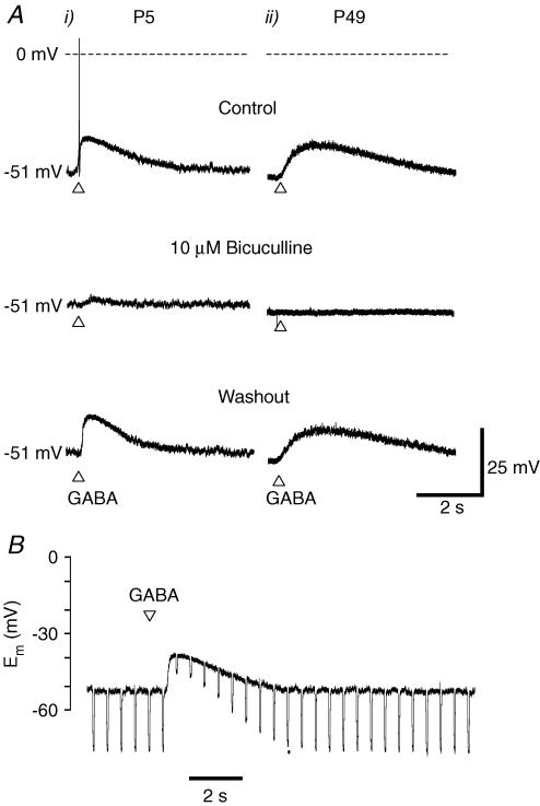
Intracellular microelectrode recording of the resting membrane potential from neurones of intact ICG of neonatal (P5) rats was −51.1 ± 1.9 mV (n = 18) and −51.8 ± 2.3 mV (n = 6) for adult (P49) rats. Focal application of GABA (100 μm) evoked transient depolarizing responses of +15.2 ± 2.0 mV (n = 18) amplitude in neonates and frequently elicited multiple, adapting, action potential discharge. The mean depolarization evoked by GABA in adult rat ICG neurones was +11.6 ± 1.5 mV (n = 6) but failed to elicit action potential firing in all neurones studied. Under current clamp conditions, the amplitude of the GABA-evoked response was enhanced upon membrane hyperpolarization (not shown). The GABA-evoked excitatory responses were inhibited by superfusion with extracellular solutions containing 10 μm bicuculline (Fig. 1A) or 100 μm picrotoxin (not shown). A marked decrease in input resistance accompanied the GABA-evoked depolarization as shown in Fig. 1B. The input resistance in neonatal rat intracardiac neurones decreased from 188 ± 39 to 68 ± 16 MΩ (n = 3) whereas, in adult neurones, GABA reduced the input resistance from 77 ± 1 to 29 ± 17 MΩ (n = 3). Bath perfusion of GABA (100 μm) reversibly attenuated action potential discharge in response to depolarizing current pulses (200 ms) in both neonatal and adult ICG neurones (not shown).
Figure 1. Intracellular microelectrode recordings obtained from intact ICG neurones.
Ai, neonatal P5 neurone: focal application of 100 μm GABA evoked membrane depolarization and multiple, adapting, action potential discharge. The excitatory response is inhibited by 10 μm bicuculline. Aii, GABA-evoked responses recorded from neurones in an adult, in situ ganglion preparation. The GABA-evoked depolarization is inhibited by bicuculline (10 μm). B, neonatal P5 neurone: conductance increase is associated with the GABA-evoked response. Cell input resistance was monitored by injecting −0.1 nA, 50 ms current pulses at 2 Hz.
GABA-evoked currents in dialysed dissociated neurones of rat ICG
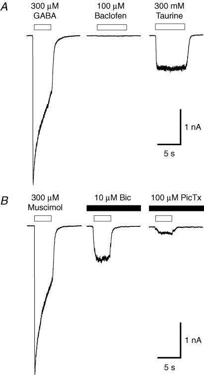
Using the dialysed whole-cell patch clamp technique and equimolar intra- and extracellular concentrations of Cl−, GABA evoked rapid, depolarizing currents in dissociated ICG neurones voltage clamped at −60 mV. This response was mimicked by the GABAA receptor agonist muscimol (Fig. 2A), whereas application of the GABAB receptor agonist, baclofen, glycine or glutamate (each 100 μm) did not evoke any membrane response. The time course of decay of GABA-evoked currents was > 8-fold slower than nicotine-evoked currents recorded from the same neurone and was inhibited by 10 μm bicuculline and 100 μm picrotoxin. GABA and muscimol (300 μm) evoked inward currents with a mean peak current density of −100.1 ± 7.4 pA pF−1 (n = 20) and −105.5 ± 8.0 pA pF−1 (n = 8), respectively, at a holding potential of −60 mV. Under similar conditions, 300 μm nicotine elicited significantly lower current densities of −75.6 ± 7.5 pA pF−1 (P < 0.01, n = 8). Muscimol (300 μm)-evoked current amplitude was reduced reversibly to 25 ± 6% (n = 6) and 5 ± 1% (n = 8) of control by 100 μm bicuculline and picrotoxin, respectively (Fig. 2B). Taurine (10 mm), a non-selective agonist for GABAA and glycine receptors, evoked small, non-desensitizing currents with a mean peak current density of −26.1 ± 5.3 pA pF−1 (n = 5) at −60 mV. These taurine-evoked currents were inhibited completely by 100 μm bicuculline or picrotoxin (not shown).
Figure 2. Effects of GABA-receptor agonists on rat neonatal ICG neurones.
A, representative whole-cell currents evoked by focal application of GABA receptor agonists, GABA, baclofen and taurine, recorded from the same dissociated neurone with equimolar internal and external Cl− concentrations. B, muscimol-evoked currents inhibited by co-application of either bicuculline (Bic) or picrotoxin (PicTx). Holding potential, −60 mV, A and B P3.
GABA-evoked responses in dissociated intracardiac neurones depends on postnatal age
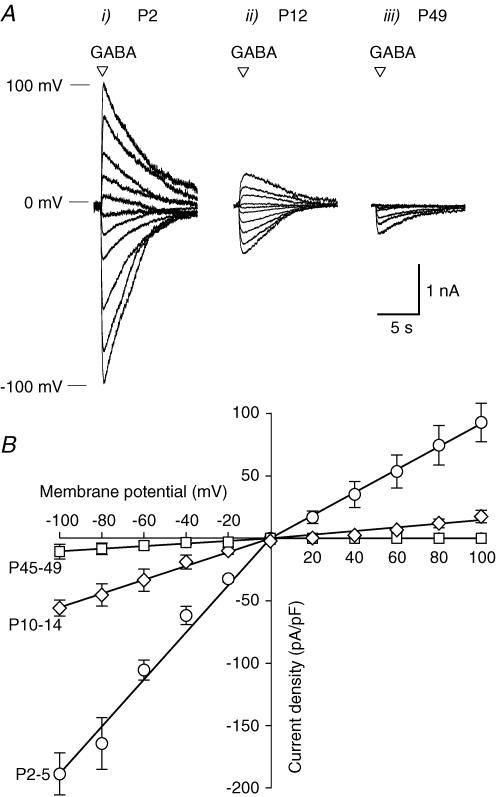
GABA-evoked responses as a function of postnatal development were investigated in dissociated neurones from neonatal (P2–5), juvenile (P12–15) and adult (P45–49) rat ICG. Figure 3A shows representative membrane currents evoked by 100 μm GABA under dialysed whole-cell recording conditions at different postnatal ages. In isolated neurones from neonatal rats, GABA evoked inwardly rectifying currents at negative holding potentials which reversed at 0 mV as expected if Cl− is the charge carrier with equimolar extra- and intracellular Cl− concentrations. Current density–voltage relationships obtained for the peak GABA-evoked current amplitudes recorded for the three age groups (n ≥ 8 each) are shown in Fig. 3B. The peak current density at −100 mV was markedly reduced in juveniles (P12–15; −55.9 ± 6.4 pA pF−1) compared with newborn (P2–5; −188.8 ± 16.8 pA pF−1) rats. GABA-evoked current amplitude recorded from neurones of adult rat ICG (P45–49) were further reduced with a peak current density of −10.6 ± 6.0 pA pF−1. The GABA-evoked current–voltage relationship obtained in dissociated intracardiac neurones exhibited marked inward rectification. The rectification of GABA-evoked current amplitude in neonatal rat intracardiac neurones was 2-fold greater at negative compared with positive membrane potentials whereas the rectification was reduced in neurones from juvenile animals.
Figure 3. Developmental regulation of GABA-induced responses in dissociated intrinsic cardiac neurones.
A, superimposed traces of GABA-evoked currents elicited by brief pulses (50 ms) of 100 μm GABA. GABA-evoked currents were recorded from rat ICG neurones at different stages of postnatal development. B, whole-cell current–voltage relationships obtained for peak current density (pA pF−1) evoked by 100 μm GABA for dissociated neurones from the different age groups: P2–5 (○), P10–14 (♦) and P45–49 (□).
Effects of GABAA receptor agonists and antagonists as a function of postnatal development
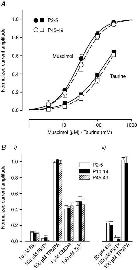
The potency of the GABA agonists tested in dissociated neurones was not altered with postnatal development. The EC50 for GABA obtained in neurones isolated from neonatal (P2–5) rats was 30.1 ± 0.9 μm (n = 6) which was not significantly different to that obtained for the GABAA agonist, muscimol (EC50 = 28.1 ± 1.2 μm, n = 8). In neurones from adult rats, the muscimol concentration–response relationship gave an EC50 of 33.6 ± 1.2 μm (n = 8), which is not significantly different from that obtained for neonatal animals. The concentration–response relationship for taurine-evoked currents exhibited an EC50 of 162 ± 46 mm and 195 ± 55 mm for neonatal and adult rats, respectively (Fig. 4A, Table 1).
Figure 4. GABAA receptor agonist potencies and effects of antagonists are independent of postnatal age.
A, concentration–response curves obtained for the GABA agonists, muscimol (circles) and taurine (squares), on dissociated ICG neurones for P2–5 (filled symbols) and P45–49 (open symbols) groups. n = 5 cells for each agonist and age group. B, effect of the GABAA receptor-channel antagonists, bicuculline (Bic) and picrotoxin (PicTx), and modulators on GABA- (Bi) and taurine- (Bii) evoked currents (10 μm GABA for Bic, DMCM, Zn2+; 100 μm GABA for PicTx and TPMPA; 50 mm taurine) recorded from dissociated ICG neurones of different postnatal age. n = 4–8 cells per GABA receptor antagonist/modulator and age group. Holding potential, −60 mV for both A and B.
Table 1.
Potency (EC50) and Hill coefficient (h) determined from concentration–response relationships for GABA-, muscimol- and taurine-evoked currents in dissociated intracardiac neurones from neonatal (P2–5) and adult (P45–49) rats
| Agonist | Neonatal | Adult |
|---|---|---|
| GABA | ||
| EC50 (μm) | 30.1 ± 0.9 | ND |
| h | 1.3 ± 0.2 | ND |
| Muscimol | ||
| EC50 (μm) | 28.1 ± 1.2 | 33.6 ± 1.2 |
| h | 1.3 ± 0.1 | 1.3 ± 0.1 |
| Taurine | ||
| EC50 (mm) | 162 ± 46 | 195 ± 55 |
| h | 1.0 ± 0.1 | 1.0 ± 0.1 |
Data expressed as mean ± s.e.m., n = 5–6 cells per age group and agonist. Membrane potential, −60 mV. ND, not determined.
Characterization of the GABA receptor type and subunit composition was carried out using selective GABAA and GABAA0 antagonists and GABAA receptor modulators. The GABAA receptor antagonist, bicuculline (10 μm), inhibited GABA (10 μm)-evoked currents to 12 ± 1% of control amplitude in P2–5, 11 ± 2% in P12–14 and 10 ± 1% in P45–49 aged rats (n = 8). Bath application of 100 μm picrotoxin reduced currents evoked by 100 μm GABA to < 5% (n = 6–8) of control amplitude in intracardiac neurones from all age groups.
The selective GABAA0 receptor antagonist, 1,2,5,6-tetrahydropyridin-4-yl)methyl phosphonic acid (TPMPA), had no effect on currents evoked by 100 μm GABA in intracardiac neurones of any age group. In contrast, diazepam (1 μm) increased the amplitude of muscimol-evoked currents in both neonatal (P5) and juvenile (P10–14) rat ICG neurones (n = 2; not shown). The inverse receptor agonist, methyl 6,7-dimethoxy-4-ethyl-b-carboline-3-carboxylate (DMCM, 1 μm), inhibited currents evoked by 10 μm GABA reversibly to 41 ± 4% of control amplitude in P2–5, 42 ± 2% in P10–14 and 45 ± 4% in P45–P49 (n = 6–8). Given that zinc has been shown to inhibit GABAA receptor-channels (Sieghart, 1995; Hosie et al. 2003), the sensitivity of GABA-activated currents to extracellular Zn2+ was examined in rat intracardiac neurones. Bath application of 100 μm Zn2+ inhibited currents evoked by 10 μm GABA to 45 ± 5% of control amplitude in P2–5, 50 ± 6% in P10–14 and to 46 ± 6% in P45–49 aged rats (n = 7–8). Taurine-evoked current amplitude was similarly inhibited by extracellular Zn2+ (Fig. 4B).
Gramicidin perforated-patch recording of GABA-induced responses in rat ICG neurones
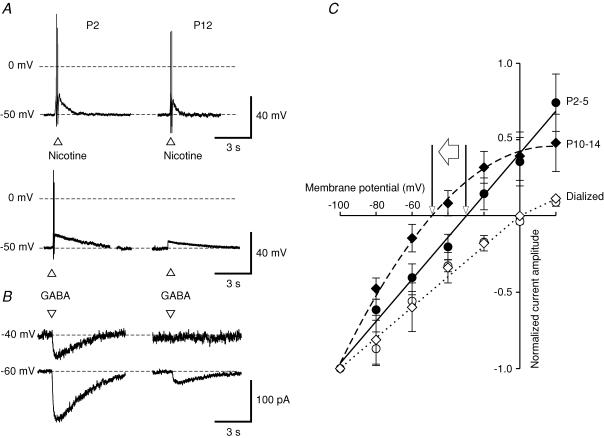
Gramicidin perforated-patch recording to maintain the intracellular Cl− concentration was carried out to investigate the GABA-induced response under physiological conditions. Under current clamp, focal application of nicotine (100 μm) elicited transient excitatory responses with multiple action potential discharge in dissociated intracardiac neurones from P2–5 and P12–14 age groups (Fig. 5A). In the same neurones, application of GABA (100 μm) evoked a depolarization and reached threshold for action potential firing in neurones isolated from P2–5 but not P12–15 rats.
Figure 5. GABAA receptor-mediated responses are reduced by postnatal changes in the intracellular Cl− concentration.
A, representative firing characteristics of P2 and P12 dissociated rat ICG neurones obtained using the gramicidin perforated-patch clamp technique. Focal application (50 ms) of 100 μm nicotine depolarizes the neurone evoking 1–3 action potentials followed by a fast repolarization phase in neurones from both age groups. Focal application (50 ms) of 100 μm GABA elicits 1–3 action potentials followed by a slow repolarization phase in neonatal rat ICG neurones. GABA does not evoke action potential firing in neurones from older rats (P12 and P49). B, GABA-evoked currents recorded using gramicidin perforated patch at −40 and −60 mV from P2 and P12 rat ICG neurones. C, current–voltage relationships obtained from gramicidin perforated-patch (filled symbols) and dialysed whole-cell (open symbols) recordings of rat ICG neurones of different age. Peak current amplitude evoked by 100 μm GABA at −100 mV normalized for each of the 3 different age groups. The reversal potential for GABA-activated Cl− currents shifts from −29.8 mV (P2–5, circles) to −49.0 mV (P10–14, diamonds), giving a calculated intracellular Cl− concentration of 45.8 mm and 21.5 mm, respectively.
The time course of decay of the GABA-evoked depolarization was significantly slower than for nicotine-evoked responses in neurones from both age groups. Under voltage clamp at −60 mV, the time constant of current decay evoked by 100 μm GABA was 4.45 ± 0.17 s (P2–5) and 4.23 ± 0.25 s (P10–14) (n = 6 each; Fig. 5B).
Focal application of 100 μm GABA evoked a transient inward current at holding potentials of −60 and −40 mV in neurones from neonatal rats but in neurones from P12–14 rats, GABA evoked inward currents only at membrane potentials ≤−60 mV. Normalized current–voltage relationships obtained for GABA-evoked currents in neurones using gramicidin perforated-patch and dialysed whole-cell recordings is shown in Fig. 5C. The reversal (zero-current) potential of the GABA-evoked current determined using gramicidin perforated-patch recording was −29.8 mV in intracardiac neurones of neonatal (P2–5) rats which shifted to −49.0 mV in juvenile (P12–14) rats, corresponding to a calculated decrease in the intracellular Cl− concentration from 45.8 mm to 21.5 mm, respectively. Subsequent cell dialysis with an equimolar intracellular Cl− concentration shifted the reversal potential of the GABA-evoked current to 0 mV.
GABA is present in neonatal rat intrinsic cardiac ganglia
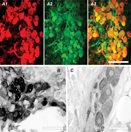
The general neuronal marker PGP-9.5 was used to identify neurones in the flattened whole-mount intracardiac ganglion plexus preparations (Fig. 6A1–3). Strong GABA immunoreactivity was observed in all neonatal principal neuronal somata and some processes (as indicated by PGP-9.5 immunoreactivity) in intracardiac ganglia of neonatal rats (Fig. 6A1 and B). Immunoreactivity appeared as an even cytoplasmic stain, with the nucleus less stained and the nucleoli unstained. The intensity of the GABA immunoreactivity in adult neurones was substantially less than for neonatal neurones labelled with the same antibody concentration and incubation time.
Figure 6. GABA in rat intracardiac neuronal somata.
A1–3, series of confocal fluorescence images of a neonatal (P2) ICG labelled for GABA (A1) and PGP-9.5 (A2). A3 shows the high co-localization of both markers. Inverted confocal fluorescence images of a neonatal (P4, B) and adult (C) ICG labelled for GABA. To generate the image shown (C), it was necessary to use higher gain settings for the adult tissue compared with neonatal tissue. Scale bars, 50 μm.
Discussion
The findings of the present study suggest that GABA and taurine may act as modulators of neurotransmission and cardiac function in the developing mammalian intrinsic cardiac nervous system. The results demonstrate that: (i) rat ICG neurones possess functional somatic GABAA receptor-channels, (ii) the GABAA receptor-channel current density is down-regulated during the first postnatal weeks, (iii) the reduction in GABA-induced current amplitude is due to a change of intracellular Cl− concentration, and (iv) GABA is present in the majority of neonatal rat intracardiac ganglion neurones.
GABAA receptor-channel expression in rat ICG neurones
Focal application of GABA evoked a transient depolarization of neurones in the intracardiac ganglion in situ of neonatal and adult rats. In dissociated intracardiac neurones, GABA and the GABAA agonists, muscimol and taurine, evoked depolarizing responses in neonatal rat ICG neurones mediated by outward Cl− flux. These currents were inhibited by picrotoxin and the specific GABAA antagonist bicuculline. Baclofen failed to evoke a response indicating the absence of functional GABAB receptors in rat intracardiac neurones. Furthermore, TPMPA did not inhibit either GABA or muscimol-evoked currents suggesting that GABAA0 receptor-channels were unlikely to mediate the GABA-evoked response in these neurones.
Previous studies of recombinant GABA receptors have shown that at least one α or β and one γ subunit are essential to form a benzodiazepine binding site (Pritchett et al. 1989) and that DMCM is an agonist at receptors composed of α, β and γ1 subunits (Puia et al. 1991). The GABA receptor-mediated current in rat ICG neurones was inhibited by the inverse agonist DMCM suggesting a contribution of γ2 or γ3 to native GABAA receptors in these neurones. However, it has been shown that recombinant GABAA receptors containing the γ3 subunits are 3- to 4-fold more sensitive to GABA than those containing γ2 (Knoflach et al. 1991). In the present study, an EC50 of ∼ 30 μm was obtained for both GABA and muscimol, suggesting a contribution of γ2 to GABAA receptor-channels in parasympathetic ICG neurones. The presence of a γ subunit in any combination with the other subunits leads to the formation of GABAA receptors that are insensitive to Zn2+ (Draguhn et al. 1990). In contrast, Zn2+ inhibited both GABA- and muscimol-evoked currents similar to DMCM, suggesting that a population of GABAA receptor-channels in rat ICG neurones may not contain γ subunits. However, further experiments using receptor subtype-specific antagonists and agonists and immunolabelling with antibodies directed against GABAA receptor subtypes will be necessary to reveal subunit composition and heterogeneity of GABAA receptor-channels in rat ICG neurones.
Postnatal regulation of GABAA receptor-channel density in rat ICG neurones
GABA-induced current amplitude decreased with increasing postnatal age whereas GABAA receptor pharmacology did not change between age groups. The potency for GABA, muscimol and taurine was relatively constant in ICG neurones for neonatal, juvenile and adult rats, suggesting a decrease in receptor number or density. GABA receptor-mediated currents exhibit marked inward rectification and the rectification observed in adult neurones could prevent the switch from excitatory to inhibitory responses in ICG neurones even if the Cl− equilibrium potential shifts to potentials more negative than the resting membrane potential.
Age-dependent reduction in GABA-induced current amplitude due to change of intracellular Cl− concentration
An age-dependent shift in the reversal potential of the GABA-evoked current due to a decrease in intracellular Cl− concentration was observed using the gramicidin perforated-patch technique in neonatal and juvenile rats. A decrease in receptor density together with the shift in intracellular Cl− concentration appears as an efficient concurrent strategy to minimize GABA receptor activation in ICG neurones of adult rats. A Cl− concentration of 21.5 mm and therefore a reversal potential of −49.0 mV, is similar to the resting membrane potential of intracardiac neurones. Activation of the GABAA receptor-channels, by either endogenous GABA or taurine, would not trigger Cl− net movement; however, the elevated intracellular Cl− concentration in neonatal rat ICG neurones ensures that GABAA receptor-channel activation leads to Cl− efflux which depolarizes the neurone beyond the firing threshold. An elevated intracellular Cl− concentration in immature neurones is common in the central nervous system (Owens & Kriegstein, 2002). It has been shown that the intracellular Cl− concentration of hippocampal GABAergic neurones is 20–40 mm higher in newborn than in adult rat (Ben-Ari, 2002) which is sufficient to shift the actions of GABA from inhibition to excitation. The delayed expression of the KCC2 Na+–K+–2Cl− co-transporter, which pumps Cl− out of these neurones and/or the early expression of Cl−-accumulating Na+–K+–2Cl− co-transporter NKCC1 appear to underlie Cl− homeostasis. NKCC1 has been suggested to play a pivotal role in the generation of GABA-mediated depolarization in immature cortical plate neurones, whereas KCC2 promotes the later maturation of GABAergic inhibition in the rat neocortex (Yamada et al. 2004). However, to date, it is not known if these cation–chloride co-transporters are expressed in ICG neurones.
The presence of GABA in neonatal rat ICG neurones
The presence of GABA in some processes but primarily in the soma of ICG neurones was demonstrated using a monoclonal antibody directed against GABA. However, there is no evidence for GABA-ergic pathways connecting the nervous system to the heart. Therefore, this suggests that putative GABA release from ICG neurones may act in an autocrine or paracrine fashion, that is, regulate intraganglionic transmission during neonatal development. Depolarization caused by the release of ACh from preganglionic nerves may trigger the somatic release of GABA to activate GABAA receptors and reinforce/amplify the depolarization of neonatal rat ICG neurones.
Sympathetic innervation of the rat heart occurs approximately 2 weeks postnatally (Robinson, 1996) during which time in development the heart rate and contraction is asymmetrically controlled largely by the parasympathetic nervous system. The expression of GABA receptor-channels in ICG neurones during that period provides a mechanism of stimulation which may contribute to the development of the mammalian heart.
Acknowledgments
This work was supported by a grant from the Australian Research Council to D.J.A. and the British Heart Foundation to A.A.H. A.A.H. thanks The Carnegie Trust for the Universities of Scotland for a Travel Research Award. We are grateful to Robert Richardson for his invaluable assistance with the whole-mount dissection and immunohistochemistry and Pankaj Sah for his constructive criticism of a draft of the manuscript.
References
- Adams DJ, Cuevas J. Electrophysiological properties of intrinsic cardiac neurons. In: Ardell JL, Armour JA, editors. Basic and Clinical Neurocardiology. Oxford: Oxford University Press; 2004. pp. 1–60. [Google Scholar]
- Akaike N. Gramicidin perforated patch recording and intracellular chloride activity in excitable cells. Prog Biophys Mol Biol. 1996;65:251–264. doi: 10.1016/s0079-6107(96)00013-2. 10.1016/S0079-6107(96)00013-2. [DOI] [PubMed] [Google Scholar]
- Akasu T, Munakata Y, Tsurusaki M, Hasuo H. Role of GABAA and GABAC receptors in the biphasic GABA responses in neurons of the rat major pelvic ganglia. J Neurophysiol. 1999;82:1489–1496. doi: 10.1152/jn.1999.82.3.1489. [DOI] [PubMed] [Google Scholar]
- Armour JA. Myocardiac ischaemia and the cardiac nervous system. Cardiovasc Res. 1999;41:41–54. doi: 10.1016/s0008-6363(98)00252-1. 10.1016/S0008-6363(98)00252-1. [DOI] [PubMed] [Google Scholar]
- Ben-Ari Y. Excitatory actions of gaba during development: the nature of the nurture. Nat Rev Neurosci. 2002;3:728–739. doi: 10.1038/nrn920. [DOI] [PubMed] [Google Scholar]
- Billingsley ML, Suria A. Effects of peripherally administered GABA and other amino acids on cardiopulmonary responses in anesthetized rats and dogs. Arch Int Pharmacodyn Ther. 1982;255:131–140. [PubMed] [Google Scholar]
- Brown DA, Adams PR, Higgins AJ, Marsh S. Distribution of GABA-receptors and GABA-carriers in the mammalian nervous system. J Physiol (Paris) 1979;75:667–671. [PubMed] [Google Scholar]
- Draguhn A, Verdorn TA, Ewert M, Seeburg PH, Sakmann B. Functional and molecular distinction between recombinant rat GABAA receptor subtypes by Zn2+ Neuron. 1990;5:781–788. doi: 10.1016/0896-6273(90)90337-f. 10.1016/0896-6273(90)90337-F. [DOI] [PubMed] [Google Scholar]
- Edwards FR, Hirst GD, Klemm MF, Steele PA. Different types of ganglion cell in the cardiac plexus of guinea-pigs. J Physiol. 1995;486:453–471. doi: 10.1113/jphysiol.1995.sp020825. [DOI] [PMC free article] [PubMed] [Google Scholar]
- Fischer H, Adams DJ. Developmental regulation of GABA-ergic responses in intrinsic cardiac neurons of the rat heart. J Mol Cell Cardiol. 2004;37:202. [Google Scholar]
- Hosie AM, Dunne EL, Harvey RJ, Smart TG. Zinc-mediated inhibition of GABAA receptors: discrete binding sites underlie subtype specificity. Nat Neurosci. 2003;6:362–369. doi: 10.1038/nn1030. 10.1038/nn1030. [DOI] [PubMed] [Google Scholar]
- Knoflach F, Rhyner T, Villa M, Kellenberger S, Drescher U, Malherbe P, Sigel E, Mohler H. The γ3-subunit of the GABAA-receptor confers sensitivity to benzodiazepine receptor ligands. FEBS Lett. 1991;293:191–194. doi: 10.1016/0014-5793(91)81184-a. 10.1016/0014-5793(91)81184-A. [DOI] [PubMed] [Google Scholar]
- Loscher W. Cardiovascular effects of GABA, GABA-aminotransferase inhibitors and valproic acid following systemic administration in rats, cats and dogs: pharmacological approach to localize the site of action. Arch Int Pharmacodyn Ther. 1982;257:32–58. [PubMed] [Google Scholar]
- Macdonald RL, Olsen RW. GABAA receptor channels. Annu Rev Neurosci. 1994;17:569–602. doi: 10.1146/annurev.ne.17.030194.003033. [DOI] [PubMed] [Google Scholar]
- Matsuyama S, Saito N, Shuntoh H, Taniyama K, Tanaka C. GABA modulates neurotransmission in sinus node via stimulation of GABAA receptor. Am J Physiol. 1993;264:H1057–1061. doi: 10.1152/ajpheart.1993.264.4.H1057. [DOI] [PubMed] [Google Scholar]
- Matsuyama S, Saito N, Taniyama K, Tanaka C. γ-Aminobutyric acid is a neuromodulator in sinus node of guinea pig heart. Am J Physiol. 1991;261:H1437–1442. doi: 10.1152/ajpheart.1991.261.5.H1437. [DOI] [PubMed] [Google Scholar]
- Owens DF, Kriegstein AR. Is there more to GABA than synaptic inhibition? Nat Rev Neurosci. 2002;3:715–727. doi: 10.1038/nrn919. 10.1038/nrn919. [DOI] [PubMed] [Google Scholar]
- Pritchett DB, Sontheimer H, Shivers BD, Ymer S, Kettenmann H, Schofield PR, Seeburg PH. Importance of a novel GABAA receptor subunit for benzodiazepine pharmacology. Nature. 1989;338:582–585. doi: 10.1038/338582a0. 10.1038/338582a0. [DOI] [PubMed] [Google Scholar]
- Puia G, Vicini S, Seeburg PH, Costa E. Influence of recombinant gamma-aminobutyric acid-A receptor subunit composition on the action of allosteric modulators of gamma-aminobutyric acid-gated Cl− currents. Mol Pharmacol. 1991;39:691–696. [PubMed] [Google Scholar]
- Reis HJ, Biscaro FV, Gomez MV, Romano-Silva MA. Depolarization-evoked GABA release from myenteric plexus is partially coupled to L-, N-, and P/Q-type calcium channels. Cell Mol Neurobiol. 2002;22:805–811. doi: 10.1023/A:1021821427540. 10.1023/A:1021821427540. [DOI] [PMC free article] [PubMed] [Google Scholar]
- Richardson RJ, Grkovic I, Anderson CR. Immunohistochemical analysis of intracardiac ganglia of the rat heart. Cell Tissue Res. 2003;314:337–350. doi: 10.1007/s00441-003-0805-2. 10.1007/s00441-003-0805-2. [DOI] [PubMed] [Google Scholar]
- Robinson RB. Autonomic receptor – effector coupling during post-natal development. Cardiovasc Res. 1996;31:E68–E76. 10.1016/0008-6363(95)00151-4. [PubMed] [Google Scholar]
- Sampaio KN, Mauad H, Spyer KM, Ford TW. Differential chronotropic and dromotropic responses to focal stimulation of cardiac vagal ganglia in the rat. Exp Physiol. 2003;88:315–327. doi: 10.1113/eph8802525. 10.1113/eph8802525. [DOI] [PubMed] [Google Scholar]
- Seabrook GR, Fieber LA, Adams DJ. Neurotransmission in neonatal rat cardiac ganglion in situ. Am J Physiol. 1990;259:H997–1005. doi: 10.1152/ajpheart.1990.259.4.H997. [DOI] [PubMed] [Google Scholar]
- Selyanko AA, Skok VI. Synaptic transmission in rat cardiac neurones. J Auton Nerv Syst. 1992;39:191–199. doi: 10.1016/0165-1838(92)90012-6. 10.1016/0165-1838(92)90012-6. [DOI] [PubMed] [Google Scholar]
- Sha L, Miller SM, Szurszewski JH. Electrophysiological effects of GABA on cat pancreatic neurons. Am J Physiol. 2001;280:G324–331. doi: 10.1152/ajpgi.2001.280.3.G324. [DOI] [PubMed] [Google Scholar]
- Sieghart W. Structure and pharmacology of gamma-aminobutyric acidA receptor subtypes. Pharmacol Rev. 1995;47:181–234. [PubMed] [Google Scholar]
- Sieghart W, Fuchs K, Tretter V, Ebert V, Jechlinger M, Hoger H, Adamiker D. Structure and subunit composition of GABAA receptors. Neurochem Int. 1999;34:379–385. doi: 10.1016/s0197-0186(99)00045-5. 10.1016/S0197-0186(99)00045-5. [DOI] [PubMed] [Google Scholar]
- Smith FM, McGuirt AS, Leger J, Armour JA, Ardell JL. Effects of chronic cardiac decentralization on functional properties of canine intracardiac neurons in vitro. Am J Physiol. 2001;281:R1474–1482. doi: 10.1152/ajpregu.2001.281.5.R1474. [DOI] [PubMed] [Google Scholar]
- Xu ZJ, Adams DJ. Resting membrane potential and potassium currents in cultured parasympathetic neurones from rat intracardiac ganglia. J Physiol. 1992;456:405–424. doi: 10.1113/jphysiol.1992.sp019343. [DOI] [PMC free article] [PubMed] [Google Scholar]
- Yamada J, Okabe A, Toyoda H, Kilb W, Luhmann HJ, Fukuda A. Cl− uptake promoting depolarizing GABA actions in immature rat neocortical neurones is mediated by NKCC1. J Physiol. 2004;557:829–841. doi: 10.1113/jphysiol.2004.062471. 10.1113/jphysiol.2004.062471. [DOI] [PMC free article] [PubMed] [Google Scholar]
- Zhou X, Galligan JJ. GABAA receptors on calbindin-immunoreactive myenteric neurons of guinea pig intestine. J Auton Nerv Syst. 2000;78:122–135. doi: 10.1016/s0165-1838(99)00065-x. 10.1016/S0165-1838(99)00065-X. [DOI] [PubMed] [Google Scholar]


