Abstract
Placement of the division site at midcell in Escherichia coli requires the MinE protein. MinE acts by imparting topological specificity to the MinCD division inhibitor, preventing the inhibitor from acting at the midcell site while permitting it to block division at other unwanted sites along the length of the cell. It was previously shown that MinE assembled into a ring structure that appeared to be localized near midcell, apparently explaining the ability of MinE to specifically counteract MinCD at midcell. We report here that the MinE ring is not fixed in position near midcell but is a dynamic structure that undergoes a repetitive cycle of movement first to one cell pole and then to the opposite pole. Taken together with studies of the dynamic behavior of the MinD protein, the results suggest that the topological specificity of division site placement may not involve a localized action of MinE to counteract the MinCD division inhibitor at midcell but rather the ability of MinE to move the division inhibitor away from midcell and to the cell poles.
Two annular structures, the septal ring and E-ring, play essential roles in the normal cell division cycle of Escherichia coli. Each of the rings is a membrane-associated structure that extends around the cell cylinder at right angles to the long axis of the cell. They have been identified by immunoelectronmicroscopy and immunofluorescence microscopy and by the use of green fluorescent protein (Gfp) fusion proteins.
The septal ring contains a number of essential division proteins and is required for ingrowth of the division septum (1). The E-ring is characterized by the presence of the MinE protein and is required for the proper placement of the division site at midcell (2). The septal ring is assembled at midcell early in the cell cycle and remains at midcell until septal invagination is completed. In this paper, we report that, in contrast to the static septal ring, the MinE ring is a mobile structure that undergoes repeated cycles of displacement, dissolution, and reformation during the course of the cell cycle.
In E. coli, placement of the division site at midcell requires the action of the three protein products of the min operon, MinC, MinD, and MinE (3). The three proteins act cooperatively to prevent division at aberrant potential division sites that are located at cell poles and perhaps elsewhere along the length of the cell. In the absence of the Min proteins, septa are frequently mislocated so that division occurs adjacent to a cell pole instead of at midcell, leading to formation of chromosomeless minicells that are unable to carry out subsequent rounds of division.
The three proteins function in the following manner. MinC is a nonspecific inhibitor of septation that is capable of blocking division at all potential division sites when expressed in the absence of MinD and MinE (4). MinD is required to bring MinC to the membrane (5, 6). However, the membrane-associated MinC division inhibitor still lacks site specificity, as shown by induction of filamentation when minC and minD are expressed in the absence of MinE. MinE gives site specificity to the MinCD division inhibitor, preventing it from blocking division at the proper midcell site, while permitting it to prevent septation at other sites. As a result, in the presence of MinC, MinD, and MinE, septation is restricted to midcell (3).
MinD is also required to permit MinE to associate with the membrane (2), suggesting that one role for MinD is to act as a membrane assembly protein for MinC and MinE. The ability of MinD to promote the membrane association of MinE is thought to explain the requirement for MinD in the topological specificity of septal placement (2)
That MinE is required to give site specificity to the division inhibitor suggests that MinE acts as a topological specificity protein, capable of recognizing the midcell site and preventing the MinC division inhibitor from acting at this site (7). Consistent with this view, it was shown that MinE-Gfp forms a ring-like structure, the E-ring, reported to be primarily located near the midpoint of the cell (2). The E-ring has been presumed to act locally at midcell to prevent the action of the MinCD division inhibitor. It is not known whether the ring is composed only of MinE or whether other molecules are also a part of the structure.
MinE also induces the redistribution of MinD and MinC so that almost all of the cellular MinD and MinC becomes localized to one end of the cell, forming a membrane-associated polar zone (6, 8, 9). Remarkably, the MinD (8) and MinC (5, 6) polar zones rapidly oscillate from pole to pole by an unknown mechanism. The rapid pole-to-pole oscillation of the division inhibitor presumably explains the fact that division is inhibited at both cell poles when MinC, MinD, and MinE are coordinately expressed.
It is not understood how MinE assembles into the ring structure nor how MinE functions to induce formation of the MinD polar zones. In the present work, we show that the MinE ring that forms near midcell is not fixed in position but is a dynamic structure that undergoes a repetitive cycle of movement to the cell pole, followed by dissolution, reformation at a new midcell site, and movement to the opposite pole. The dynamic movement of the MinE ring sharply distinguishes it from the FtsZ-based septal ring that remains at its original midcell site until septation is concluded. The studies further show that MinE molecules that are not present in the E-ring are localized in a membrane-associated polar zone at one end of the cell, whose medial boundary is formed by the MinE ring. The MinE polar zones undergo pole-to-pole oscillation synchronously with the MinE ring, in a process that is similar to that of MinD and MinC. The results have important implications for the behavior of membrane-associated organelles and the process of selection of the site of cell division.
Materials and Methods
Bacterial Strains.
E. coli RC1 [ΔminCDE Δ(araAB01C-leu)] (9), PB103 [minCDE+ dadR trpE trpA tna] (3), and PB103(λCH50) [minCDE+(Plac-zipA∷gfpS65T)] (10) have been described. E. coli SJ307 [ΔlacX74 galE galK thi rpsL ΔphoA(PvuII) Δara714] was kindly provided by Sheryl Justice (University of Connecticut School of Medicine).
Plasmids.
pAS73 [Para-minD] (9), pDB175 [pMLB1113∷Plac-minD minE] (3), and pSLR22 [Para-gfpmut2∷minD] (9) have been previously described.
pSY1083 [Plac-minE].
pDB170 (3) was used as template in a PCR to generate a minCDE fragment with an EcoRI site upstream of minC, and with BamHI and XbaI cleavage sites positioned upstream and downstream of the minE stop codon. Plasmid pKENgfpmut2 (11) was used as template in a PCR to generate a fragment containing gfpmut2, with a HindIII site positioned downstream of the stop codon and with sequential XbaI and BamHI sites positioned immediately 5′ to the gfp start codon, which was changed from ATG (Met) to ATC (Ile). The EcoRI/XbaI-digested minCDE fragment and the XbaI/HindIII-digested gfp fragment were ligated into EcoRI/HindIII-digested pMLB1113 vector (3) in a three-fragment ligation reaction. The resulting plasmid (pSY1083) contains a TAA stop codon at the normal termination position of minE, immediately downstream of a BamHI site. pSY1083 codes for the complete MinE protein with an additional Gly-Ser at the C terminus. The pSY1083 MinE product was indistinguishable from normal MinE in ability to restore a wild-type division pattern to a Δmin strain when expressed coordinately with minC and minD, consistent with the fact that the C terminus of minE is unstructured (12) and that the seven C-terminal amino acids of MinE can be removed without affecting MinE function (13). The coding sequence of gfpmut2 is located downstream of minE, with XbaI and BamHI sites located between the minE stop codon and the gfp initiation codon. The gfp message is not translated because of lack of an initiator Met codon and lack of a suitably placed ribosome-binding site. Cleavage of this plasmid with BamHI and subsequent religation generates an in-frame minE∷gfpmut2 fusion.
pSY1083G [Plac-minC minD minE∷gfpmut2].
pSY1083 was cleaved with BamHI and religated to generate pSY1083G. The plasmid codes for a MinE-Gfpmut2 fusion protein in which Gly-Ser-Ile is inserted between the end of MinE and the second amino acid of Gfp.
pFX7 [Plac-minD minE∷gfpmut2].
A minD minE∷gfpmut2 fragment was obtained by XbaI (Klenow enzyme-treated)/HindIII treatment of pAS82 (9) and was inserted into SmaI/HindIII-digested pMLB1113.
pFX37 [Plac-minE∷gfpmut2].
A minE∷gfpmut2 fragment was obtained by XmnI/HindIII digestion of pAS82 (9) and was ligated into SmaI/HindIII-digested pMLB1113. The minE∷gfp gene is preceded by its normal ribosome-binding site.
pFX8 [Plac-gfpmut2∷minD].
The EcoRI/HindIII gfpmut2∷minD fragment from pSLR22 (9) was ligated into EcoRI/HindIII-digested pMLB1113.
pFX9 [Plac-gfpmut2∷minD minE].
The EcoRI/HindIII gfpmut2∷minD minE fragment from pSLR23 (9) was ligated into EcoRI/HindIII-digested pMLB1113.
Growth Conditions.
Strains were grown overnight in LB medium with 1% glucose, and 50 μg/ml ampicillin or 25 μg/ml chloramphenicol. Cells were harvested by centrifugation, washed twice with fresh LB, and then suspended to a density (A600) of 0.03–0.05 in Medium A (M9 minimal medium containing 0.2% maltose, 0.005% tryptophan, and 0.2% casamino acids) or Medium B [Medium A/L-broth (1:1)], plus the indicated antibiotics. Medium A was used for all experiments unless otherwise noted. After adding isopropyl β-d-thiogalactoside (IPTG) and/or arabinose, the cultures were grown at 30°C with shaking for 4–6 h, to an A600 of approximately 0.5. The cultures were either fixed with formaldehyde and glutaraldehyde (9) or were directly applied to slides coated with 0.1% polylysine.
Microscopy and Data Analysis.
Samples were examined by using an Olympus BX60 microscope (New Hyde Park, NY). Images were collected at intervals and were processed by using Adobe photoshop. For measurement of oscillation rates (expressed as mean ±1 SD), strain PB103/pFX7 was grown for 5 h at 30°C in Medium B containing 10 μM IPTG. Samples were examined at 15-s intervals. One cycle was defined as the time from appearance of the MinE ring near midcell to the time of its reappearance at the same position, after intermediate stages of movement to the pole and formation and displacement of a new ring in the opposite half of the cell. A total of 25 cells were analyzed.
Immunoblot Analysis.
Western blot analysis was performed as previously described (14) with antibody directed against MinE (2–19) or directed against the whole wild-type MinD protein (13).
Results
The E-Ring Cycle.
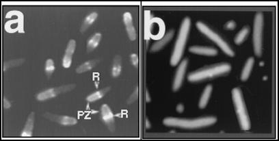
In cells that coexpress minD and minE∷gfp, MinE rings (R in Fig. 1a) were identified as bright fluorescent bands that extended at right angles to the long axis of the cell or as fluorescent spots that were present as doublets on opposite sides of the cell cylinder. Similar doublets are characteristic of other division-related ring structures in E. coli (10, 15). In addition, the cells also contained membrane-associated polar zones (PZ in Fig. 1a), in which most of the remaining membrane-associated MinE-Gfp fluorescence was located in a single zone that extended from the E-ring to a cell pole. Raskin and de Boer (2) had also noted that MinE-Gfp fluorescence that was not present in the E-ring was often asymmetrically distributed in the two halves of the cell.
Figure 1.
MinE-Gfp rings and polar zones. Strains RC1/pFX7 [Δmin/Plac-minD minE∷gfp] (a) and RC1/pFX37 [Δmin/Plac-minE∷gfp] (b) were grown for 4 h in the presence of 10 μM IPTG. Cells were then fixed and examined by fluorescence microscopy. R, MinE ring; PZ, MinE polar zone.
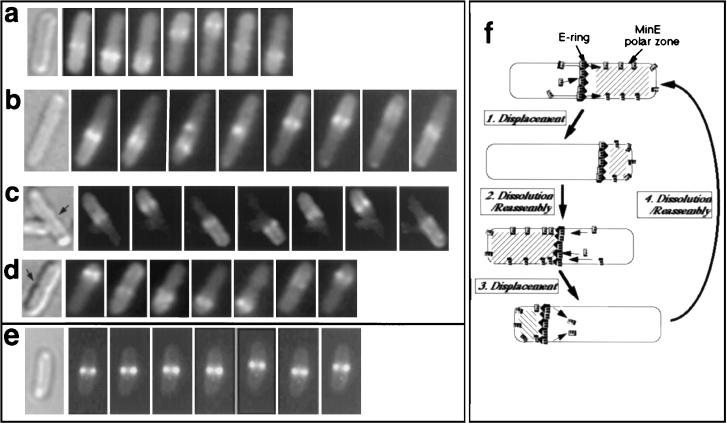
It was apparent from examination of the MinE-Gfp fluorescence patterns in fixed cells that the E-rings were not limited to positions near the midpoint of the cell, as had originally been described (2). Examination of living cells showed that this variable distribution pattern reflected the fact that the E-rings were not static structures, but instead underwent a topologically defined process of movement within the cell. Time-lapse photography revealed the following sequence of events (summarized in Fig. 2f).
Figure 2.
Time-lapse analysis of MinE-Gfp and Gfp-ZipA. (a–f) Cells of the following strains were grown for 4 h in the presence of 10 μM IPTG before fluorescence microscopy of unfixed cells. (Left) Nomarski. (Right) Fluorescence micrographs at 20-s intervals (left to right). Arrow indicates nascent septum. (a) RC1/pFX7 [Δmin/Plac-minD minE∷gfp]. (b) PB103/pFX7 [minCDE+/Plac-minD minE∷gfp]. (c) RC1/pSY1083G [Δmin/Plac-minC minD minE∷gfp]. (d) RC1/pSY1083G [Δmin/Plac-minC minD minE∷gfp]. (e) PB103(λCH50) [minCDE +(Plac-zipA∷gfp)]. (e) Diagram of cyclic behavior of MinE-Gfp rings and polar zones, based on time-lapse fluorescence microscopy (a–d).
The E-ring first appeared near midcell and then was displaced progressively toward the nearest pole (Fig. 2 a–d). This was associated with a progressive shrinking of the MinE polar zone, with the E-ring remaining associated with the medial edge of the polar zone. On approaching the pole, the MinE polar zone and ring disappeared and a new E-ring and polar zone were formed in the opposite half of the cell. In approximately 25% of the cells, the new ring appeared before disappearance of the original E-ring from the other cell pole (panel 4 in Fig. 2b; panel 5 in Fig. 4a). The newly formed E-ring was then displaced from midcell toward the pole, repeating the movement pattern of the original ring but moving in the opposite direction. The ring and polar zone then disappeared and a new cycle was initiated by formation of a new midcell ring oriented toward the other half of the cell. The cycle continued to repeat itself until photobleaching reduced the fluorescence to low levels that were difficult to follow.
Figure 4.
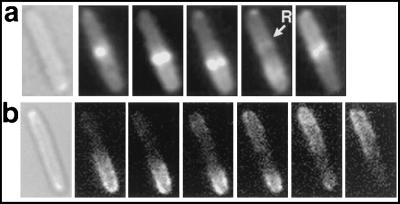
Comparison of MinE and MinD polar zones. Strains RC1/pFX7 [Δmin/Plac-minD minE∷gfp] (a) and SJ307/pFX8 [minCDE+/Plac-gfp∷minD minE] were grown for 5 h in 10 μM IPTG. Unfixed cells were examined by Nomarski microscopy (Left) and time-lapse fluorescence microscopy (Right). Micrographs were collected every 20 s (a) or every 15 s (b). R, MinE ring.
The repetitive end-to-end oscillation phenomenon was not prevented by the nascent septum of dividing cells (Fig. 2 c and d). In these cells, the new MinE ring appeared to form on one side of the ingrowing septum, and then to move in the usual manner to the proximal cell pole. This was followed by repetition of the process in the opposite half of the dividing cell. We presume that, either shortly before or after cell separation, the midpoint of the new daughter cell is then established as the reference point for formation of E-rings during subsequent oscillation events.
The average time required for a single oscillation cycle was 128 ± 39 s (see Materials and Methods). The cycling time was affected by the length of the cell being observed, being shortest in the shorter cells.
The surprising observation that the MinE ring structures were capable of a systematic cycle of migration, dissolution, and reformation raised the possibility that other ring structures associated with the bacterial division site might behave similarly. To ask whether the septal ring underwent a similar process, we examined the behavior of ZipA, a component of the septal ring (10), by using a ZipA-Gfp fusion protein. This revealed that the ZipA ring failed to move from its original position at midcell (Fig. 2e), suggesting that the septal ring remains in place during the division cycle, in contrast to the behavior of the E-ring.
The MinE rings and polar zones were present in 80–90% of cells from cultures of RC1/pFX7 [ΔminCDE/Plac-minD minE∷gfp] grown in L-broth for 3–4 h at 30°C in the presence of 10 μM IPTG. The polar zones appeared to be associated with the membrane or cell envelope as shown by the concentration of the fluorescence in a thin zone at the periphery of the cell (Fig. 1a), although MinE-Gfp fluorescence was also visible elsewhere in the cell. It was striking that the medial edge of the MinE polar zone was almost always delimited by a MinE ring.
MinD was required for formation of both the E-ring and the MinE polar zone. Evidence that formation of the MinE polar zones required MinD came from the observation that MinE polar zones were present when minD and minE∷gfp were coexpressed in a strain that contained a chromosomal deletion of the minCDE operon (Figs. 1a and 2a), but were absent when minE∷gfp was expressed in the same strain in the absence of MinD (Fig. 1b). In the latter case, the MinE-Gfp was present diffusely within the cytoplasmic region of the cell. The requirement for MinD in formation of the MinE ring and polar zone presumably reflects the fact that MinD is required for cytoplasmic MinE to assemble into the membrane (2).
In the experiments described above, minD and minE∷gfp were coexpressed in a ΔminCDE strain. As a result, the cells showed a minicelling phenotype resulting from the absence of the MinC division inhibitor. Similar results were obtained when minD and minE∷gfp were coexpressed in cells that expressed minC, minD, and minE from the chromosomal minCDE locus (Fig. 2b); in this case, the cells were somewhat longer than wild-type cells, and occasional minicells were present. Evidence that the formation of MinE rings and polar zones and their oscillatory behavior was also characteristic of normally dividing cells was obtained by studying the distribution of MinE-Gfp in ΔminCDE cells that expressed minC, minD, and minE∷gfp from plasmid pSY1083G (Fig. 2 c and d). Under these conditions, the division pattern of the cells was essentially wild type. We conclude from the experiments described in Figs. 1 and 2 that the ability of MinE-Gfp to form both the E-ring and the MinE polar zone, and their pole-to-pole oscillation, requires MinD but is independent of the presence of MinC, and is seen in normally dividing cells as well as in cells from minicelling cultures.
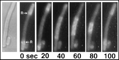
It was previously noted in fixed preparations that MinE-Gfp in filamentous cells often showed a “zebra” pattern (2), where fluorescence alternated between bright and less bright domains along the length of the filaments, with each internal domain usually delimited by MinE rings at both ends. In the present experiments, time-lapse photography showed that the internal membrane-associated MinE zones along the length of the filaments were not fixed in position but underwent oscillatory behavior that resembled that of the MinE polar zones in wild-type cells. This is illustrated in Fig. 3, where disappearance of the two fluorescent polar zones was associated with the appearance of a fluorescent zone in the middle of the filament. This was followed by disappearance of the central zone and reappearance of the original fluorescent MinE polar zones. The oscillatory behavior of the ring-delimited MinE-Gfp structures in filamentous cells supports the view that these structures are analogous to the oscillating MinE polar zones of shorter cells (Fig. 2 a–d).
Figure 3.
Cyclic redistribution of Gfp-MinE in filamentous cells. Strain PB103/pFX7 [minCDE +/Plac-minD minE∷gfp] was grown for 4 h in the presence of 10 μM IPTG. Unfixed cells were examined. The culture contained minicells and short- and medium-length filaments, because of overexpression of minDE. A medium-length filament was examined. (Left) Nomarski micrograph. (Right) Fluorescence micrographs taken at 20-s intervals. Polar zones are initially present at both ends of the cell, delimited by MinE rings. Each zone is then displaced toward the cell poles, together with the ring. The polar zones then disappear, and a single long MinE zone appears at midcell. The central zone then collapses toward midcell and the polar zones reappear. R, MinE ring.
Relation of MinE Polar Zones to MinD Polar Zones.
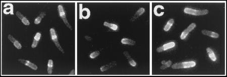
In their cellular distribution and oscillatory behavior, the MinE-Gfp polar zones resembled the previously described MinD and MinC polar zones, whose presence at the ends of the cell required the presence of MinE. Time-lapse experiments showed that Gfp-MinD polar zones (Fig. 4b) behaved like the MinE zones (Fig. 4a). In both cases, the polar zones initially extend from midcell to the cell pole. The polar zones then retract toward the pole before disappearing from the polar region and reappearing in the opposite half of the cell. However, in contrast to MinE polar zones, the Gfp-MinD polar zones rarely contained visible fluorescent rings at their trailing edge.
Because of the similarities between the MinE and MinD polar zones, we attempted to determine whether the MinE zones and MinD zones were located at the same end or at opposite ends of the cell. We have not yet succeeded in using fluophores with different emission spectra to unequivocally identify MinD and MinE in individual cells. We therefore studied cells that simultaneously expressed gfp∷minD and minE∷gfp, by growth of strain SJ307/pFX7/pSLR22 [minCDE+/Plac-minD minE∷gfp/Para-gfp∷minD] in the presence of IPTG and arabinose (Fig. 5c). This revealed that most cells in the population contained a clear ring structure, presumed to represent the E-ring, and a single polar zone that presumably represented a mixture of Gfp-MinD and MinE-Gfp. Strikingly, no cells contained polar zones at both ends of the cell. Control experiments in which minE∷gfp was replaced by normal minE showed that Gfp-MinD polar zones were clearly visible (Fig. 5b). Similarly, MinE-Gfp rings and polar zones were present in cells in which gfp∷minD was replaced by normal minD (Fig. 5a). Western blot analysis with anti-MinD and MinE antibodies showed that the concentrations of Gfp-MinD and/or MinE-Gfp in the experimental (Fig. 5c) and control samples (Figs. 5 a and b) were the same, thereby excluding the possibility that the failure to see separate MinD polar zones in the doubly labeled cells might have been because of decreased expression of gfp∷minD. The failure to see any polar zones in the cell end that did not contain the E-rings suggests that the MinE and MinD polar zones were probably present at the same end of the cell.
Figure 5.
Coexpression of Gfp-MinD and MinE-Gfp in the same cell. Strains SJ307/pFX7/pAS73 [minCDE+/Plac-minD minE∷gfp/Para -minD] (a), SJ307/pDB175/pSLR22 [minCDE+/Plac-minD minE/Para -gfp∷minD] (b), and SJ307/pFX7/pSLR22 [minCDE+/Plac-minD minE∷gfp/Para -gfp∷minD] (c) were grown in Medium B in the presence of 10 μM IPTG and 0.0005% arabinose for 5 h. Generation times were 72, 62, and 61 min, and cells were harvested at OD600 0.86, 1.06, and 1.0 for panels a, b, and c, respectively. Unfixed cells were examined by using identical exposure times (0.4 s) for capture of the fluorescent images.
Discussion
The MinE ring is a membrane-associated structure that prevents the MinCD division inhibitor from blocking septation at midcell. As a result, the division inhibitor prevents septation at aberrant division sites elsewhere along the cell cylinder without interfering with septation at the normal midcell site. The original observation that the E-ring appeared to be primarily located near the middle of the cell and to remain at this site until septal invagination was underway had suggested that the ring represented an independent organelle whose maintenance at midcell was required to prevent the local action of the division inhibitor (2).
The present work shows that the E-ring is not fixed in position at midcell but is a mobile structure that undergoes a repetitive cycle of assembly, movement to the cell pole, dissolution, and reassembly (Fig. 2f).† The cycle is repeated many times within each division cycle. The E-ring therefore differs from the other division-related structure at midcell, the septal ring, which assembles and remains at the midcell site until the division cycle is completed (16). The ability of the E-ring to undergo a defined pattern of topological reorganization and relocalization raises the question of whether other well-defined membrane-associated organelles might also alternate between different topologically defined sites as part of normal developmental processes.
Coincident with formation of the central E-ring, most or all of the remaining membrane-associated MinE-Gfp accumulated in a polar zone that extended from the E-ring to the nearest cell pole. It is striking that the E-ring and polar zone behave as a single unit, with the polar zone becoming smaller as the E-ring that forms its medial boundary moves toward the pole. The MinE polar zones closely resemble the membrane-associated polar zones of Gfp-MinD, which also formed in one half of the cell and retracted to the cell pole before undergoing dissolution (Fig. 4b). The MinE and MinD zones are probably located in the same half of the cell because only a single polar zone was visible in cells that coexpressed Gfp-MinD and MinE-Gfp. It will be necessary to more rigorously test this important point and also to establish whether MinD and MinE oscillate coordinately by experiments that simultaneously define the positions of MinD and MinE within the same cell.
It is necessary to reconcile the present observations with the fact that the biological role of MinE is to prevent the MinCD division inhibitor from acting at midcell.
The idea that the MinE ring acts locally to directly counteract the activity of the division inhibitor at midcell could be reconciled with the present observation that the E-ring is only intermittently present at midcell by assuming that the aggregate residency time at midcell is the relevant factor in MinE action, even if residency were discontinuous. On the other hand, instead of acting directly near midcell to counteract the division inhibitor, the sole role of MinE may be to move MinD and MinC away from the middle of the cell, thereby avoiding the risk that the division inhibitor would block septation at the midcell site. This would be consistent with the observation that the MinD polar zone retracts away from midcell in the presence of MinE and would explain all of the current observations. It is also possible that the two mechanisms operate together to prevent MinCD-mediated division inhibition at midcell.
Current models to explain MinE action have assumed that the E-ring is an independent structure. If the E-ring is indeed a separate structure, a topological target or receptor for MinE presumably exists at midcell to explain assembly of the E-ring at this site (12, 13). The present work does not exclude this possibility. However, the present observation that the E-ring and MinE polar zone are contiguous structures that always move together suggests that the E-ring may not be a structurally autonomous element. Instead, the E-ring may represent MinE molecules that associate with the trailing (medial) edge of the MinD-MinE polar zone to form a collar in which the high density of MinE molecules gives the appearance of a separate MinE ring structure. If this is correct, there will be no need for a separate midcell target for E-ring assembly.
The three-dimensional structure of the C-terminal topological specificity domain (TSD) of MinE indicates that the two anti-MinCD domains of the MinE homodimer project from opposite sides of the TSD (12). This raised the possibility that MinE may be oriented so that the anti-MinCD domains project in opposite directions from the E-ring at midcell toward the two cell poles, permitting the E-ring to counteract the MinCD division inhibitor as it approached the midcell ring from either direction (12). The idea that the opposing anti-MinCD domains within the ring counteract MinCD molecules that approach the midcell ring from both poles now seems unlikely in view of the present demonstration that the E-ring does not remain at midcell.
It is striking that MinE is required for formation of MinD polar zones (8, 9), and that MinD is required for formation of the MinE polar zone (2). How might this be explained? In one model, MinE and MinD would enter the polar zone as a membrane-associated MinD-MinE complex. This would explain the similar distribution pattern of the MinD and MinE zones and the observation that neither MinD nor MinE forms polar zones in the absence of the other partner. The binding sites within the polar membrane could be proteins or could be regions in which the molecular organization of the membrane lipids or the local submembranous environment was altered. Alternatively, instead of entering the membrane as a MinD-MinE complex, MinE might catalytically convert MinD to a state that can interact with the polar binding sites, thereby explaining the MinE requirement in formation of MinD polar zones. If this were correct, the entry of MinE molecules into the polar zone would be an independent process, perhaps reflecting secondary MinE–MinD interactions. This would be compatible with the observation (discussed below) that MinD can form polar zones in the absence of detectable MinE polar zones.
The mechanism responsible for the shrinkage of the polar zones and the concomitant movement of the E-ring to the pole is not known. It is possible that the ring moves from midcell to the pole as an intact membrane-associated structure that acts like a piston to progressively compress the polar MinDE domains. However, it is equally plausible that the lateral movement of the E-ring is accomplished by its repeated disassembly and reassembly at the free edge of the shrinking polar zone, with retraction of the polar zone providing the driving force for E-ring movement. Retraction of the polar zone might be accomplished, for example, by a progressive loss of affinity of the polar docking sites for MinD–MinE or MinE, with the loss of affinity beginning at midcell and then extending to the cell poles. The decay in the predicated high affinity state of the polar membrane could be initiated by the binding of the Min protein(s) to the membrane sites. The later reestablishment of the high affinity state would permit the later reformation of the polar MinD–MinE zone at the original end of the cell, thereby explaining the oscillatory behavior of the polar zones.
It should be noted that several additional observations do not readily fit into the present models. The ability to directly counteract the local action of the MinCD division inhibitor, perhaps by dissociating MinD–MinC complexes (17), has been thought to be an intrinsic property of MinE. Therefore, if membrane-associated MinE molecules are actually capable of counteracting the local division inhibitory action of MinCD, and if it proves correct that the polar zones of MinCD and MinE are located in the same half of the cell, it would be expected that the division inhibitor would be prevented from acting at the cell poles. This would lead to minicell formation in wild-type cells, which does not occur. This apparent paradox might be explained if the concentration of MinE in the polar zone were too low to effectively counter the division inhibitor.
In addition, any model to explain the interdependency of MinD and MinE in formation of the polar zones must also account for the fact that truncated MinE proteins that do not form visible E-rings or MinE polar zones are still capable of supporting formation of MinD polar zones (9). Therefore, if formation of the MinD polar zone is brought about by the interaction of MinD–MinE complexes with high affinity sites in one half of the cell, the mutant MinE molecules would have to dissociate from the complex after they initiated formation of the MinD polar zone, presumably because of a decreased affinity for MinD or for the membrane attachment sites. This would explain the fact that E-rings and MinE polar zones are not seen when the mutant MinE molecules are studied. In addition, any model will also have to account for the fact that the mutant MinE proteins induce minicelling when substituted for wild-type MinE. Because of these complexities, validation of these or other plausible models must await further experimentation.
Acknowledgments
This work was supported by Grants GM41978 and GM53276 from the National Institutes of Health.
Abbreviations
- Gfp
green fluorescent protein
- IPTG
isopropyl β-d-thiogalactoside
Footnotes
Article published online before print: Proc. Natl. Acad. Sci. USA, 10.1073/pnas.031549298.
Article and publication date are at www.pnas.org/cgi/doi/10.1073/pnas.031549298
Similar observations have been made by Hale, Raskin, and de Boer (presented at EMBO workshop on Cell Cycle and Nucleoid Organization in Bacteria, Sept. 4, 2000).
References
- 1.Rothfield L, Justice S, Garcia-Lara J. Annu Rev Genet. 1999;33:423–448. doi: 10.1146/annurev.genet.33.1.423. [DOI] [PubMed] [Google Scholar]
- 2.Raskin D, de Boer P. Cell. 1997;91:685–694. doi: 10.1016/s0092-8674(00)80455-9. [DOI] [PubMed] [Google Scholar]
- 3.de Boer P A J, Crossley R E, Rothfield L I. Cell. 1989;56:641–649. doi: 10.1016/0092-8674(89)90586-2. [DOI] [PubMed] [Google Scholar]
- 4.de Boer P, Crossley R E, Rothfield L I. J Bacteriol. 1992;174:63–70. doi: 10.1128/jb.174.1.63-70.1992. [DOI] [PMC free article] [PubMed] [Google Scholar]
- 5.Raskin D, de Boer P. J Bacteriol. 1999;181:6419–6424. doi: 10.1128/jb.181.20.6419-6424.1999. [DOI] [PMC free article] [PubMed] [Google Scholar]
- 6.Hu Z, Lutkenhaus J. Mol Microbiol. 1999;34:82–90. doi: 10.1046/j.1365-2958.1999.01575.x. [DOI] [PubMed] [Google Scholar]
- 7.Rothfield L I, Zhao C R. Cell. 1996;84:183–186. doi: 10.1016/s0092-8674(00)80971-x. [DOI] [PubMed] [Google Scholar]
- 8.Raskin D, de Boer P. Proc Natl Acad Sci USA. 1999;96:4971–4976. doi: 10.1073/pnas.96.9.4971. [DOI] [PMC free article] [PubMed] [Google Scholar]
- 9.Rowland S L, Fu X, Sayed M A, Zhang Y, Cook W R, Rothfield L I. J Bacteriol. 2000;182:613–619. doi: 10.1128/jb.182.3.613-619.2000. [DOI] [PMC free article] [PubMed] [Google Scholar]
- 10.Hale C, de Boer P. Cell. 1997;88:175–185. doi: 10.1016/s0092-8674(00)81838-3. [DOI] [PubMed] [Google Scholar]
- 11.Cormack B P, Valdivia R H, Falkow S. Gene. 1996;173:33–38. doi: 10.1016/0378-1119(95)00685-0. [DOI] [PubMed] [Google Scholar]
- 12.King G F, Shih Y-L, Maciejewski M W, Bains N P S, Pan B, Rowland S, Mullen G P, Rothfield L I. Nat Struct Biol. 2000;7:1013–1017. doi: 10.1038/80917. [DOI] [PubMed] [Google Scholar]
- 13.Zhao C-R, de Boer P, Rothfield L. Proc Natl Acad Sci USA. 1995;92:4313–4317. doi: 10.1073/pnas.92.10.4313. [DOI] [PMC free article] [PubMed] [Google Scholar]
- 14.Zhang Y, Rowland S, King G, Rothfield L. Mol Microbiol. 1998;30:265–273. doi: 10.1046/j.1365-2958.1998.01059.x. [DOI] [PubMed] [Google Scholar]
- 15.Addinall S, Bi E, Lutkenhaus J. J Bacteriol. 1996;178:3877–3884. doi: 10.1128/jb.178.13.3877-3884.1996. [DOI] [PMC free article] [PubMed] [Google Scholar]
- 16.Lutkenhaus J, Addinall S. Annu Rev Biochem. 1997;66:93–116. doi: 10.1146/annurev.biochem.66.1.93. [DOI] [PubMed] [Google Scholar]
- 17.Huang J, Lutkenhaus J. J Bacteriol. 1996;178:5080–5085. doi: 10.1128/jb.178.17.5080-5085.1996. [DOI] [PMC free article] [PubMed] [Google Scholar]


