Abstract
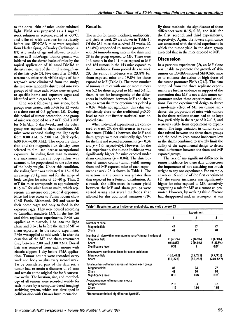
Three independent experiments involving a total of 288 SENCAR mice were used to study the effects of 60-Hz magnetic fields on the growth and development of skin tumors. Given the constraints imposed by the experimental design, the results did not support a role for magnetic fields as a tumor co-promoter. This negative finding could also be interpreted to mean that the SENCAR mouse skin tumor model was not sensitive enough to detect the action of a weak co-promoter. The two-stage (initiation/promotion) model was used to assess the genotoxic potential of magnetic fields because it had been widely used to evaluate chemical carcinogens. This model, however, lacks the sensitivity to detect all but the most potent direct-acting carcinogens, and the tumor response to the action of low doses of promoter results in large random fluctuations in tumor incidence, yield, and multiplicity. The need to limit tumor incidence in the sham is a necessary condition to ensure that a magnetic field-induced effect on tumorigenesis would have a reasonable chance of being detected. This requirement, and the variability in tumor development between and within experiments, increases the level of uncertainty in the system and makes a weak response to the magnetic field difficult to detect and interpret.
Full text
PDF


Selected References
These references are in PubMed. This may not be the complete list of references from this article.
- Bull R. J., Robinson M., Laurie R. D. Association of carcinoma yield with early papilloma development in SENCAR mice. Environ Health Perspect. 1986 Sep;68:11–17. doi: 10.1289/ehp.866811. [DOI] [PMC free article] [PubMed] [Google Scholar]
- Cohen M. M., Kunska A., Astemborski J. A., McCulloch D., Paskewitz D. A. Effect of low-level, 60-Hz electromagnetic fields on human lymphoid cells: I. Mitotic rate and chromosome breakage in human peripheral lymphocytes. Bioelectromagnetics. 1986;7(4):415–423. doi: 10.1002/bem.2250070409. [DOI] [PubMed] [Google Scholar]
- Feinendegen L. E., Mühlensiepen H. Effect of static magnetic field on cellular metabolism in the living mouse. Endeavour. 1988;12(3):119–123. doi: 10.1016/0160-9327(88)90132-9. [DOI] [PubMed] [Google Scholar]
- Harkins T. T., Grissom C. B. Magnetic field effects on B12 ethanolamine ammonia lyase: evidence for a radical mechanism. Science. 1994 Feb 18;263(5149):958–960. doi: 10.1126/science.8310292. [DOI] [PubMed] [Google Scholar]
- Holmberg B. Magnetic fields and cancer: animal and cellular evidence--an overview. Environ Health Perspect. 1995 Mar;103 (Suppl 2):63–67. doi: 10.1289/ehp.95103s263. [DOI] [PMC free article] [PubMed] [Google Scholar]
- Juutilainen J., Liimatainen A. Mutation frequency in Salmonella exposed to weak 100-Hz magnetic fields. Hereditas. 1986;104(1):145–147. doi: 10.1111/j.1601-5223.1986.tb00527.x. [DOI] [PubMed] [Google Scholar]
- McLean J. R., Stuchly M. A., Mitchel R. E., Wilkinson D., Yang H., Goddard M., Lecuyer D. W., Schunk M., Callary E., Morrison D. Cancer promotion in a mouse-skin model by a 60-Hz magnetic field: II. Tumor development and immune response. Bioelectromagnetics. 1991;12(5):273–287. doi: 10.1002/bem.2250120503. [DOI] [PubMed] [Google Scholar]
- McLean J., Thansandote A., Lecuyer D., Goddard M., Tryphonas L., Scaiano J. C., Johnson F. A 60-Hz magnetic field increases the incidence of squamous cell carcinomas in mice previously exposed to chemical carcinogens. Cancer Lett. 1995 Jun 8;92(2):121–125. doi: 10.1016/0304-3835(95)03766-p. [DOI] [PubMed] [Google Scholar]
- Mevissen M., Stamm A., Buntenkötter S., Zwingelberg R., Wahnschaffe U., Löscher W. Effects of magnetic fields on mammary tumor development induced by 7,12-dimethylbenz(a)anthracene in rats. Bioelectromagnetics. 1993;14(2):131–143. doi: 10.1002/bem.2250140206. [DOI] [PubMed] [Google Scholar]
- Milham S., Jr Mortality from leukemia in workers exposed to electrical and magnetic fields. N Engl J Med. 1982 Jul 22;307(4):249–249. doi: 10.1056/nejm198207223070412. [DOI] [PubMed] [Google Scholar]
- Rannug A., Holmberg B., Ekström T., Mild K. H., Gimenez-Conti I., Slaga T. J. Intermittent 50 Hz magnetic field and skin tumor promotion in SENCAR mice. Carcinogenesis. 1994 Feb;15(2):153–157. doi: 10.1093/carcin/15.2.153. [DOI] [PubMed] [Google Scholar]
- Savitz D. A., Loomis D. P. Magnetic field exposure in relation to leukemia and brain cancer mortality among electric utility workers. Am J Epidemiol. 1995 Jan 15;141(2):123–134. doi: 10.1093/oxfordjournals.aje.a117400. [DOI] [PubMed] [Google Scholar]
- Scaiano J. C., Cozens F. L., McLean J. Model for the rationalization of magnetic field effects in vivo. Application of the radical-pair mechanism to biological systems. Photochem Photobiol. 1994 Jun;59(6):585–589. doi: 10.1111/j.1751-1097.1994.tb09660.x. [DOI] [PubMed] [Google Scholar]
- Stuchly M. A., Lecuyer D. W., McLean J. Cancer promotion in a mouse-skin model by a 60-Hz magnetic field: I. Experimental design and exposure system. Bioelectromagnetics. 1991;12(5):261–271. doi: 10.1002/bem.2250120502. [DOI] [PubMed] [Google Scholar]
- Stuchly M. A., McLean J. R., Burnett R., Goddard M., Lecuyer D. W., Mitchel R. E. Modification of tumor promotion in the mouse skin by exposure to an alternating magnetic field. Cancer Lett. 1992 Jul 31;65(1):1–7. doi: 10.1016/0304-3835(92)90205-a. [DOI] [PubMed] [Google Scholar]
- Takatsuji T., Sasaki M. S., Takekoshi H. Effect of static magnetic field on the induction of chromosome aberrations by 4.9 MeV protons and 23 MeV alpha particles. J Radiat Res. 1989 Sep;30(3):238–246. doi: 10.1269/jrr.30.238. [DOI] [PubMed] [Google Scholar]


