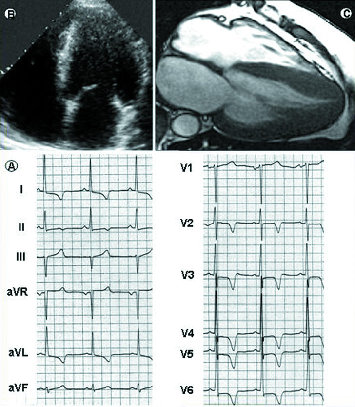
Fig 2.

Diagnostic role of cardiac magnetic resonance in hypertrophic cardiomyopathy. In a 33 year old asymptomatic patient, the 12 lead electrocardiogram (bottom left, A) is grossly abnormal, with increased R wave voltages and marked S-T segment alterations in the precordial leads. The two dimensional echocardiogram (top left, B), however, cannot visualise morphological abnormalities and, in particular, does not provide clear images of the apical portion of the left ventricle. Cardiac magnetic resonance (top right, C) shows high resolution images of the heart and marked thickening of the left ventricular wall, which is principally confined to the apical portion of the ventricle. Left ventricular mass is 156 g/m (normal values 83 g/m). The magnetic resonance image is shown courtesy of Massimo Lombardi, MRI Laboratory, Istituto di Fisiologia Clinica, CNR, Pisa, Italy
