Abstract
This lab reported previously that the plating efficiency of a herpes simplex virus type 1 ICP0-null mutant was enhanced upon release from an isoleucine block which synchronizes cells to G1 phase (W. Cai and P. A. Schaffer, J. Virol. 65:4078-4090, 1991). Peak plating efficiency occurred as cells cycled out of G1 and into S phase, suggesting that the enhanced plating efficiency was due to cellular activities present in late G1/early S phase. We have found, however, that the enhanced plating efficiency did not occur when cells were synchronized by alternative methods. We now report that the plating efficiency of ICP0− viruses is not enhanced at a particular stage of the cell cycle but rather is enhanced by specific cellular stresses. Both the plating and replication efficiencies of ICP0− viruses were enhanced as much as 25-fold to levels similar to that of wild-type virus when monolayers were heat shocked prior to infection. In addition to heat shock, UV-C irradiation but not cold shock of monolayers prior to infection resulted in enhanced plating efficiency. We further report that the effect of cellular stress is transient and that cell density rather than age of the monolayers is the primary determinant of ICP0− virus plating efficiency. As both cell stress and ICP0 are required for efficient reactivation from latency, the identification of cellular activities that complement ICP0− viruses may lead to the identification of cellular activities that are important for reactivation from neuronal latency.
In the human host, herpes simplex virus type 1 (HSV-1) establishes both acute productive infections and lifelong latent infections which reactivate periodically in response to stress. During productive infection of cycling epithelial cells and fibroblasts, all viral genes are expressed, new virus is synthesized, and cell death ensues. In contrast, latent infection of noncycling neurons is characterized by minimal viral gene expression, the absence of detectable viral proteins, the absence of new virus synthesis, and neuron viability. An important distinction exists between the initiation of productive infection and the initial stages of reactivation from latency. During the initiation of productive infection of cycling cells, viral regulatory proteins ICP0, ICP4, and VP16, which are potent transactivators of viral and cellular genes, are present and active. Therefore, through the activities of these regulatory proteins, the virus actively controls the expression of viral and cellular proteins that are important for virus replication during productive infection. In contrast, the only viral gene products expressed at high levels in latently infected neurons are the latency-associated transcripts, which do not appear to encode proteins (21, 33). Virally encoded transactivators are absent or are not present in abundance. Therefore, reactivation of latent viral genomes depends upon cellular activities that are induced in response to stressful stimuli to induce viral gene expression. These cellular activities have not yet been identified.
ICP0 is one of five immediate-early (IE) regulatory proteins of HSV-1 whose activities induce a cellular environment that is favorable for viral replication. Although ICP0 is not essential for viral replication in vitro or in vivo (39), it is essential for efficient plaque formation (3), replication at low multiplicities of infection (MOIs) (3, 4, 15, 40, 45, 51), and reactivation from latency (5, 18, 24). At low MOIs, ICP0− viruses form plaques ∼25- to 100-fold less efficiently than does wild-type virus on most noncomplementing cell types (3, 15, 45). Additionally, as cell monolayers age and grow to higher densities, the plating efficiency of ICP0− viruses decreases such that, by 96 h postplating, these viruses no longer form plaques (3). ICP0− viruses also exhibit an MOI-dependent replication defect, replicating very poorly at low MOIs but as efficiently as the wild-type virus at high MOIs (∼>5) (15, 40, 45). These plating and replication defects are not due to impaired cell entry as wild-type and ICP0− viruses induce similar levels of ICP4 (3), viral DNA, and DNA encapsidation (45). ICP0− viruses also form plaques and replicate as efficiently as wild-type virus on monolayers of the osteosarcoma cell line U2OS (51; our unpublished observations) and the ICP0-expressing Vero-derived cell lines 0-28 (3, 4, 51) and L7 (41; our unpublished observations). Consequently, the defect in plating and replication efficiencies of ICP0− viruses at low MOIs is likely due to differences in the cellular proteome following infection with these viruses relative to wild-type virus. In addition to plating and replication defects, ICP0− viruses reactivate from latently infected mouse trigeminal ganglia far less efficiently than does wild-type virus when mutant and wild-type genome loads in trigeminal ganglia are equal (18). In transient-expression assays, ICP0 functions as a potent, global transcriptional activator of HSV-1 genes (2, 4, 7, 9-11, 17, 20, 28, 35, 36), genes of other viruses (29, 30), and mammalian genes (8), but interestingly, it is not a DNA-binding protein (12, 13). The mechanisms by which ICP0 activates transcription, enhances the plating efficiency of wild-type virus at low MOIs, and increases the efficiency of reactivation from latency are not understood.
Supporting the concept that the plating defect of ICP0− viruses is due to differences in the cellular proteome, this defect can be partially complemented by certain treatments of the cell monolayer prior to infection. Following trypsinization/replating or isoleucine (Ile) deprivation/refeeding of Vero cell monolayers, the plating efficiency of an ICP0− virus (7134) is enhanced approximately two- or sixfold, respectively (3). Following cell synchronization in G1 phase by Ile deprivation/refeeding, peak plating efficiency of the ICP0− virus occurs as the cells exit G1 phase and enter S phase. However, as the synchronized cells progress from G1 to S phase in the second cycle, the plating efficiency of the ICP0− virus is no longer enhanced. Many papers, including some from this lab, cited the paper of Cai and Schaffer (3) as evidence relating the plating and replication efficiencies of ICP0− viruses to late G1/early S phase of the cell cycle. Herein we present evidence that this is not the case.
In initial attempts to confirm the connection between the enhanced plating efficiency of ICP0− viruses and the G1/S phase of the cell cycle, we used multiple methods to synchronize cells and found that although cells were synchronized following all treatments, the plating efficiency was not enhanced in late G1/S phase following some treatments. This indicates that the enhanced plating efficiency of an ICP0− virus is not associated with a specific phase of the cell cycle, although cell cycle-associated proteins are likely involved in the complementation of ICP0− viruses (42). Rather, the enhanced plating efficiency of ICP0− viruses is due to the cellular response to stress. Specifically, we show that treating Vero cell monolayers with heat shock or UV-C irradiation but not cold shock prior to infection resulted in the enhanced plating efficiency of two ICP0− viruses. For heat shock, the maximum plating efficiency of the ICP0− viruses approached wild-type levels. Heat shock was further found to enhance the replication efficiency of an ICP0− virus at low multiplicities of infection to near wild-type levels. Our findings indicate that trypsinization/replating and Ile deprivation/refeeding as well as heat stress and UV-C irradiation likely induce a cellular stress response that complements the plating and replication efficiencies of ICP0− viruses.
MATERIALS AND METHODS
Cells and viruses.
Vero cells, derived from African green monkey kidney cells (CCC-81; American Type Culture Collection, Manassas, Va.), and L7 cells, a derivative of Vero cells stably transformed with ICP0 (41), were propagated in Dulbecco's modified Eagle's medium (DMEM) with high glucose (4,500 mg/liter), 4 mM glutamine (Gln), and no sodium pyruvate (catalog no. 11965; Invitrogen, Carlsbad, CA) supplemented with 10% fetal bovine serum (FBS) (Biomeda, Foster City, CA), 2 mM Gln (6 mM final concentration), penicillin G (100 units/ml), and streptomycin (100 μg/ml). This medium was used for all experiments except the Ile deprivation and serum starvation experiments. Vero cells were passaged at least twice per week to a maximum of 25 passages after receipt from the ATCC. Cells were incubated at 37°C in a humidified incubator with 5% CO2.
HSV-1 strain KOS was the wild-type virus used in all experiments. KOS-derived ICP0− viruses included 7134, containing lacZ in place of ICP0 (2), and n212, containing nonsense mutations in all three reading frames at nucleotide 212 (2, 5).
Cell synchronization. (i) Ile deprivation and serum starvation.
DMEM was prepared by dissolving all components, except Ile and Gln, individually in water according to Invitrogen's formulation for DMEM (catalog no. 11965) with three substitutions: 200 mg/liter calcium chloride (anhydrous) for 264.92 mg/liter calcium chloride dihydrate, 48 mg/liter l-cystine for 63 mg/liter l-cystine dihydrochloride, and 72 mg/liter l-tyrosine for 104 mg/liter l-tyrosine disodium salt dihydrate. Medium was sterilized by passage through a 0.22-μm filter. Ile and Gln were added as needed, not more than 2 days prior to use, at a final concentration of 0.802 or 2 mM, respectively.
Sixty-millimeter petri plates were seeded with 5 × 10⁵ cells/plate and incubated for 24 h at 37°C. For Ile deprivation experiments, medium was removed and monolayers were washed with warm Hanks' balanced salt solution (HBSS) (37°C) and overlaid with 4 ml warm Ile-free DMEM with 10% dialyzed FBS and 0 or 2 mM Gln. For serum starvation experiments, medium was removed and monolayers were washed with warm HBSS and overlaid with 4 ml warm serum-free DMEM with 0.802 mM Ile and 0 or 2 mM Gln. Monolayers in Ile- or serum-free medium were incubated for 42 h at 37°C. Media were replaced with warm DMEM containing 10% nondialyzed FBS, 0.802 Ile, and 2 mM Gln. At 3-h intervals, monolayers were infected with wild-type or ICP0− virus to determine plating efficiency or analyzed for cellular DNA content by flow cytometry as described below.
(ii) Double thymidine block.
Sixty-millimeter petri plates were seeded with 5 × 10⁵ cells/plate and incubated at 37°C for 24 h. Thymidine was added to the medium to a final concentration of 2 mM, and cells were incubated for 10 h at 37°C. Thymidine-containing medium was removed, monolayers were washed three times with warm HBSS, and 4 ml warm thymidine-free medium was added to each plate. After incubation at 37°C for 14 h, thymidine was again added to the medium at a final concentration of 2 mM, and cells were incubated at 37°C for 10 h. Thymidine-containing medium was removed, monolayers were washed three times with warm HBSS, and 4 ml warm thymidine-free medium was added to each plate. At 3-h intervals, monolayers were infected with wild-type or ICP0− virus to determine plating efficiency or analyzed for DNA content by flow cytometry as described below.
Flow cytometry to determine cellular DNA content.
Cells in 60-mm petri plates were harvested by trypsinization, pelleted, resuspended in 300 μl of cold phosphate-buffered saline (PBS), and transferred to a microcentrifuge tube containing 700 μl of cold ethanol. Cells were stored at 4°C until analyzed (less than 7 days). Cells were pelleted, washed once with PBS, and resuspended in 500 μl propidium iodide solution consisting of 50 μg/ml propidium iodide, 0.58 mg/ml sodium chloride, 1.065 mg/ml sodium citrate dihydrate, 0.06% NP-40, and 0.4 mg/ml RNase A. Stained cells were analyzed with a FACSCalibur flow cytometer (BD Biosciences, San Jose, CA). The percentage of cells in each phase of the cell cycle was determined using the ModFit LT software package (Verity Software House, Inc., Topsham, ME).
Heat shock and cold shock.
Thirty-five-millimeter petri plates were seeded with 1.5 × 10⁵ Vero cells/plate and incubated at 37°C for 24 h. For heat shock, monolayers were transferred to incubators set to 37°C, 41°C, 43°C, or 44°C and containing 5% CO2. For cold shock, monolayers were transferred to incubators set to 34°C or 37°C and containing 5% CO2 or the plates were sealed with Parafilm (Pechiney Plastic Packaging, Inc., Chicago, IL) and incubated at 4°C, 22.5°C, or 37°C in atmospheric CO2. Monolayers were incubated for 1 to 5 h and infected with wild-type or ICP0− virus to determine plating efficiency as described below.
UV-C irradiation.
Six-well plates (35-mm wells) were seeded with 1.5 × 10⁵ Vero cells/well and incubated for 24 h at 37°C. Medium was aspirated in groups of six plates for individual time points, and cells were washed once with 2 ml/well warm PBS (37°C). PBS was aspirated, and 2 ml warm PBS was added to each well. Plates were placed in a UV cross-linker (UV Stratalinker 1800; Stratagene, La Jolla, CA) and exposed to 0, 1, 2, 3, 4, or 6 mJ/cm2 of UV-C irradiation. For all time points except 0 h postirradiation, PBS was replaced with warm medium and cells were incubated at 37°C for 3, 6, 9, or 12 h prior to infection with wild-type or ICP0− virus to determine plating efficiency. For the 0-h time point, PBS was aspirated, and monolayers were infected immediately.
Infections to determine plating efficiency.
Vero cell monolayers in 35-, 60-, and 100-mm plates were infected with volumes of 200 μl, 400 μl, or 2 ml/plate, respectively, with 5, 10, 20, 40, or 100 PFU/plate. For all experiments except Ile deprivation and serum starvation, titers were determined on 24-h-old Vero cell monolayers that had been seeded at a concentration of 1.5 × 10⁵ Vero cells/35-mm plate and incubated at 37°C. For Ile deprivation and serum starvation, a higher titer of ICP0− virus was used such that 100 plaques were produced at the 0-h time point for Ile deprivation/refeeding with 2 mM Gln (Fig. 1F). Plates were incubated at 37°C and shaken every 15 min for 1 h. Two, 4, or 10 ml of DMEM containing 5% FBS and 0.5% methyl cellulose was added to 35-, 60-, and 100-mm plates, respectively, and monolayers were incubated for 3 to 5 days at 37°C to allow plaques to form. Medium was aspirated, cells were stained with crystal violet, and plaques were counted. The number of plaques was normalized to 100 PFU by multiplying the resulting number of plaques by 20, 10, 5, or 2.5 for infections of 5, 10, 20, or 40 PFU/plate, respectively. These dilutions were necessary to allow for sufficient separation of plaques. For example, in Fig. 2, incubation of 4 h at 43°C produced ∼2,000 plaques of the ICP0− viruses, which are too numerous to count on a 35-mm plate. Therefore, this infection was performed by infecting with 10 PFU and multiplying the resulting ∼200 plaques by 10.
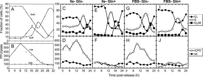
FIG. 1.
The plating efficiency of an ICP0− virus is not enhanced at a specific stage of the cell cycle. (A and B) As previously reported (3), 24-h-old Vero cell monolayers were incubated for 42 h in Ile-free medium and then released from the block by growth in medium containing isoleucine. At 3-h intervals postrelease of the block, cells were harvested and subjected to analysis by flow cytometry to determine the stage of the cell cycle or infected with 7134 or KOS to determine plating efficiency. The panels are reprinted from Cai and Schaffer (3) with permission. Twenty-four-hour-old Vero cell monolayers were washed once and incubated for 42 h in Ile-free medium containing 0 or 2 mM Gln (Ile− Gln− or Ile− Gln+, respectively) or in serum-free medium with 0 or 2 mM Gln (FBS− Gln− or FBS− Gln+, respectively). Ile-free media were supplemented with 10% dialyzed FBS. Cells were released from the G1 block by replacing Ile- or serum-free medium with medium supplemented with 10% nondialyzed FBS, 0.802 mM Ile, and 2 mM Gln. At 3-h intervals, cells were harvested and analyzed for DNA content by flow cytometry (C, E, G, and I) or replicate monolayers were infected with 100 PFU wild-type (KOS) or 40 or 100 PFU (Gln− or Gln+, respectively) ICP0− virus (n212) (D, F, H, and J). Input multiplicities were adjusted such that the 0-h time point in Ile-free Gln-containing medium (F) produced 100 plaques. Plaque counts for monolayers infected with 40 PFU were multiplied by 2.5 to normalize to an input of 100 PFU/plate. Each experiment was performed at least twice, and the determination of plating efficiencies at the 9-h time point for Ile deprivation/refeeding (C through F) was performed five times. This figure represents the results of one experiment.
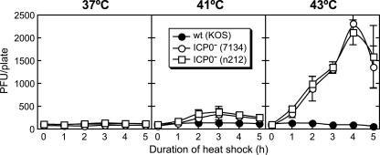
FIG. 2.
The plating efficiencies of ICP0− viruses are enhanced when cells are heat shocked prior to infection. Twenty-four-hour-old Vero cell monolayers in 35-mm plates were incubated for 1 to 5 h at 37°C, 41°C, or 43°C, and at hourly intervals following heat stress, replicate monolayers were infected with ∼100 PFU/plate wild-type (KOS) or ICP0− (7134 or n212) virus. The average number of plaques/plate is shown, and error bars indicate the standard deviations of four independent experiments.
One-step growth curves to determine replication efficiency.
Thirty-five-millimeter petri plates were seeded with 1.5 × 10⁵ Vero cells/plate and incubated at 37°C for 24 h. Cells from three plates were harvested by trypsinization, pooled, pelleted, resuspended, and counted. MOIs were calculated for the average number of cells per plate. Monolayers were heat shocked at 43°C for 4 h immediately prior to infection or remained at 37°C. Monolayers were infected at MOIs of 0.1, 1, and 5 PFU/cell in a volume of 100 μl/plate. Titers for KOS and n212 were determined on 24-h-old monolayers of Vero and L7 cells, respectively. Cells were incubated at 37°C with shaking every 15 min for 1 h. Immediately following infection, monolayers were washed three times with warm HBSS (37°C) and 1 ml of warm medium was added to each plate. At 1 h postinfection and at 3-h intervals postinfection, one plate from each combination of virus (KOS or n212), incubation temperature (37°C or 43°C), and MOI (0.1, 1, or 5) was frozen at −80°C. After all samples were frozen, cells were thawed, removed from the plate by repeated pipetting with a micropipetter, lysed by sonication, and assayed for virus titer by standard plaque assay. KOS and n212 were assayed on Vero and L7 cells, respectively. After cells were infected, titers of the KOS and n212 inocula were verified by standard plaque assay and were designated the 0-h time point (see Fig. 4).
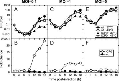
FIG. 4.
Cellular heat shock enhances the replication efficiency of an ICP0− virus in an MOI-dependent manner. Twenty-four-hour-old Vero cell monolayers were infected at MOIs of 0.1, 1, and 5 PFU/cell of wild-type (KOS) or ICP0− (n212) virus. Viral titers for wild-type and ICP0− viruses were determined on Vero and L7 cells, respectively. Infected cells were incubated at 37°C and harvested at 3-h intervals. Virus titers were determined by standard plaque assay, with wild-type virus titrated on Vero cells and ICP0− virus titrated on ICP0-expressing L7 cells. The titers of the inocula were also determined by standard plaque assay and are represented as the 0-h time points. The top row (A, C, and E) represents viral yields, and the bottom row (B, D, and F) represents the change (n-fold) in replication efficiency following heat shock, calculated by dividing the number of PFU/cell following heat shock by the number of PFU/cell without heat shock. The figure is representative of two experiments which produced similar results.
RESULTS
The stage of the cell cycle is not responsible for the enhanced plating efficiency of ICP0− viruses.
Cai and Schaffer reported previously that Ile deprivation/refeeding results in a sixfold enhancement of the plating efficiency of an ICP0− virus (7134) (3). While attempting to repeat the experiment shown in Fig. 3 of that publication (reprinted here as Fig. 1A and B), we were able to synchronize cells consistently, but the plating efficiency of the ICP0− viruses was highly variable, with maximum increases (n-fold) ranging from none to sixfold (data not shown). Because there appeared to be a trend toward greater plating efficiency with older medium and because high levels of Gln appeared to prevent the enhanced plating efficiency of the virus, we reasoned that the variability in plating efficiency was due to the instability of Gln in the Ile-free medium (26, 37, 48). We therefore repeated the experiment using Ile-free medium containing 0 or 2 mM Gln (Fig. 1C through F). In the absence of Gln, the growth of cells for 42 h in Ile-free medium resulted in the synchronization of 60% of cells to G1 phase (Fig. 1C) and a maximum sixfold enhancement in plating efficiency of an ICP0− virus (n212) at the time that cells transitioned from G1 to S phase (Fig. 1D). These results are similar to those reported previously (Fig. 1A and B). The presence of 2 mM Gln in Ile-free medium resulted in improved synchrony, with >90% of cells in G1 phase at the time of the release of the block (Fig. 1E); however, the enhanced plating efficiency of the ICP0− virus was eliminated (Fig. 1F).
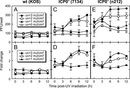
FIG. 3.
UV-C irradiation of Vero cells prior to infection enhances the plating efficiencies of ICP0− viruses. Twenty-four-hour-old Vero cell monolayers in six-well plates (35-mm diameter wells) were subjected to 0, 1, 2, 3, 4, or 6 mJ/cm2 of UV-C irradiation. Monolayers at the 0-h time point were infected immediately. All others were incubated at 37°C until the time of infection as indicated on the x axis. Replicate monolayers were infected with 100 PFU/well of wild-type (KOS) or ICP0− virus (7134 or n212). (A, C, and E) Plaques were counted, and (B, D, and F) plaque numbers were divided by the number of plaques on mock-irradiated monolayers to determine the change (n-fold) in plating efficiency. The average number of plaques/well is shown, and error bars indicate the standard deviations of two independent measurements.
In addition to Ile deprivation/refeeding, we used a second method that is commonly used to synchronize cells to G1 phase, serum starvation, in a similar experiment using serum-free media containing 0 or 2 mM Gln (Fig. 2G through J). In the absence of Gln, 70% of cells were synchronized to G1 phase at the time of readdition of serum (Fig. 1G) and accompanied by a fivefold enhancement in plating efficiency of the ICP0− virus (Fig. 1H). In the presence of Gln, serum starvation resulted in 80% of cells synchronized to G1 phase (Fig. 1I) but the enhanced plating efficiency was again eliminated (Fig. 1J). The plating efficiency of wild-type virus was unaffected by any of the treatments. Because cells can be synchronized without resulting in enhanced plating efficiency of an ICP0− virus at a specific stage of the cell cycle, the enhanced plating efficiency following Ile deprivation/refeeding is not associated specifically with late G1/early S phase of the cell cycle as previously reported (Fig. 1A and B) (3). Studies are currently ongoing to assess the role of Gln in the enhanced plating efficiency of ICP0− viruses as observed in these experiments.
(It should be noted in this and subsequent experiments that ICP0− viruses do not plate more efficiently than the wild-type virus. For Vero cells seeded at the density used in these experiments, the particle-to-PFU ratio is ∼25 times higher for ICP0− viruses than for the wild-type virus [3]. Consequently, enhancement in plating efficiency of ∼25-fold would indicate that ICP0− and wild-type viruses have approximately equal plating efficiencies.)
We also synchronized cells to S phase by a double thymidine block and examined the plating efficiencies of ICP0− and wild-type viruses. In this experiment, we hypothesized that if an ICP0− virus is able to initiate plaque formation only in cells in late G1/early S phase, then ICP0− viruses plated on cells synchronized by a double thymidine block should also show an enhanced plating efficiency when cells progress from G1 to S phase. Upon release of the second thymidine block, replicate monolayers were infected with ICP0− or wild-type viruses to determine plating efficiencies or analyzed for DNA content by flow cytometry at 3-h intervals to determine the phase of the cell cycle and level of cell synchrony. At the time of release of the block, approximately 80% of cells were in S phase, but the plating efficiency of the ICP0− virus (7134) was not enhanced as cells cycled from G1 to S phase (data not shown). As for experiments involving Ile deprivation/refeeding, these results indicate that the plating efficiency of ICP0− viruses is not associated specifically with late G1/early S phase or any other phase of the cell cycle.
Heat shock prior to infection enhances the plating efficiencies of ICP0− viruses.
Because the studies described above revealed that the plating efficiency of an ICP0− virus is not enhanced at a specific stage of the cell cycle, we propose an alternative hypothesis. Because ICP0 is necessary for efficient reactivation from latency and because cellular stress induces reactivation, we hypothesize that the enhanced plating efficiencies of ICP0− viruses observed following Ile deprivation/refeeding and serum starvation/refeeding in the absence of Gln (Fig. 1D and H) are due to the induction of a cellular stress response. To test whether a recognized cellular stressor, heat, can induce enhanced plating efficiency of an ICP0− virus, 24-h-old Vero cell monolayers were incubated for 1 to 5 h at 37°C, 41°C, or 43°C (Fig. 2). At hourly intervals, monolayers incubated at each temperature were infected at 37°C with wild-type or ICP0− virus, with continued incubation at 37°C, to determine plating efficiencies. Whereas the wild-type virus showed no enhanced plating efficiency on monolayers that had been heat shocked at any temperature for any length of time tested, the plating efficiencies of two ICP0− viruses peaked at ∼4-fold and ∼23-fold above their plating efficiencies on nonstressed monolayers following incubation at 41°C and 43°C, respectively. (As mentioned previously, ICP0− viruses have particle-to-PFU ratios that are ∼25 times higher than that of the wild-type virus; thus, the ∼23-fold enhancement in plating efficiency following heat shock indicates that the ICP0− viruses plated nearly as well as the wild-type virus.) Incubation for 5 h at 43°C caused the death of many cells in the monolayer, as indicated by lightly stained monolayers and the observation of fewer cells per monolayer by light microscopy. Following 5 h of heat shock at 43°C, variability in the extent of cell death is likely the cause of the decreased plating efficiency, variability in cell death between experiments, and decreased reproducibility at this time point. This experiment was also performed with cells incubated at 44°C prior to infection (data not shown). Here we found that the plating efficiency of ICP0− viruses peaked following a 3-h incubation, yielding an ∼20-fold enhancement above the plating efficiency on nonstressed monolayers. Four- and 5-h incubations at 44°C caused complete detachment of the monolayers. In summary, the greater the stress applied to monolayers prior to infection without inducing widespread cell death, the greater the enhancement in plating efficiency for ICP0− viruses.
To determine the validity of infecting cells with diluted ICP0− virus inocula and normalizing to 100 PFU/plate after counting plaques, the experiment shown in Fig. 2 was repeated in 100-mm plates. For this experiment, all monolayers were infected with 100 PFU (as determined on untreated 24-h-old Vero cell monolayers) of virus and therefore required no normalization. This experiment produced results similar to those shown in Fig. 2, that is, ∼100 wild-type virus plaques for all treatments and time points and ∼100, ∼350, and ∼2,000 plaques for the ICP0− viruses following 4 h of incubation at 37°C, 41°C, and 43°C, respectively (data not shown). Thus, infection with less than 100 PFU/plate followed by normalization to 100 PFU/plate produces valid results.
Cold shock did not enhance the plating efficiency of ICP0− viruses.
To determine if cells treated by a related stress, cold shock, would also result in enhanced plating efficiency of ICP0− viruses, 24-h-old monolayers of Vero cells were incubated at 37°C, 34°C, 22.5°C, or 4°C for 3 h and infected with 100 PFU/plate of wild-type or ICP0− virus (data not shown). No change was observed in the plating efficiencies of wild-type or ICP0− virus following any of the cold shock treatments. We conclude that the cellular activities altered by heat shock that complement the plating defect of ICP0− viruses are not induced by cold shock, suggesting that these activities are not part of a response to all stresses but are specific for certain types of stress.
UV-C irradiation of cells prior to infection enhances the plating efficiency of ICP0− viruses.
After we demonstrated that the plating efficiency of ICP0− viruses is enhanced following heat shock but not cold shock, cells were treated with a third recognized cellular stressor, UV-C irradiation, to determine if the plating efficiency of ICP0− viruses would be enhanced. Twenty-four-hour-old Vero cell monolayers in six-well plates (35-mm diameter wells) were overlaid with 2 ml/well PBS, exposed to UV-C radiation in a UV cross-linker at levels of 0, 1, 2, 3, 4, or 6 mJ/cm2, and infected with 100 PFU/well wild-type or ICP0− virus at 3-h intervals postirradiation. Following exposure of monolayers to 1 mJ/cm2, the plating efficiencies of wild-type and ICP0− viruses were similar to their plating efficiencies on untreated monolayers (Fig. 3). Following exposures to 2 or 3 mJ/cm2, ICP0− viruses showed maximum increases in plating efficiency of approximately threefold at 9 and 12 h postirradiation, whereas the wild-type virus showed a decrease of about twofold. The decreased plating efficiency of the wild-type virus was more pronounced following exposure to increasing intensities of UV-C irradiation. Increasing intensities of UV-C also caused the death of many cells in the monolayer such that monolayers stained very lightly and fewer cells were observed by light microscopy. Thus, the decreased plating efficiency of wild-type virus was likely due to an increase in cell death when infected cells detached from the plate, taking with them infectious particles that would otherwise have initiated plaque formation. Importantly, UV-C irradiation, like heat shock, results in enhanced plating efficiency of ICP0− viruses, further implicating a role for stress-induced cellular activities in the complementation of ICP0− viruses. Possible explanations for the 2.5- to 3-fold maximum enhancement in plating efficiency of ICP0− viruses observed following UV-C irradiation rather than the nearly 25-fold increase seen following heat shock are considered in Discussion.
Heat shock prior to infection results in an MOI-dependent enhancement in replication efficiency of an ICP0− virus.
After observing that the plating efficiency of ICP0− viruses is enhanced by the application of heat shock to cells prior to infection (Fig. 2), we asked if replication efficiency at low MOIs would be similarly enhanced. Monolayers of 24-h-old Vero cells were incubated at 37°C or 43°C for 4 h and infected for 1 h with either the wild-type or an ICP0− virus (n212) at an MOI of 0.1, 1, or 5 (Fig. 4). The top row of Fig. 4 (panels A, C, and E) shows virus replication as numbers of PFU/cell, and the bottom row (panels B, D, and F) shows the same data plotted as a change (n-fold) (the number of PFU/cell for heat-shocked cells divided by the number of PFU/cell for non-heat-shocked cells). On non-heat-shocked monolayers, the MOI-dependent replication defect of the ICP0− virus relative to that of the wild type at 18 h postinfection (Fig. 4A, C, and E, filled symbols) was similar to that reported previously (2, 15, 40). At an MOI of 0.1 PFU/cell (Fig. 4A and B), the amount of infectious virus present between 1 and 9 h postinfection was similar for both viruses in heat-shocked and non-heat-shocked cells. From 12 to 18 h postinfection, the amount of infectious ICP0− virus in non-heat-shocked cells was reduced ∼150-fold relative to that in the wild-type virus. In heat-shocked cells, wild-type replication was decreased approximately sixfold, whereas ICP0− virus replication was enhanced nearly 25-fold such that the two viruses exhibited similar replication efficiencies. At an MOI of 1, the pattern of virus production was similar to that observed at the lower MOI, except that the replication defect of the ICP0− virus was less severe. Heat shock produced enhancement of approximately fivefold for the ICP0− virus (Fig. 4C and D) such that the two viruses exhibited similar replication efficiencies. As mentioned in the introduction, at high MOIs (∼>5), ICP0− viruses replicate as well as the wild-type virus. Accordingly, at an MOI of 5, little or no enhancement in the replication of the ICP0− virus occurred following heat shock (Fig. 4E and F). These results demonstrate that the replication defect of an ICP0− virus at low MOIs can be entirely overcome by subjecting cells to heat shock prior to infection.
The enhanced plating efficiency of ICP0− viruses on heat-shocked monolayers is transient following removal of the heat stress.
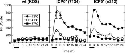
Heat shock induces the expression/activation (or repression/inactivation) of certain cellular activities resulting in enhanced plating and replication efficiencies of ICP0− viruses. To better define the nature of the alteration in cellular activities that is responsible for the enhanced plating and replication efficiencies, we examined the kinetics of the decline of the plating efficiency of ICP0− viruses following heat shock at 41°C or 43°C. For this purpose, 24-h-old Vero cell monolayers were incubated at 37°C, 41°C, or 43°C for 3 h, followed by incubation at 37°C for up to 21 h (Fig. 5). At 3-h intervals, monolayers were infected with wild-type or ICP0− virus to determine plating efficiency. The plating efficiency of wild-type virus was not significantly altered by any treatment. In contrast, following 3 h of incubation at 41°C and 43°C, the plating efficiencies of the ICP0− viruses peaked at three- and ninefold, respectively, at the time of removal from heat stress. For monolayers incubated at 41°C, the plating efficiencies of ICP0− viruses declined for 6 h after removal of the stress (the 9-h point in Fig. 5) until they were similar to the plating efficiency on non-heat-shocked monolayers. For monolayers incubated at 43°C, the enhanced plating efficiency declined for 6 h after removal of the stress at approximately the same rate as that for monolayers incubated at 41°C. After 6 h, however, the plating efficiency declined at a reduced rate and remained detectable throughout the duration of the experiment. In summary, the heat shock-inducible cellular factors that are responsible for the complementation of the ICP0− viruses are expressed transiently after removal of the heat stress.
FIG. 5.
The heat shock-induced increase in plating efficiency of ICP0− viruses on Vero cell monolayers is transient. Twenty-four-hour-old Vero cell monolayers in 35-mm plates were incubated at 37°C, 41°C, or 43°C for 3 h (from 0 to 3 h on the x axis, as indicated by the bars under the axis) and at 37°C for the remainder of the experiment. At 3-h intervals, replicate monolayers from each treatment group were infected with ∼100 PFU/plate wild-type or ∼20 or ∼100 PFU/plate ICP0− virus. If monolayers infected with ∼100 PFU/plate ICP0− virus resulted in too many plaques to count, those infected with ∼20 PFU were counted instead, and the result was multiplied by 5. Infected cells were incubated at 37°C for 4 days and stained, and plaques were counted. Times shown on the x axis are the numbers of hours following transfer to the elevated temperature. This experiment was performed twice for KOS and 7134 and once for n212. For KOS and 7134, the figure represents the average results for the two experiments and error bars represent the standard deviations.
High density rather than age of the Vero cell monolayer inhibits plaque formation of ICP0− viruses.
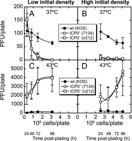
As noted in the introduction, it has been reported that the plating efficiency of ICP0− viruses decreases as cellular monolayers age and grow to high densities (3). We sought to determine first whether the age or the density of the monolayer is the factor that results in reduced plating efficiency and second whether these aged/dense monolayers can be induced by heat shock to support efficient ICP0− virus plaque formation. At 24-h intervals postseeding at a low (1.5 × 10⁵ cells/35-mm plate) or high (1.5 × 10⁶ cells/35-mm plate) initial density, Vero cell monolayers were incubated at 37°C or heat shocked at 43°C for 3 h. Monolayers were then infected with 100 PFU/plate of wild-type or ICP0− virus on non-heat-shocked monolayers or 5 PFU/plate for ICP0− viruses on heat-shocked monolayers (Fig. 6). Plaque counts for infections of 5 PFU/plate were multiplied by 20 to normalize to an input of 100 PFU/plate.
FIG. 6.
The plating efficiency of ICP0− viruses decreases with increasing cell density rather than increasing age of the monolayers, and heat shock greatly enhances plating efficiency on very dense monolayers. Thirty-five-millimeter plates were seeded with Vero cells at 1.5 × 10⁵ cells/plate (A and C) or 1.5 × 10⁶ cells/plate (B and D). At 24-h intervals, monolayers were heat shocked for 3 h at 43°C (C and D) or incubated at 37°C (A and B). Cells were either harvested and counted or replicate monolayers were infected with wild-type (KOS) or ICP0− virus (7134 or n212) at the times indicated and incubated at 37°C for 4 days. The number of PFU/plate was plotted against the number of cells/plate at the time of infection. The number of hours postplating, though not used to generate the plots, is also shown and corresponds to the time of infection. The average number of plaques/plate corresponds to the cell number at the indicated times; error bars indicate the standard deviations of three independent experiments.
On monolayers seeded at a low initial density, we confirmed the previously described decrease in ICP0− virus plating efficiency resulting in the nearly complete absence of plaques on monolayers incubated continuously at 37°C for 96 h (Fig. 6A). In contrast, wild-type virus showed only a moderate decrease (approximately twofold) in plating efficiency on similar monolayers. On monolayers seeded at a high initial density, the decrease in wild-type plating efficiency was similar to that observed at a low initial density (Fig. 6B). For the ICP0− viruses, however, very few plaques formed at any time postplating. A comparison of the plating efficiencies of the ICP0− viruses at similar densities (2.5 × 10⁶ cells, near 96 h postplating for low initial density [Fig. 6A] and 24 h postplating for high initial density [Fig. 6B]) revealed that very few plaques formed on monolayers at either initial density. In contrast, a comparison of the plating efficiencies of the ICP0− viruses at 24 h postplating revealed that, at a low initial density, 150 plaques formed, whereas at a high initial density, very few plaques formed. These observations suggest that cell density rather than age of the monolayer is the predominant factor underlying the reduced plating efficiency of ICP0− viruses.
Heat shock of dense monolayers prior to infection allows ICP0− viruses to plaque efficiently.
As described above, monolayers seeded at low or high initial densities were heat shocked or remained at 37°C for 3 h and were infected with wild-type or ICP0− virus (Fig. 6C and D). Heat shock prior to infection did not significantly alter the plating efficiency of wild-type virus, whereas the plating efficiency of the ICP0− viruses was greatly enhanced, with the maximum change being an increase from 0 to >4,000 plaques at 96 h postplating for monolayers seeded at both initial densities (Fig. 6C and D). This observation suggests that the decrease in ICP0− virus plating efficiency on dense monolayers is likely due to the loss of heat shock-inducible cellular activities present in low-density, but not high-density, monolayers (or conversely, heat shock-labile/-repressible activities that inhibit plaque formation by ICP0− viruses and accumulate in high-density monolayers). In summary, as monolayers age and become more dense, the decrease in plating efficiency of ICP0− viruses is due to increased cell density and this effect can be overcome by cellular heat shock prior to infection.
DISCUSSION
This lab previously demonstrated that Ile deprivation/refeeding results in complementation of an ICP0− virus at a time corresponding to the cellular transition from G1 to S phase (3); however, while attempting to repeat the experiment shown in Fig. 1A and B, we were able to reproduce the level of cell synchrony consistently but not the enhanced plating efficiency. Moreover, the variability in plating efficiencies was eliminated by preparing Gln-free DMEM and adding 2 mM Gln within 2 days of use. This led us to conclude that the plating efficiencies of ICP0− viruses are enhanced when Ile-free medium does not contain Gln but are not enhanced when 2 mM Gln is present. We obtained similar results when cells were synchronized to G1 phase by serum starvation in the presence or absence of 2 mM Gln. Importantly, the presence of Gln enhanced the synchrony of the cells while eliminating the enhanced plating efficiency. Although Cai and Schaffer reported the presence of 0.03% Gln (∼2 mM) in the Ile-free medium (3), it is probable that due to the instability of Gln in growth medium (26, 37, 48), little or no Gln was present at the time that the medium was used. The role of Gln in the complementation of ICP0− viruses is currently being investigated. For the purpose of this study, however, it is sufficient to note that cells can be synchronized without resulting in enhanced plating efficiency of ICP0− viruses. This conclusion was further confirmed by synchronizing cells to S phase by a double thymidine block, which did not result in enhanced plating efficiency at late G1/early S phase. Therefore, a factor(s) other than stage of the cell cycle must be responsible for the enhanced plating efficiency.
If ICP0− viruses are not complemented at a specific stage of the cell cycle, why does Ile/Gln deprivation/refeeding result in enhanced plating efficiency? Since ICP0 is needed for efficient reactivation from latency and stressful stimuli induce reactivation, we hypothesize that Ile/Gln deprivation/refeeding induces a cellular stress response that causes the enhanced plating efficiency. This hypothesis was tested by treating Vero cells with three known cellular stressors, heat shock, cold shock, and UV-C irradiation, to determine which, if any, would result in the enhanced plating efficiency of ICP0− viruses. Both heat shock and UV-C irradiation resulted in enhanced plating efficiency, whereas cold shock did not; therefore, the enhanced plating efficiency is due to the cellular response to certain types of stress and is not a general response common to all types of stress. Importantly, on monolayers treated with heat shock or UV-C irradiation, the plating efficiency of ICP0− viruses increases with increasing cell stress.
Heat shock resulted in a much greater enhancement in ICP0− virus plating efficiency than did UV-C irradiation. One reason for the difference may be associated with the effects of administration of the stressful stimulus. Heat shock for up to 4 h resulted in minimal cell death, whereas doses of UV-C greater than 3 mJ/cm2, delivered within a few seconds, were of sufficient intensity to kill all cells in the monolayer. The cellular adaptation to heat shock was ongoing during the incubation at elevated temperatures, whereas monolayers subjected to UV-C irradiation received the entire dose followed by recovery. Therefore, it is possible that very low intensity UV-C irradiation applied to monolayers for hours would result in an enhancement similar to that from heat shock. Conversely, heat shock for several seconds at a temperature nearly high enough to cause cell death would likely decrease the maximum level of enhancement.
Many studies have shown that cellular stress and infection with several DNA-containing viruses, including HSV-1, have similar effects on the cell. Specifically, infection with these viruses induces the expression of heat shock proteins. For example, infection with HSV-1 or adenovirus type 5, but not simian virus 40 or vaccinia virus, results in an increased rate of transcription of some members of the HSP70 and HSP110 families (22, 38, 46). The induction of members of the HSP70 family is due to the E1A protein in adenovirus type 5 infection (31, 49, 50) and is likely due to an IE protein(s) in HSV-1 infection (22). ICP0 is an IE protein that is functionally similar to E1A (43) and hence is a strong candidate for an inducer of these HSP70 proteins, although other IE and cellular proteins may also be involved. The possible role of ICP0 in the induction of heat shock proteins is further supported by the results of a recent microarray study in which infection of human embryonic lung cells with an HSV-1 virus expressing only ICP0 and none of the other IE genes activated the expression of three of five heat shock transcripts represented on the array; Hsp60, Hsp70, and Hsp90 were upregulated, but Hsp27 and Hsp40 were not (19).
In addition to heat shock protein induction, cellular stress and ICP0 induce similar effects on cells. ICP0 and cellular stress both cause perturbation of the cell cycle, causing an apparent block or delay in progression at G1/S (16, 19, 23, 27, 32, 44) and G2/M (1, 6, 19, 23, 27). Also similar to the effect of ICP0, hyposmotic shock, another form of cellular stress, induces the degradation of cell cycle-associated proteins and blocks cell cycle progression through proteasome activation (47). Additionally, both ICP0 and heat shock cause the dissociation or dispersion of ND10 structures (14). Thus, consistent with the ability to complement ICP0− viruses, ICP0 and cellular stress may induce similar changes in cellular gene expression and protein activities.
Though several groups have reported the upregulation of heat shock proteins by HSV-1 infection, the roles of stress response proteins per se have not been investigated. Of particular interest is the role of Hsp90. It has been shown that geldanamycin, a specific inhibitor of Hsp90 (34), inhibits the replication of HSV-1 in cell culture (25); therefore, Hsp90 and at least one of its downstream targets are necessary for HSV-1 replication. Geldanamycin also inhibits the replication of HSV-2, vesicular stomatitis virus, coxsackie B3, human immunodeficiency virus type 1, and severe acute respiratory virus, but not influenza virus type A or B (25). Because Hsp90 is critical for the replication of many viruses and is an important component of the cellular response to stress, which induces HSV-1 reactivation from latency, Hsp90 is a potentially important factor in HSV-1 reactivation.
The study of cellular activities involved in the complementation of ICP0− viruses may lead to the identification of cellular activities that trigger reactivation from latency. There are three findings that relate ICP0− virus infection of monolayers in cell culture to the reactivation of wild-type virus from latency: (i) ICP0 is required for efficient reactivation from latency; (ii) ICP0 is not present during latency or productive infection with an ICP0− virus; and (iii) heat shock and UV-C irradiation cause enhanced plating and replication efficiencies of ICP0− viruses in cell culture and induce reactivation of wild-type virus from latency. Therefore, the stress-induced activities in cells that enhance plating and replication of ICP0− viruses may also be involved in the early steps of reactivation from latency.
Although heat stress is sufficient to enhance ICP0− virus plating and replication efficiencies to near wild-type levels, the stress is not sufficient to induce efficient reactivation of an ICP0− virus from latently infected mouse trigeminal ganglia (18). Therefore, ICP0 provides functions required for efficient reactivation from latency that are not induced by stress in trigeminal ganglia cell culture.
Acknowledgments
This work was supported by National Institutes of Health grants RO1CA20260 from the National Cancer Institute and PO1NS35138 from the National Institute of Neurological Disorders and Stroke. R.M.B. was supported in part by grant T32AI706126 from the National Institute of Allergy and Infectious Disease.
We thank members of the Schaffer lab for helpful discussions and suggestions.
REFERENCES
- 1.Bulavin, D. V., Y. Higashimoto, I. J. Popoff, W. A. Gaarde, V. Basrur, O. Potapova, E. Appella, and A. J. Fornace, Jr. 2001. Initiation of a G2/M checkpoint after ultraviolet radiation requires p38 kinase. Nature 411:102-107. [DOI] [PubMed] [Google Scholar]
- 2.Cai, W., and P. A. Schaffer. 1989. Herpes simplex virus type 1 ICP0 plays a critical role in the de novo synthesis of infectious virus following transfection of viral DNA. J. Virol. 63:4579-4589. [DOI] [PMC free article] [PubMed] [Google Scholar]
- 3.Cai, W., and P. A. Schaffer. 1991. A cellular function can enhance gene expression and plating efficiency of a mutant defective in the gene for ICP0, a transactivating protein of herpes simplex virus type 1. J. Virol. 65:4078-4090. [DOI] [PMC free article] [PubMed] [Google Scholar]
- 4.Cai, W., and P. A. Schaffer. 1992. Herpes simplex virus type 1 ICP0 regulates expression of immediate-early, early, and late genes in productively infected cells. J. Virol. 66:2904-2915. [DOI] [PMC free article] [PubMed] [Google Scholar]
- 5.Cai, W., T. L. Astor, L. M. Liptak, C. Cho, D. M. Coen, and P. A. Schaffer. 1993. The herpes simplex virus type 1 regulatory protein ICP0 enhances virus replication during acute infection and reactivation from latency. J. Virol. 67:7501-7512. [DOI] [PMC free article] [PubMed] [Google Scholar]
- 6.Dmitrieva, N. I., D. V. Bulavin, A. J. Fornace, Jr., and M. B. Burg. 2002. Rapid activation of G2/M checkpoint after hypertonic stress in renal inner medullary epithelial (IME) cells is protective and requires p38 kinase. Proc. Natl. Acad. Sci. USA 99:184-189. [DOI] [PMC free article] [PubMed] [Google Scholar]
- 7.Everett, R. D. 1984. Trans activation of transcription by herpes virus products: requirement for two HSV-1 immediate-early polypeptides for maximum activity. EMBO J. 3:3135-3141. [DOI] [PMC free article] [PubMed] [Google Scholar]
- 8.Everett, R. D. 1985. Activation of cellular promoters during herpes virus infection of biochemically transformed cells. EMBO J. 4:1973-1980. [DOI] [PMC free article] [PubMed] [Google Scholar]
- 9.Everett, R. D. 1986. The products of herpes simplex virus type 1 (HSV-1) immediate early genes 1, 2 and 3 can activate HSV-1 gene expression in trans. J. Gen. Virol. 67:2507-2513. [DOI] [PubMed] [Google Scholar]
- 10.Everett, R. D. 1987. A detailed mutational analysis of Vmw110, a trans-acting transcriptional activator encoded by herpes simplex virus type 1. EMBO J. 6:2069-2076. [DOI] [PMC free article] [PubMed] [Google Scholar]
- 11.Everett, R. D. 1988. Analysis of the functional domains of herpes simplex virus type 1 immediate-early polypeptide Vmw110. J. Mol. Biol. 202:87-96. [DOI] [PubMed] [Google Scholar]
- 12.Everett, R. D., A. Orr, and M. Elliott. 1991. High level expression and purification of herpes simplex virus type 1 immediate early polypeptide Vmw110. Nucleic Acids Res. 19:6155-6161. [DOI] [PMC free article] [PubMed] [Google Scholar]
- 13.Everett, R. D., P. Barlow, A. Milner, B. Luisi, A. Orr, G. Hope, and D. Lyon. 1993. A novel arrangement of zinc-binding residues and secondary structure in the C3HC4 motif of an alpha herpes virus protein family. J. Mol. Biol. 234:1038-1047. [DOI] [PubMed] [Google Scholar]
- 14.Everett, R. D., P. Lomonte, T. Sternsdorf, R. van Driel, and A. Orr. 1999. Cell cycle regulation of PML modification and ND10 composition. J. Cell Sci. 112:4581-4588. [DOI] [PubMed] [Google Scholar]
- 15.Everett, R. D., C. Boutell, and A. Orr. 2004. Phenotype of a herpes simplex virus type 1 mutant that fails to express immediate-early regulatory protein ICP0. J. Virol. 78:1763-1774. [DOI] [PMC free article] [PubMed] [Google Scholar]
- 16.Fuse, T., K. Yamada, K. Asai, T. Kato, and M. Nakanishi. 1996. Heat shock-mediated cell cycle arrest is accompanied by induction of p21 CKI. Biochem. Biophys. Res. Commun. 225:759-763. [DOI] [PubMed] [Google Scholar]
- 17.Gelman, I. H., and S. Silverstein. 1985. Identification of immediate early genes from herpes simplex virus that transactivate the virus thymidine kinase gene. Proc. Natl. Acad. Sci. USA 82:5265-5269. [DOI] [PMC free article] [PubMed] [Google Scholar]
- 18.Halford, W. P., and P. A. Schaffer. 2001. ICP0 is required for efficient reactivation of herpes simplex virus type 1 from neuronal latency. J. Virol. 75:3240-3249. [DOI] [PMC free article] [PubMed] [Google Scholar]
- 19.Hobbs, W. E., II, and N. A. DeLuca. 1999. Perturbation of cell cycle progression and cellular gene expression as a function of herpes simplex virus ICP0. J. Virol. 73:8245-8255. [DOI] [PMC free article] [PubMed] [Google Scholar]
- 20.Jordan, R., and P. A. Schaffer. 1997. Activation of gene expression by herpes simplex virus type 1 ICP0 occurs at the level of mRNA synthesis. J. Virol. 71:6850-6862. [DOI] [PMC free article] [PubMed] [Google Scholar]
- 21.Kent, J. R., W. Kang, C. G. Miller, and N. W. Fraser. 2003. Herpes simplex virus latency-associated transcript gene function. J. Neurovirol. 9:285-290. [DOI] [PubMed] [Google Scholar]
- 22.Kobayashi, K., E. Ohgitani, Y. Tanaka, M. Kita, and J. Imanishi. 1994. Herpes simplex virus-induced expression of 70 kDa heat shock protein (HSP70) requires early protein synthesis but not viral DNA replication. Microbiol. Immunol. 38:321-325. [DOI] [PubMed] [Google Scholar]
- 23.Kühl, N. M., and L. Rensing. 2000. Heat shock effects on cell cycle progression. Cell. Mol. Life Sci. 57:450-463. [DOI] [PMC free article] [PubMed] [Google Scholar]
- 24.Leib, D. A., D. M. Coen, C. L. Bogard, K. A. Hicks, D. R. Yager, D. M. Knipe, K. L. Tyler, and P. A. Schaffer. 1989. Immediate-early regulatory gene mutants define different stages in the establishment and reactivation of herpes simplex virus latency. J. Virol. 63:759-768. [DOI] [PMC free article] [PubMed] [Google Scholar]
- 25.Li, Y. H., P. Z. Tao, Y. Z. Liu, and J. D. Jiang. 2004. Geldanamycin, a ligand of heat shock protein 90, inhibits the replication of herpes simplex virus type 1 in vitro. Antimicrob. Agents Chemother. 48:867-872. [DOI] [PMC free article] [PubMed] [Google Scholar]
- 26.Lin, A., and P. Agrawal. 1988. Glutamine decomposition in DMEM: effect of pH and serum concentration. Biotechnol. Lett. 10:695-698. [Google Scholar]
- 27.Lomonte, P., and R. D. Everett. 1999. Herpes simplex virus type 1 immediate-early protein Vmw110 inhibits progression of cells through mitosis and from G1 into S phase of the cell cycle. J. Virol. 73:9456-9467. [DOI] [PMC free article] [PubMed] [Google Scholar]
- 28.Mavromara-Nazos, P., S. Silver, J. Hubenthal-Voss, J. L. McKnight, and B. Roizman. 1986. Regulation of herpes simplex virus 1 genes: alpha gene sequence requirements for transient induction of indicator genes regulated by beta or late (gamma 2) promoters. Virology 149:152-164. [DOI] [PubMed] [Google Scholar]
- 29.Mosca, J. D., D. P. Bednarik, N. B. Raj, C. A. Rosen, J. G. Sodroski, W. A. Haseltine, G. S. Hayward, and P. M. Pitha. 1987. Activation of human immunodeficiency virus by herpesvirus infection: identification of a region within the long terminal repeat that responds to a trans-acting factor encoded by herpes simplex virus 1. Proc. Natl. Acad. Sci. USA 84:7408-7412. [DOI] [PMC free article] [PubMed] [Google Scholar]
- 30.Nabel, G. J., S. A. Rice, D. M. Knipe, and D. Baltimore. 1988. Alternative mechanisms for activation of human immunodeficiency virus enhancer in T cells. Science 239:1299-1302. [DOI] [PubMed] [Google Scholar]
- 31.Nevins, J. R. 1982. Induction of the synthesis of a 70,000 dalton mammalian heat shock protein by the adenovirus E1A gene product. Cell 29:913-919. [DOI] [PubMed] [Google Scholar]
- 32.Nitta, M., H. Okamura, S. Aizawa, and M. Yamaizumi. 1997. Heat shock induces transient p53-dependent cell cycle arrest at G1/S. Oncogene 15:561-568. [DOI] [PubMed] [Google Scholar]
- 33.Nsiah, Y. A., and F. Rapp. 1991. Role of latency-associated transcript in herpes simplex virus infection. Intervirology 32:101-115. [DOI] [PubMed] [Google Scholar]
- 34.Ochel, H. J., K. Eichhorn, and G. Gademann. 2001. Geldanamycin: the prototype of a class of antitumor drugs targeting the heat shock protein 90 family of molecular chaperones. Cell Stress Chaperones 6:105-112. [DOI] [PMC free article] [PubMed] [Google Scholar]
- 35.O'Hare, P., and G. S. Hayward. 1985. Evidence for a direct role for both the 175,000- and 110,000-molecular-weight immediate-early proteins of herpes simplex virus in the transactivation of delayed-early promoters. J. Virol. 53:751-760. [DOI] [PMC free article] [PubMed] [Google Scholar]
- 36.O'Hare, P., and G. S. Hayward. 1985. Three trans-acting regulatory proteins of herpes simplex virus modulate immediate-early gene expression in a pathway involving positive and negative feedback regulation. J. Virol. 56:723-733. [DOI] [PMC free article] [PubMed] [Google Scholar]
- 37.Ozturk, S. S., and B. O. Palsson. 1990. Chemical decomposition of glutamine in cell culture media: effect of media type, pH, and serum concentration. Biotechnol. Prog. 6:121-128. [DOI] [PubMed] [Google Scholar]
- 38.Phillips, B., K. Abravaya, and R. I. Morimoto. 1991. Analysis of the specificity and mechanism of transcriptional activation of the human hsp70 gene during infection by DNA viruses. J. Virol. 65:5680-5692. [DOI] [PMC free article] [PubMed] [Google Scholar]
- 39.Roizman, B., and D. M. Knipe. 2001. Herpes simplex viruses and their replication, p. 2399-2459. In D. M. Knipe, P. M. Howley, D. E. Griffin, R. A. Lamb, M. A. Martin, B. Roizman, and S. E. Straus (ed.), Fields virology, 4th ed., vol. 2. Lippincott Williams and Wilkins, Philadelphia, Pa. [Google Scholar]
- 40.Sacks, W. R., and P. A. Schaffer. 1987. Deletion mutants in the gene encoding the herpes simplex virus type 1 immediate-early protein ICP0 exhibit impaired growth in cell culture. J. Virol. 61:829-839. [DOI] [PMC free article] [PubMed] [Google Scholar]
- 41.Samaniego, L. A., N. Wu, and N. A. DeLuca. 1997. The herpes simplex virus immediate-early protein ICP0 affects transcription from the viral genome and infected-cell survival in the absence of ICP4 and ICP27. J. Virol. 71:4614-4625. [DOI] [PMC free article] [PubMed] [Google Scholar]
- 42.Schang, L. M., J. Phillips, and P. A. Schaffer. 1998. Requirement for cellular cyclin-dependent kinases in herpes simplex virus replication and transcription. J. Virol. 72:5626-5637. [DOI] [PMC free article] [PubMed] [Google Scholar]
- 43.Shenk, T. 2001. Adenoviridae: the viruses and their replication, p. 2265-2300. In D. M. Knipe, P. M. Howley, D. E. Griffin, R. A. Lamb, M. A. Martin, B. Roizman, and S. E. Straus (ed.), Fields virology, 4th ed., vol. 2. Lippincott Williams and Wilkins, Philadephia, Pa. [Google Scholar]
- 44.Song, B., J. J. Liu, K. C. Yeh, and D. M. Knipe. 2000. Herpes simplex virus infection blocks events in the G1 phase of the cell cycle. Virology 267:326-334. [DOI] [PubMed] [Google Scholar]
- 45.Stow, N. D., and E. C. Stow. 1986. Isolation and characterization of a herpes simplex virus type 1 mutant containing a deletion within the gene encoding the immediate early polypeptide Vmw110. J. Gen. Virol. 67:2571-2585. [DOI] [PubMed] [Google Scholar]
- 46.Taddeo, B., A. Esclatine, and B. Roizman. 2002. The patterns of accumulation of cellular RNAs in cells infected with a wild-type and a mutant herpes simplex virus 1 lacking the virion host shutoff gene. Proc. Natl. Acad. Sci. USA 99:17031-17036. [DOI] [PMC free article] [PubMed] [Google Scholar]
- 47.Tao, G. Z., L. S. Rott, A. W. Lowe, and M. B. Omary. 2002. Hyposmotic stress induces cell growth arrest via proteasome activation and cyclin/cyclin-dependent kinase degradation. J. Biol. Chem. 277:19295-19303. [DOI] [PubMed] [Google Scholar]
- 48.Tritsch, G. L., and G. E. Moore. 1962. Spontaneous decomposition of glutamine in cell culture media. Exp. Cell Res. 28:360-364. [DOI] [PubMed] [Google Scholar]
- 49.Williams, G. T., T. K. McClanahan, and R. I. Morimoto. 1989. E1a transactivation of the human HSP70 promoter is mediated through the basal transcriptional complex. Mol. Cell. Biol. 9:2574-2587. [DOI] [PMC free article] [PubMed] [Google Scholar]
- 50.Wu, B. J., H. C. Hurst, N. C. Jones, and R. I. Morimoto. 1986. The E1A 13S product of adenovirus 5 activates transcription of the cellular human HSP70 gene. Mol. Cell. Biol. 6:2994-2999. [DOI] [PMC free article] [PubMed] [Google Scholar]
- 51.Yao, F., and P. A. Schaffer. 1995. An activity specified by the osteosarcoma line U2OS can substitute functionally for ICP0, a major regulatory protein of herpes simplex virus type 1. J. Virol. 69:6249-6258. [DOI] [PMC free article] [PubMed] [Google Scholar]


