Abstract
Viruses classified in the order Mononegavirales have a single nonsegmented RNA molecule as the genome and employ similar strategies for genome replication and gene expression. Infectious particles of Measles virus (MeV), a member of the family Paramyxoviridae in the order Mononegavirales, with two or three RNA genome segments (2 seg- or 3 seg-MeV) were generated using a highly efficient reverse genetics system. All RNA segments of the viruses were designed to have authentic 3′ and 5′ self-complementary termini, similar to those of negative-stranded RNA viruses that intrinsically have multiple RNA genome segments. The 2 seg- and 3 seg-MeV were viable and replicated well in cultured cells. 3 seg-MeV could accommodate up to six additional transcriptional units, five of which were shown to be capable of expressing foreign proteins efficiently. These data indicate that the MeV genome can be segmented, providing an experimental insight into the divergence of the negative-stranded RNA viruses with nonsegmented or segmented RNA genomes. They also illustrate a new strategy to develop mononegavirus-derived vectors harboring multiple additional transcriptional units.
RNA viruses are classified as positive-, negative-, and double-stranded RNA viruses, according to their genome structure. Retroviruses are the fourth type of RNA viruses, which reverse transcribe their positive-stranded RNA genomes into DNAs and integrate them into the host genome. The negative-stranded RNA viruses share several common features and employ similar strategies of genome replication. These viruses have self-complementary 3′ and 5′ termini, regardless of whether the genomes are nonsegmented or divided into multiple segments. In view of these similarities, the segmented and nonsegmented negative-stranded RNA viruses are believed to have a common ancestor (28). Among the negative-stranded RNA viruses, those classified in the order Mononegavirales have nonsegmented negative-stranded RNA genomes, in which genes are arranged in a highly conserved order. The mononegavirus genes (transcriptional units) are tandemly linked and generally separated by nontranscribed intergenic sequences. Viral RNA-dependent RNA polymerase enters the single promoter at the 3′ terminus of the genome and transcribes respective genes sequentially toward the 5′ terminus, by recognizing gene start (GS) and gene end (GE) sequences at each gene boundary. Since the reinitiation of transcription at each gene boundary is efficient but not perfect, the mRNA levels of the downstream genes become less abundant than those of the upstream genes. This transcriptional gradient (or polar attenuation) is a common feature of mononegavirus gene expression (14). Previous studies showed that some paramyxoviruses in the order Mononegavirales produce significant amounts of polyploid particles (4, 7, 11, 22). Rager et al. and others have suggested that segmented RNA genomes may have evolved from such nonsegmented genomes incorporated in the polyploid particles by the reduction of redundant genetic regions (4, 7, 11, 22).
Reverse genetics has allowed the mononegaviruses to be used as gene expression vectors, and many studies have proved the high potential of mononegavirus-based vectors in vaccine development and virotherapy (2, 5, 8, 12, 17, 24). The highest expression levels of foreign genes are achieved when the transcriptional units are inserted at the 3′-proximal first locus of the virus genome (9, 13, 25, 33, 35, 36). However, insertion of additional transcriptional units generally incurs a cost in virus multiplication, and this effect becomes more severe when the 3′-proximal locus is used for the insertion (1, 3, 9, 13, 16, 19, 23, 25, 33, 35, 37). Although insertion of the foreign gene into more distal positions results in smaller effects on virus multiplication, the expression level of the inserted genes decreases (23, 33, 35, 37). Genome length is also a restriction factor for the generation of mononegavirus-derived vectors, as longer gene insertions usually reduce virus replication more severely (25, 27). In theory, segmentation of a long nonsegmented RNA genome into short RNA segments may raise the limitation to the genome length and weaken the effect of the polar attenuation in transcription. It is therefore of great interest to generate and characterize recombinant mononegaviruses with segmented RNA genomes.
MATERIALS AND METHODS
Cell cultures.
Vero cells constitutively expressing a measles virus (MeV) receptor, human signaling lymphocyte activation molecule (hSLAM) (Vero/hSLAM) (21, 34) were maintained in Dulbecco's modified Eagle's medium (DMEM; ICN Biomedicals, Aurora, Ohio) supplemented with 7.5% fetal bovine serum (FBS) and 500 μg of Geneticin (G418; Nacalai Tesque, Tokyo, Japan) per ml. CHO cells constitutively expressing hSLAM (CHO/hSLAM) (34) were maintained in RPMI medium (ICN Biomedicals) supplemented with 7.5% FBS and 500 μg of G418 (Nacalai Tesque) per ml. B95a cells were maintained in RPMI medium supplemented with 7.5% FBS (15).
Generation of recombinant MeVs from clone cDNAs.
We recently reported an efficient system to generate MeV from clone cDNAs (29). The system was further improved by using the caspase inhibitor Apoptame Q (ICN Biochemicals, Inc.) (Y. Nakatsu et al., unpublished data). CHO/hSLAM cells were infected with the vaccinia virus encoding T7 RNA polymerase, vTF7-3 (a gift from B. Moss) and then transfected with the full-length genome plasmids encoding the antigenomes of MeV and three support plasmids, pCAG-T7-IC-N, pCAG-T7-IC-PΔC, and pGEMCR-9301B-L (29). The cells were then incubated in Opti-MEM supplemented with 20 μM of Apoptame Q. On the following day, the CHO/hSLAM cells were cocultured with B95a cells to amplify the recombinant MeV rescued from the transfected full-length genome plasmids.
Plasmid constructions and recombinant viruses.
Recombinant MeVs, IC323, IC323-EGFP, and IC323-lacZ were reported previously (10, 29, 31). To generate the full-length genome plasmid p(+)MV323-DsRed2, the coding sequence of a red fluorescent protein (DsRed2) (BD Biosciences Clontech, Palo Alto, Calif.) was inserted into the same locus as those of enhanced green fluorescent protein (EGFP) and β-galactosidase (β-Gal) in p(+)MV323-EGFP and p(+)MV323-lac Z, respectively, using the AscI and AatII recognition sites as reported previously (10, 29). IC323-DsRed virus was generated from p(+)MV323-DsRed2. To make the plasmid p(+)MV-DsRed-N-P-M-F for the first RNA genome segment of the two RNA genome segments of MeV (2 seg-MeV), a PacI-ClaI fragment at nucleotide (nt) positions 7,238 to 14,936 was removed from the p(+)MV323-DsRed2 and replaced by the short linker sequence 5′-TTAATTAAAACTTAGGGTGCAAGATCATCCACAATGTGATAATCGAT-3′. In this paper, nucleotide position numbers are shown in accordance with the sequence of the parental IC-B strain genome (GenBank accession number NC_001498) (32). To make the plasmid p(+)MV-EGFP-H-L for the second RNA genome segment of 2 seg-MeV, a BstBI-PacI fragment was removed from p(+)MV323-EGFP and replaced by the short linker sequence 5′-TTCGAACGAGATGGCCAACGATATCGGTAGTTAATTAA-3′. The BstBI site is an artificially created site for the insertion of the additional transcriptional unit just upstream of the open reading frame (ORF) of the nucleocapsid (N) protein (10); the PacI site is at nt 7,238. This plasmid was also used for the third RNA genome segment of 3 seg-MeV. 2 seg-MeV was generated using p(+)MV-DsRed-N-P-M-F and p(+)MV-EGFP-H-L. To make the plasmid p(+)MV-lacZ-N-P for the first RNA genome segment of 3 seg-MeV, the SalI-PmlI fragment at nt 3,364 to 14,795 was removed from p(+)MV323-lac Z and replaced by the short linker sequence 5′-GTCGACCTAATTAGTGGCCCGAAGTCACGTG-3′. To make the plasmid p(+)MV-DsRed-M-F for the second RNA genome segment of 3 seg-MeV, a BlpI-BlpI fragment at nt 127 to 3,146 was removed from p(+)MV-DsRed-N-P-M-F and replaced by the short linker sequence 5′-GCTTAGCATAGTTCTAATGAAACAAGCTCAGC-3′. 3 seg-MeV was generated using p(+)MV-lacZ-N-P, p(+)MV-DsRed-M-F, and p(+)MV-EGFP-H-L. To make the plasmid p(+)MV-DsRed-CAT-M-F for 3 seg-MeV-chloramphenicol acetyltransferase (CAT) (designated 3 seg-MeV-CAT), the coding sequence of CAT was inserted into the p(+)MV-DsRed-M-F plasmid using the BstBI and BlpI sites. The BstBI site is an artificially created site for the insertion of the additional transcriptional unit just upstream of the N ORF (10), and the BlpI site is at nt 3,146. To make the plasmid p(+)MV-DsRed-CAT-M-F-SEAP, the human secreted alkaline phosphatase (SEAP) ORF sequence with the short sequence 5′-TTAATTAAAACTTAGGGTGCAAGATCACGCGAATTCGCCCACC-3′ was inserted into p(+)MV-DsRed-CAT-M-F using the PacI and Eco47III sites at nt 7,238 and 15,765, respectively.
Plaque assay.
Monolayers of Vero/hSLAM cells on six-well cluster plates were infected with serially diluted virus samples and incubated for 1 h at 37°C. The inoculum was then removed, and the cells were washed with phosphate-buffered saline. The cells were overlaid with DMEM containing 5% FBS and 0.75% agarose. At 3 days postinfection (p.i.), plaques were observed and the number of PFU was determined by counting the number of plaques under a fluorescence microscope. Neutral red staining was also performed at 5 days p.i. to analyze the size of plaques.
Reverse transcription and quantitative PCR.
Purified mRNAs from virus-infected cells were reverse transcribed into cDNAs with an iScript cDNA synthesis kit (Promega, Madison, Wis.). PCR (40 to 60 cycles, each cycle consisting of 95°C for 5 s and 60°C for 20 s) was then performed with SYBR Premix Ex Taq (TaKaRa Bio, Inc., Otsu, Japan) in capillary tubes. Primers used for the MeV genes were described previously (30). Primers for the EGFP gene were 5′-GACCACATGAAGCAGCACGA-3′ and 5′-TTGCCGTCCTCCTTGAAGTC-3′. Fluorescence of SYBR green was monitored at the end of each PCR cycle with the LightCycler instrument (Roche Diagnostics, Indianapolis, Ind.). Serially diluted p(+)MV323-EGFP plasmid was amplified in parallel with samples as a standard. Glyceraldehyde-3-phosphate dehydrogenase (GAPDH) mRNA was also quantified as an internal control as described previously (30). Data were analyzed with LightCycler software, version 3.5 (Roche Diagnostics).
Protein labeling, immunoprecipitation, and SDS-PAGE.
Monolayers of Vero/hSLAM cells on 6-cm dishes were infected with recombinant MeVs. At 24 h p.i., the cells were cultured in methionine- and cysteine-deficient medium for 1 h. The cells were pulse labeled with [35S]methionine and cysteine using Pro-Mix [35S] In Vitro Cell Labeling mixture (Amersham Biosciences, Piscataway, NJ), and then lysed in radioimmunoprecipitation buffer (0.15 mM NaCl-0.05 mM Tris-HCl, pH 7.2-1% Triton X-100-1% sodium deoxycholate-0.1% sodium dodecyl sulfate [SDS]). Polypeptides in the cell lysate were immunoprecipitated with a rabbit polyclonal antibody (Ab) raised against the MeV Toyoshima stain (a gift from T. Kohama) or a cocktail of Abs against EGFP (JL-8; BD Biosciences Clontech), DsRed2 (rabbit polyclonal Ab; BD Biosciences Clontech), and β-galactosidase (GAL-40; Sigma-Aldrich, St. Louis, Mo.); separated by SDS-polyacrylamide gel electrophoresis (SDS-PAGE); and then detected by Fuji BioImager 1000 (Fuji, Tokyo, Japan).
Reporter assays.
Enzymatic activities of β-galactosidase in cells and SEAP in culture medium were analyzed by a β-Gal reporter gene assay, chemiluminescent kit (Roche), and BD Great EscAPe SEAP kit (BD Biosciences Clontech), respectively, according to the manufacturers' recommendations. Chemiluminescence was measured using Mithras LB940 (Berthold Technologies, Pforzheim, Germany). Enzymatic activity of CAT in cells was analyzed with the FAST CAT assay kit (Molecular Probes), according to the manufacturer's recommendation. Autofluorescence of EGFP was measured with Mithras LB940 (Berthold Technologies).
RESULTS
Rescue of recombinant MeVs with nonsegmented or segmented RNA genomes harboring additional transcriptional units.
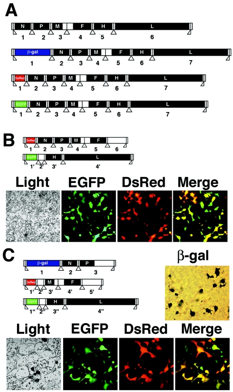
We generated recombinant MeVs with a nonsegmented RNA genome with a single transcriptional unit insertion at the 3′-proximal first locus of the genome encoding EGFP (10), β-galactosidase (29), and DsRed2 (in this paper), respectively (Fig. 1A). The viruses (IC323-EGFP, IC323-lac Z, and IC323-DsRed) were recovered from the respective cDNA plasmids using the method reported previously (29). Next, we generated the recombinant MeV with two RNA genome segments (2 seg-MeV) (Fig. 1B). 2 seg-MeV was recovered from p(+)MV-DsRed-N-P-M-F and p(+)MV-EGFP-H-L by a modified protocol in which cells were treated with caspase inhibitor (29; Nakatsu et al., unpublished). The modified protocol allowed us to generate 2 seg-MeV efficiently. We further tried to generate recombinant MeV with three RNA genome segments (3 seg-MeV) (Fig. 1C). 3 seg-MeV was recovered from p(+)MV-lac Z-N-P, p(+)MV-DsRed-M-F, and p(+)MV-EGFP-H-L by the modified rescue protocol. The modified protocol also allowed us to generate 3 seg-MeV efficiently.
FIG. 1.
Diagram of the genome organization of the recombinant viruses. (A) Recombinant MeVs, wt (IC323), IC323-lacZ, IC323-DsRed, and IC323-EGFP with nonsegmented RNA genomes with six or seven transcriptional units. (B) Recombinant MeV with 2 RNA genome segments (2 seg-MeV). One RNA genome segment encodes N, P, M, and F proteins and DsRed2. In addition, the segment has another transcriptional unit at the sixth locus (6) providing a second insertion site for a foreign gene. The sixth transcriptional unit has the authentic GS sequence and the initiation codon of the H gene, followed by two tandem termination codons to stop unwanted translation. After ∼800 base-noncoding sequences, the unit ends with the authentic GE sequence of the L gene. The other RNA genome segment encodes H and L proteins and EGFP. The segment also has another transcriptional unit at the second locus (2′), providing a second insertion site for a foreign gene. The second transcriptional unit starts with the authentic GS sequence of the P gene, combined with the noncoding sequence and the initiation codon of the N gene, and ends with the authentic GE sequence of the F gene after 17 nucleotides. (C) Recombinant MeV with three RNA genome segments (3 seg-MeV). The first RNA genome segment encodes N and P proteins and β-galactosidase. The second RNA genome segment encodes M and F proteins and DsRed2. In addition, the segment has two extra transcriptional units at the second (2′) and fifth (5′) loci, providing additional insertion sites for the foreign genes. The second transcriptional unit has the authentic GS sequence of the P gene combined with the noncoding sequence and the initiation codon of the N gene, followed by tandem termination codons after 24 nucleotides, and ends with the authentic GE sequence of the P gene. The fifth transcriptional unit has the same structure as that of the sixth unit of the first RNA genome segment of 2 seg-MeV. The third RNA genome segment has the same structure as that of the second RNA genome segment of 2 seg-MeV. Filled boxes show ORFs in which encoded proteins are shown by white characters. Open boxes show untranslated regions, and vertical lines with open triangles show the positions of the gene junctions. Panels show light and fluorescence microscopic images of the 2 seg-MeV- or 3 seg-MeV-infected B95a cells. X-Gal staining was performed for 3 seg-MeV-infected cells (results shown in panel C, β-gal).
The 2 seg- and 3 seg-MeVs recovered from the plasmids were propagated in B95a cells (Fig. 1B and C). The negative-stranded RNA virus genome needs to form ribonucleocapsid complex to function as an active template for either transcription or replication. Since the MeV genome requires N, phospho (P), and large (L) proteins to form the ribonucleocapsid complex, each of the two segments of 2 seg-MeV is essential for transcription and genome replication to occur. On the other hand, 3 seg-MeV needs only the first and third segments for transcription and genome replication, while matrix (M) and fusion (F) proteins encoded in the second segment are required for virion assembly and cell-to-cell fusion (Table 1). Accordingly, 2 seg-MeV replicated in B95a cells, producing the multinucleated giant cells that express both EGFP and DsRed2 autofluorescence. Intensity of DsRed2 autofluorescence was weaker than that of EGFP, but all syncytia were shown to express both EGFP and DsRed2 autofluorescence by fluorescence microscopy. 3 seg-MeV also replicated in B95a cells, producing multinucleated giant cells that expressed EGFP and DsRed2 autofluorescence and β-galactosidase activity (Fig. 1C).
TABLE 1.
Predicted phenotypes of 3 seg-MeV and derivative particles
| Particle type | Segment incorporated
|
Transcription and genome replication | Reporter protein expression
|
Particle production | Syncytium or plaque formation | ||||
|---|---|---|---|---|---|---|---|---|---|
| 1 | 2 | 3 | β-Gal | DsRed2 | EGFP | ||||
| 1 | + | + | + | + | + | + | + | + | + |
| 2 | + | − | − | − | − | − | − | − | − |
| 3 | − | + | − | − | − | − | − | − | − |
| 4 | − | − | + | − | − | − | − | − | − |
| 5 | + | + | − | − | − | − | − | − | − |
| 6 | + | − | + | + | + | − | + | − | − |
| 7 | − | + | + | − | − | − | − | − | − |
Plaque phenotype and replication kinetics of recombinant viruses.
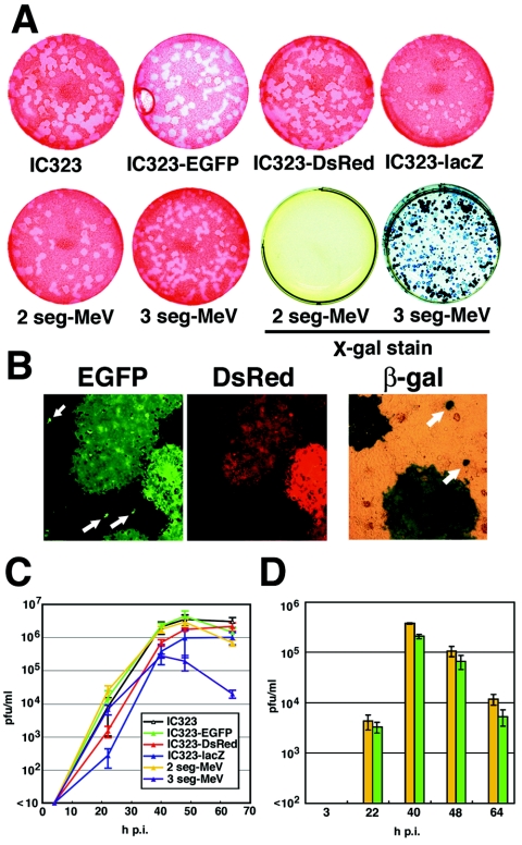
We compared the plaque phenotype of the recombinant wild-type (wt) MeV (IC323) (31), MeVs with a single unit insertion (IC323-EGFP, IC323-DsRed, and IC323-lac Z), 2 seg-MeV, and 3 seg-MeV. Plaque sizes of IC323-EGFP, IC323-DsRed, and 2 seg-MeV were comparable to that of wt IC323, while those of IC323-lac Z and 3 seg-MeV were smaller than that of wt IC323 (Fig. 2A). As expected, all plaques of the 2 seg-MeV expressed both green and red autofluorescence, and all plaques of 3 seg-MeV expressed β-galactosidase, as well as green and red autofluorescence (Fig. 2A and B; Table 1; and data not shown). When 3 seg-MeV was plaqued, some individual cells were found to express green autofluorescence and β-galactosidase but not red autofluorescence (in this paper, we designated these cells satellites) (Fig. 2B, arrows; Table 1, particle type 6). This was what we expected, since cells infected with particles containing only the first and third segments but not the second segment should replicate and transcribe the viral genome efficiently but fail to produce progeny infectious particles and form plaques, due to the lack of the M and F proteins encoded in the second segment (Table 1, particle type 6).
FIG. 2.
Plaque formation and replication kinetics of recombinant viruses. (A) Neutral red and X-Gal staining of plaques of recombinant MeVs on Vero/hSLAM cells. (B) Plaques and EGFP- and β-galactosidase-expressing individual cells (satellites are indicated by arrows) produced by 3 seg-MeV. (C) Replication kinetics. Vero/hSLAM cells were infected with viruses at a multiplicity of infection of 0.01. At various intervals, cells were harvested with culture medium, and the number of PFU was determined for Vero/hSLAM cells. (D) Production of satellites and plaques by 3 seg-MeV. The number of satellites (green bars) in the samples shown in panel C was counted and compared to the number of plaques (yellow bars).
Growth kinetics of the viruses was analyzed in Vero/hSLAM cells. Similar to the findings of many previous papers (3, 9, 10, 13, 16, 19, 23, 25, 33, 35, 37), the IC323-DsRed and IC323-lacZ viruses that have a single unit insertion into the authentic nonsegmented RNA genome replicated less efficiently than the wt IC323 virus, while IC323-EGFP replicated as efficiently as the wt virus (Fig. 2C). At 24 h p.i., virus titers of the IC323-DsRed and IC323-lac Z were ∼10 and ∼100 times lower, respectively, than that of the wt virus (Fig. 2C). On the other hand, the growth kinetics of 2 seg-MeV was comparable to that of the wt IC323 virus, in spite of the double insertions of EGFP and DsRed2 genes (Fig. 2C). 3 seg-MeV replicated efficiently until 24 h p.i., but thereafter the virus titers were not as high as those of the wt virus (Fig. 2C). However, it was noteworthy that the virus titer of the 3 seg-MeV that harbors three additional units encoding EGFP, DsRed2, and β-galactosidase at 24 h p.i. was ∼50 times higher than that of the singly inserted IC323-lac Z virus (Fig. 2C).
As described above, 3 seg-MeV produced satellites, which are cells infected with the virus particles with only two genome segment species encoding β-galactosidase and EGFP (Table 1). At various times postinfection, the numbers of the satellites were compared to those of plaques, which corresponded to the numbers of infectious particles containing all three genome segment species (Fig. 2D). At all of these times (22, 40, 48, and 64 h p.i.), the numbers of plaques were about twice those of the satellites (Fig. 2D). Presumably, 3 seg-MeV incorporated genome segments randomly, and some particles that produced plaques contained more than three RNA segments. Similarly, particles that produced satellites could contain more than two segments. These data suggested that the 3 seg-MeV particles incorporated multiple RNA segments efficiently, consistent with a previous paper showing that MeV particles can contain at least two genomes (22).
Viral and reporter gene expression of recombinant viruses.
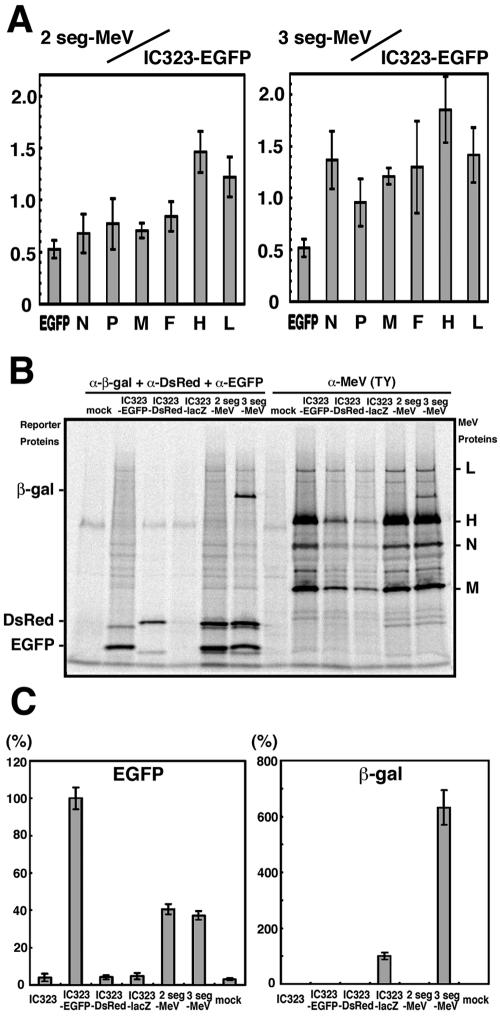
We analyzed levels of gene expression of the recombinant viruses at 24 h p.i. In the 2 seg-MeV-infected cells, levels of the N, P, M, and F mRNAs transcribed from the first segment were ∼0.7 fold, compared with those in the IC323-EGFP-infected cells, while the levels of the hemagglutinin (H) and L mRNAs from the second segment were ∼1.4 fold (Fig. 3A). The gene expression level was affected by several factors including the location of the gene in the genome, replication rate of the virus, and each segment's efficiency of incorporation into budding particles. The increase in H and L mRNA levels could be explained partially by the fact that the H and L genes were moved to the 3′ proximal loci in the segmented RNA genome of the 2 seg-MeV (Fig. 1A and B). On the other hand, in the 3 seg-MeV-infected cells, the relative expression levels of the EGFP gene decreased compared to levels for other genes (Fig. 3A). We speculate that the replication and/or incorporation of the third segment might be somewhat less efficient than those of the other two segments, as the third segment is longer than the other two segments (Fig. 1C). Immunoprecipitation of proteins from virus-infected cells clearly showed that production of the viral proteins (N, M, H, and L) increased greatly in 2 seg-MeV- and 3 seg-MeV-infected cells, compared with that in IC323-DsRed- or IC323-lac Z-infected cells (Fig. 3B). Accordingly, 2 seg-MeV and 3 seg-MeV produced reporter proteins in infected cells at much higher levels than in IC323-DsRed or IC323-lac Z (Fig. 3B). Expression levels of EGFP and β-galactosidase were also quantified by fluorescent and chemiluminescent detection, respectively. Expression of EGFP in the 2 seg-MeV- and 3 seg-MeV-infected cells were ∼40% of that in IC323-EGFP-infected cells (Fig. 3C). These results were consistent with the data shown in Fig. 3A measuring mRNA levels. This lowered (∼40%) EGFP expression is likely attributable to difference in virus growth. 2 seg- and 3 seg-MeV grew somewhat less efficiently than the IC323-EGFP virus (Fig. 2C). On the other hand, expression of β-galactosidase in 3 seg-MeV-infected cells was much higher than that in IC323-lacZ-infected cells (Fig. 3C). Thus, these data suggest that genome segmentation could be a useful strategy for the generation of mononegavirus-derived vectors when the single insertion (especially a long gene insertion, such as the 3.2-kb lacZ gene) into the nonsegmented genome affects virus replication significantly.
FIG. 3.
Gene expression of recombinant viruses. (A) Levels of viral mRNAs. Vero/hSLAM cells were infected with IC323-EGFP, 2 seg-MeV, and 3 seg-MeV. At 24 h p.i., viral mRNAs in the 2 seg-MeV- or 3 seg-MeV-infected cells were quantified by reverse transcription and quantitative PCR and compared with those in the IC323-EGFP-infected cells. Relative amounts of them were shown (2 seg-MeV/IC323-EGFP and 3 seg-MeV/IC323-EGFP, respectively). (B) Synthesis of reporter and viral proteins. Cells at 24 h p.i. were pulse-labeled for 1 h, and reporter and viral proteins were immunoprecipitated and analyzed by SDS-PAGE. (C) Quantification of reporter proteins. EGFP autofluorescence and enzymatic activity of β-galactosidase in cells at 24 h p.i. were quantified as described in Materials and Methods. Levels of reporter proteins in the IC323-EGFP- or IC323-lacZ-infected cells were set at 100%.
Tolerance of 3 seg-MeV to multiple foreign gene insertions.
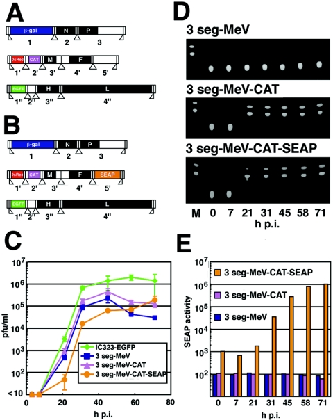
Finally, we analyzed the tolerance of 3 seg-MeV to more foreign gene insertions. We inserted the ORF of CAT in the second locus (2′) of the second RNA genome segment (Fig. 4A). In addition, the ORF of SEAP was inserted in the fifth locus (5′) of the second segment (Fig. 4B). These viruses were generated by the vaccinia virus-infected CHO cell-based reverse genetics method using caspase inhibitors. They were designated 3 seg-MeV-CAT and 3 seg-MeV-CAT-SEAP, respectively. 3 seg-MeV-CAT-SEAP replicated less efficiently than 3 seg-MeV, whereas 3 seg-MeV-CAT grew slightly more efficiently than 3 seg-MeV (Fig. 4C). These viruses produced clear plaques on Vero/hSLAM cells, though their sizes were smaller than those of IC323-EGFP (data not shown). We confirmed the expression of CAT and SEAP by detecting their enzymatic activities. Increased enzymatic activity of CAT was observed with cells infected with either 3 seg-MeV-CAT or 3 seg-MeV-CAT-SEAP in a time course experiment (Fig. 4D). Similarly, increased SEAP enzymatic activity was detected in cells infected with 3 seg-MeV-CAT-SEAP (Fig. 4E). These data showed that 3 seg-MeV functioned as a mononegavirus-derived vector expressing up to five foreign proteins.
FIG. 4.
Multiple foreign gene insertion into the 3 seg-MeV genome. (A) Diagram of the 3 seg-MeV-CAT genome. (B) Diagram of the 3 seg-MeV-CAT-SEAP genome. (C) Replication kinetics. Vero/hSLAM cells were infected with IC323-EGFP, 3 seg-MeV, 3 seg-MeV-CAT, and 3 seg-MeV-CAT-SEAP at a multiplicity of infection of 0.005. At various intervals, cells were harvested with culture medium, and the number of PFU was determined with Vero/hSLAM cells. (D and E) Analysis of CAT and SEAP enzymatic activities. In parallel to the results of PFU analysis shown in panel C, CAT activity in cells (D) and SEAP activity in culture medium (E) were quantified as described in Materials and Methods. SEAP activity is expressed in chemiluminescent units per second.
DISCUSSION
There are seven families of negative-stranded RNA viruses. Members of the four families (Paramyxoviridae, Filoviridae, Rhabdoviridae, and Bornaviridae) in the order Mononegavirales have a single nonsegmented RNA molecule as the genome, while those in the other three families (Orthomyxoviridae, Bunyaviridae, and Arenaviridae) have multiple RNA segments as their genome. Our data showed that the overall genome structure (whether it is nonsegmented or segmented) is not critical for MeV and presumably for other paramyxoviruses to replicate in cultured cells. The viruses therefore may have the potential to convert the nonsegmented genome into several segments when it is required for their survival in nature. Since short genomes (or segments) replicate more rapidly than long ones, such a genome segmentation strategy may be advantageous for virus multiplication. In addition, since each segment needs to encode only a few viral genes, the polar attenuation of the transcription becomes minimized. Our data indeed showed that 3 seg-MeV exhibited a greatly expanded coding capacity with a high gene expression competence. The extra task required for the viruses that have the segmented genome is to package all segments into each budding virion. The recombinant MeVs reported in this paper likely packaged their genome segments randomly, yet still replicated reasonably well. Once the viruses develop a strategy to package all segments selectively, their replication rate should become very fast. In this regard, it is interesting that influenza virus in the family Orthomyxoviridae is shown to have a selective packaging strategy of eight RNA genome segments (6).
Expansion of the coding capacity is advantageous for the virus vectors. The initial report of the generation of a mononegavirus from plasmid DNAs appeared in 1994; it used rabies virus and provided a way to manipulate mononegaviruses by design (26). This powerful technology has been applied to many mononegavirus species, covering many important human and animal mononegavirus pathogens, as well as model viruses used to study mononegavirus biology (18, 20). Originally virus rescue from cloned cDNAs was inefficient, and approaches to the engineering of mononegaviruses were rather restricted. Recently, we reported a vaccinia virus-infected CHO cell-based rescue system of MeV that exhibits a remarkably high rescue efficiency (29). This system greatly improved our ability to engineer the MeV genome, and further improvement of the rescue system using caspase inhibitors allowed recovery of virus even after drastic alterations of the virus genome, such as segmentation of the MeV genome, as described here. Our data showed that MeV with three RNA genome segments could harbor up to six additional transcriptional units, five of which were shown to be capable of encoding and expressing foreign proteins efficiently. We believe that the segmentation strategy is also applicable at least to Sendai and Newcastle disease viruses, since they were shown to incorporate multiple RNA genomes like MeV (4, 7, 11, 22). These viruses or vaccine strains of MeV would be more suitable as vectors in clinical use than our MeV based on a clinical isolate, and experiments to apply this genome segmentation strategy to Sendai virus are in progress. Importantly, the segmentation strategy can be used to overcome limitations on insertion of long foreign genes, since the 3 seg-MeV carrying the lacZ gene (∼3.2 kb), as well as two fluorescent protein genes, replicated much faster than the virus with the nonsegmented RNA genome carrying the lacZ gene. The 3′-proximal first locus is the most desirable position to achieve high gene expression, and each segment of the segmented genome vectors can accommodate the foreign gene in its 3′-proximal first locus. Therefore, these vectors are useful when high-level expression of the three genes is required. The 3 seg-MeV with the three reporter genes could be passaged 20 times successfully in cultured cells. We are planning to verify the stability of the segmented genome viruses in animal models.
In conclusion, infectious MeVs with segmented RNA genomes could be rescued. They replicated well in cultured cells, accommodating up to six additional transcriptional units. These data add further experimental insight into evolution of the negative-stranded RNA viruses and also illustrate a new design of mononegavirus-derived vectors harboring multiple additional transcriptional units.
Acknowledgments
We thank H. Minagawa and K. Takeuchi for helpful discussions and kind supports and T. Kohama and B. Moss for providing the anti-measles virus antibody and the recombinant vaccinia virus, respectively. We are grateful to A. P. Schmitt for critical reading and helpful comments.
This work was supported by grants from the Ministry of Education, Science, and Culture and the Ministry of Health, Labor, and Welfare of Japan; the Japan Society for the Promotion of Science; and Takeda Science Foundation.
REFERENCES
- 1.Biacchesi, S., M. H. Skiadopoulos, K. C. Tran, B. R. Murphy, P. L. Collins, and U. J. Buchholz. 2004. Recovery of human metapneumovirus from cDNA: optimization of growth in vitro and expression of additional genes. Virology 321:247-259. [DOI] [PubMed] [Google Scholar]
- 2.Bitzer, M., S. Armeanu, U. M. Lauer, and W. J. Neubert. 2003. Sendai virus vectors as an emerging negative-strand RNA viral vector system. J. Gene Med. 5:543-553. [DOI] [PubMed] [Google Scholar]
- 3.Bukreyev, A., E. Camargo, and P. L. Collins. 1996. Recovery of infectious respiratory syncytial virus expressing an additional, foreign gene. J. Virol. 70:6634-6641. [DOI] [PMC free article] [PubMed] [Google Scholar]
- 4.Dahlberg, J. E., and E. H. Simon. 1969. Physical and genetic studies of Newcastle disease virus: evidence for multiploid particles. Virology 38:666-678. [DOI] [PubMed] [Google Scholar]
- 5.Fielding, A. K. 2005. Measles as a potential oncolytic virus. Rev. Med. Virol. 15:135-142. [DOI] [PubMed] [Google Scholar]
- 6.Fujii, Y., H. Goto, T. Watanabe, T. Yoshida, and Y. Kawaoka. 2003. Selective incorporation of influenza virus RNA segments into virions. Proc. Natl. Acad. Sci. USA 100:2002-2007. [DOI] [PMC free article] [PubMed] [Google Scholar]
- 7.Granoff, A. 1959. Studies on mixed infection with Newcastle disease virus. II. The occurrence of Newcastle disease virus heterozygotes and study of phenotypic mixing involving serotype and thermal stability. Virology 9:649-670. [DOI] [PubMed] [Google Scholar]
- 8.Griesenbach, U., M. Inoue, M. Hasegawa, and E. W. Alton. 2005. Sendai virus for gene therapy and vaccination. Curr. Opin. Mol. Ther. 7:346-352. [PubMed] [Google Scholar]
- 9.Hasan, M. K., A. Kato, T. Shioda, Y. Sakai, D. Yu, and Y. Nagai. 1997. Creation of an infectious recombinant Sendai virus expressing the firefly luciferase gene from the 3′ proximal first locus. J. Gen. Virol. 78:2813-2820. [DOI] [PubMed] [Google Scholar]
- 10.Hashimoto, K., N. Ono, H. Tatsuo, H. Minagawa, M. Takeda, K. Takeuchi, and Y. Yanagi. 2002. SLAM (CD150)-independent measles virus entry as revealed by recombinant virus expressing green fluorescent protein. J. Virol. 76:6743-6749. [DOI] [PMC free article] [PubMed] [Google Scholar]
- 11.Hosaka, Y., H. Kitano, and S. Ikeguchi. 1966. Studies on the pleomorphism of HVJ virons. Virology 29:205-221. [DOI] [PubMed] [Google Scholar]
- 12.Huang, Z., S. Elankumaran, A. Panda, and S. K. Samal. 2003. Recombinant Newcastle disease virus as a vaccine vector. Poult. Sci. 82:899-906. [DOI] [PubMed] [Google Scholar]
- 13.Huang, Z., S. Krishnamurthy, A. Panda, and S. K. Samal. 2001. High-level expression of a foreign gene from the most 3′-proximal locus of a recombinant Newcastle disease virus. J. Gen. Virol. 82:1729-1736. [DOI] [PubMed] [Google Scholar]
- 14.Iverson, L. E., and J. K. Rose. 1981. Localized attenuation and discontinuous synthesis during vesicular stomatitis virus transcription. Cell 23:477-484. [DOI] [PubMed] [Google Scholar]
- 15.Kobune, F., H. Sakata, and A. Sugiura. 1990. Marmoset lymphoblastoid cells as a sensitive host for isolation of measles virus. J. Virol. 64:700-705. [DOI] [PMC free article] [PubMed] [Google Scholar]
- 16.Krishnamurthy, S., Z. Huang, and S. K. Samal. 2000. Recovery of a virulent strain of Newcastle disease virus from cloned cDNA: expression of a foreign gene results in growth retardation and attenuation. Virology 278:168-182. [DOI] [PubMed] [Google Scholar]
- 17.McKenna, P. M., J. P. McGettigan, R. J. Pomerantz, B. Dietzschold, and M. J. Schnell. 2003. Recombinant rhabdoviruses as potential vaccines for HIV-1 and other diseases. Curr. HIV Res. 1:229-237. [DOI] [PubMed] [Google Scholar]
- 18.Nagai, Y., and A. Kato. 1999. Paramyxovirus reverse genetics is coming of age. Microbiol. Immunol. 43:613-624. [DOI] [PubMed] [Google Scholar]
- 19.Nakaya, T., J. Cros, M. S. Park, Y. Nakaya, H. Zheng, A. Sagrera, E. Villar, A. Garcia-Sastre, and P. Palese. 2001. Recombinant Newcastle disease virus as a vaccine vector. J. Virol. 75:11868-11873. [DOI] [PMC free article] [PubMed] [Google Scholar]
- 20.Neumann, G., M. A. Whitt, and Y. Kawaoka. 2002. A decade after the generation of a negative-sense RNA virus from cloned cDNA—what have we learned? J. Gen. Virol. 83:2635-2662. [DOI] [PubMed] [Google Scholar]
- 21.Ono, N., H. Tatsuo, Y. Hidaka, T. Aoki, H. Minagawa, and Y. Yanagi. 2001. Measles viruses on throat swabs from measles patients use signaling lymphocytic activation molecule (CDw150) but not CD46 as a cellular receptor. J. Virol. 75:4399-4401. [DOI] [PMC free article] [PubMed] [Google Scholar]
- 22.Rager, M., S. Vongpunsawad, W. P. Duprex, and R. Cattaneo. 2002. Polyploid measles virus with hexameric genome length. EMBO J. 21:2364-2372. [DOI] [PMC free article] [PubMed] [Google Scholar]
- 23.Roberts, A., and J. K. Rose. 1998. Recovery of negative-strand RNA viruses from plasmid DNAs: a positive approach revitalizes a negative field. Virology 247:1-6. [DOI] [PubMed] [Google Scholar]
- 24.Russell, S. J. 2002. RNA viruses as virotherapy agents. Cancer Gene Ther. 9:961-966. [DOI] [PubMed] [Google Scholar]
- 25.Sakai, Y., K. Kiyotani, M. Fukumura, M. Asakawa, A. Kato, T. Shioda, T. Yoshida, A. Tanaka, M. Hasegawa, and Y. Nagai. 1999. Accommodation of foreign genes into the Sendai virus genome: sizes of inserted genes and viral replication. FEBS Lett. 456:221-226. [DOI] [PubMed] [Google Scholar]
- 26.Schnell, M. J., T. Mebatsion, and K. K. Conzelmann. 1994. Infectious rabies viruses from cloned cDNA. EMBO J. 13:4195-4203. [DOI] [PMC free article] [PubMed] [Google Scholar]
- 27.Skiadopoulos, M. H., S. R. Surman, A. P. Durbin, P. L. Collins, and B. R. Murphy. 2000. Long nucleotide insertions between the HN and L protein coding regions of human parainfluenza virus type 3 yield viruses with temperature-sensitive and attenuation phenotypes. Virology 272:225-234. [DOI] [PubMed] [Google Scholar]
- 28.Strauss, E. G., J. H. Strauss, and A. J. Levine. 1996. Virus evolution, p. 153-171. In B. N. Fields, D. M. Knipe, and P. M. Howley (ed.), Fields virology, 3rd ed. Lippincott-Raven Publishers, Philadelphia, Pa.
- 29.Takeda, M., S. Ohno, F. Seki, K. Hashimoto, N. Miyajima, K. Takeuchi, and Y. Yanagi. 2005. Efficient rescue of measles virus from cloned cDNA using SLAM-expressing Chinese hamster ovary cells. Virus Res. 108:161-165. [DOI] [PubMed] [Google Scholar]
- 30.Takeda, M., S. Ohno, F. Seki, Y. Nakatsu, M. Tahara, and Y. Yanagi. 2005. Long untranslated regions of measles virus M and F genes control virus replication and cytopathogenicity. J. Virol. 79:14346-14354. [DOI] [PMC free article] [PubMed] [Google Scholar]
- 31.Takeda, M., K. Takeuchi, N. Miyajima, F. Kobune, Y. Ami, N. Nagata, Y. Suzaki, Y. Nagai, and M. Tashiro. 2000. Recovery of pathogenic measles virus from cloned cDNA. J. Virol. 74:6643-6647. [DOI] [PMC free article] [PubMed] [Google Scholar]
- 32.Takeuchi, K., N. Miyajima, F. Kobune, and M. Tashiro. 2000. Comparative nucleotide sequence analysis of the entire genomes of B95a cell-isolated and Vero cell-isolated measles viruses from the same patient. Virus Genes 20:253-257. [DOI] [PubMed] [Google Scholar]
- 33.Tang, R. S., J. H. Schickli, M. MacPhail, F. Fernandes, L. Bicha, J. Spaete, R. A. Fouchier, A. D. Osterhaus, R. Spaete, and A. A. Haller. 2003. Effects of human metapneumovirus and respiratory syncytial virus antigen insertion in two 3′ proximal genome positions of bovine/human parainfluenza virus type 3 on virus replication and immunogenicity. J. Virol. 77:10819-10828. [DOI] [PMC free article] [PubMed] [Google Scholar]
- 34.Tatsuo, H., N. Ono, K. Tanaka, and Y. Yanagi. 2000. SLAM (CDw150) is a cellular receptor for measles virus. Nature 406:893-897. [DOI] [PubMed] [Google Scholar]
- 35.Tokusumi, T., A. Iida, T. Hirata, A. Kato, Y. Nagai, and M. Hasegawa. 2002. Recombinant Sendai viruses expressing different levels of a foreign reporter gene. Virus Res. 86:33-38. [DOI] [PubMed] [Google Scholar]
- 36.Wertz, G. W., V. P. Perepelitsa, and L. A. Ball. 1998. Gene rearrangement attenuates expression and lethality of a nonsegmented negative strand RNA virus. Proc. Natl. Acad. Sci. USA 95:3501-3506. [DOI] [PMC free article] [PubMed] [Google Scholar]
- 37.Zhao, H., and B. P. Peeters. 2003. Recombinant Newcastle disease virus as a viral vector: effect of genomic location of foreign gene on gene expression and virus replication. J. Gen. Virol. 84:781-788. [DOI] [PubMed] [Google Scholar]


