

Abstract
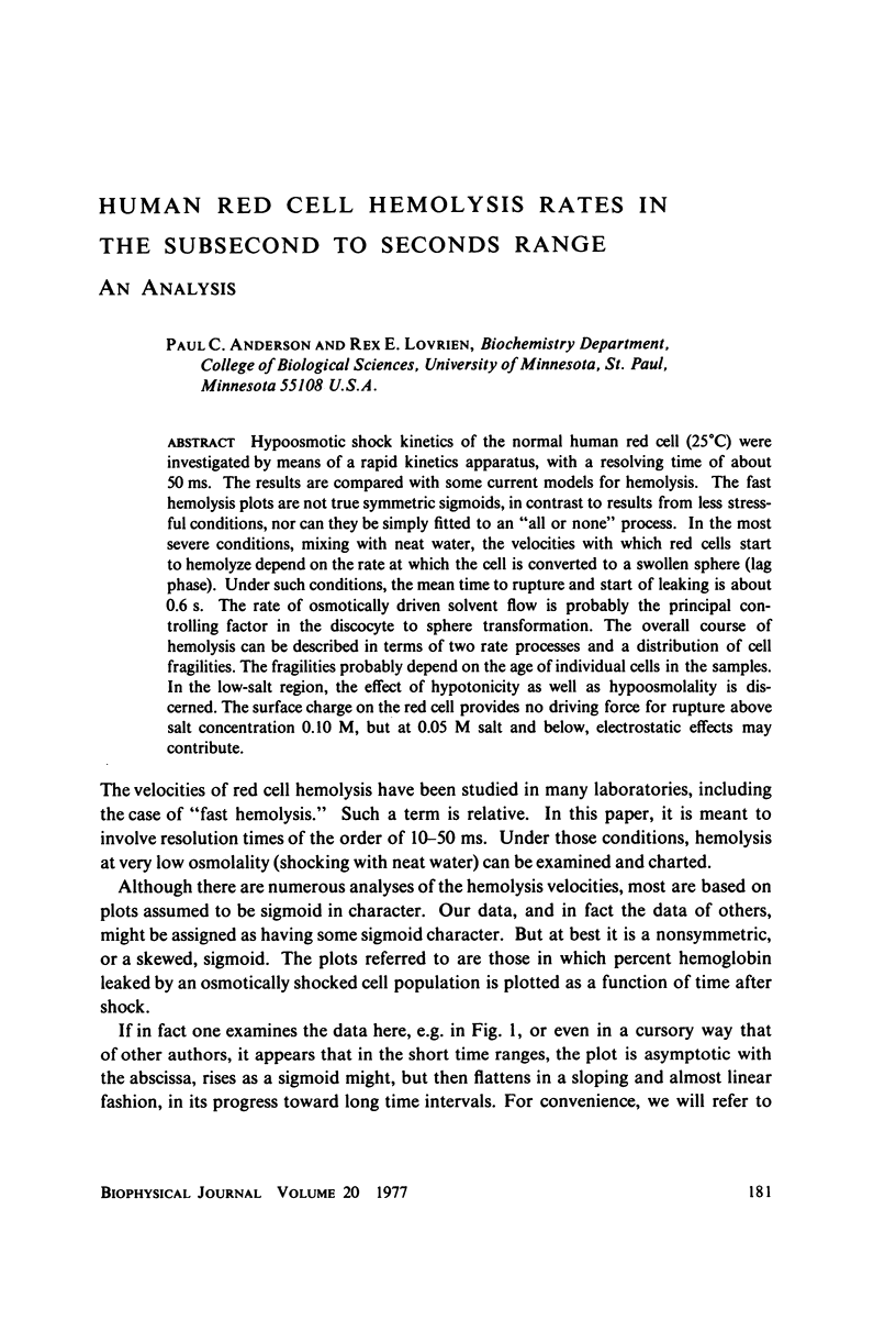
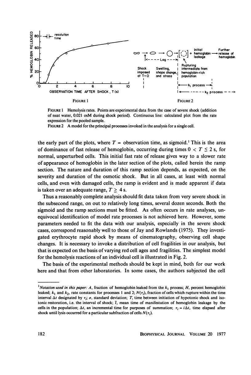
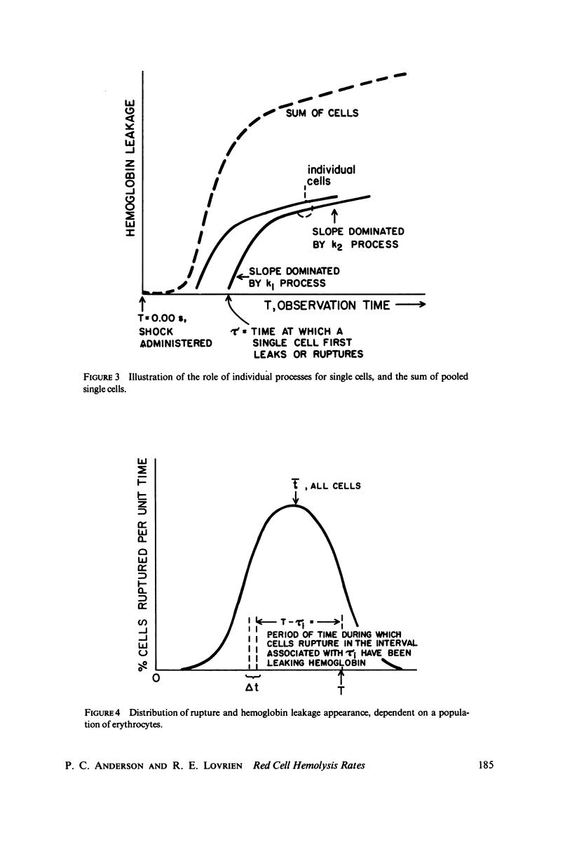
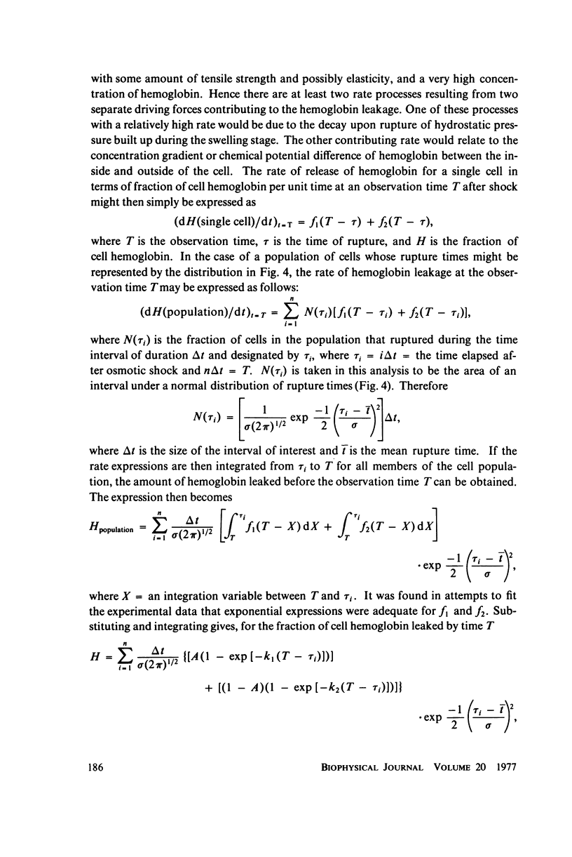
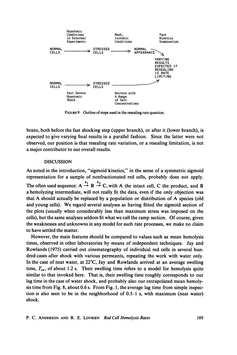
Hypoosmotic shock kinetics of the normal human red cell (25 degrees C) were investigated by means of a rapid kinetics apparatus, with a resolving time of about 50 ms. The results are compared with some current models for hemolysis. The fast hemolysis plots are not true symmetric sigmoids, in contrast to results from less stressful conditions, nor can they be simply fitted to an "all or none" process. In the most severe conditions, mixing with neat water, the velocities with which red cells start to hemolyze depend on the rate at which the cell is converted to a swollen sphere (lag phase). Under such conditions, the mean time to rupture and start of leaking is about 0.6 s. The rate of osmotically driven solvent flow is probably the principal controlling factor in the discocyte to sphere transformation. The overall course of hemolysis can be described in terms of two rate processes and a distribution of cell fragilities. The fragilities probably depend on the age of individual cells in the samples. In the low-salt region, the effect of hypotonicity as well as hypoosmolality is discerned. The surface charge on the red cell provided no driving force for rupture above salt concentration 0.10M, but at 0.05 M salt and below, electrostatic effects may contribute.
Full text
PDF

Selected References
These references are in PubMed. This may not be the complete list of references from this article.
- Lovrien R., Tisel W., Pesheck P. Stoichiometry of compounds bound to human erythrocytes in relation to morphology. J Biol Chem. 1975 Apr 25;250(8):3136–3141. [PubMed] [Google Scholar]
- Rojas E., Taylor R. E. Simultaneous measurements of magnesium, calcium and sodium influxes in perfused squid giant axons under membrane potential control. J Physiol. 1975 Oct;252(1):1–27. doi: 10.1113/jphysiol.1975.sp011131. [DOI] [PMC free article] [PubMed] [Google Scholar]