Abstract
At the nerve terminal, both N- and P/Q-type Ca2+ channels mediate synaptic transmission, with their relative contribution varying between synapses and with postnatal age. To clarify functional significance of different presynaptic Ca2+ channel subtypes, we recorded N-type and P/Q-type Ca2+ currents directly from calyces of Held nerve terminals in α1A-subunit-deficient mice and wild-type (WT) mice, respectively. The most prominent feature of P/Q-type Ca2+ currents was activity-dependent facilitation, which was absent for N-type Ca2+ currents. EPSCs mediated by P/Q-type Ca2+ currents showed less depression during high-frequency stimulation compared with those mediated by N-type Ca2+ currents. In addition, the maximal inhibition by the GABAB receptor agonist baclofen was greater for EPSCs mediated by N-type channels than for those mediated by P/Q-type channels. These results suggest that the developmental switch of presynaptic Ca2+ channels from N- to P/Q-type may serve to increase synaptic efficacy at high frequencies of activity, securing high-fidelity synaptic transmission.
Transmitter release is triggered by Ca2+ entry through presynaptic voltage-dependent Ca2+ channels (Katz, 1969). In the mammalian CNS, multiple types of high-voltage-activated Ca2+ channels mediate synaptic transmission (Luebke et al. 1993; Takahashi & Momiyama, 1993). Among them N-type Ca2+ channels widely mediate synaptic transmission at immature synapses, but their contribution decreases with postnatal development, being replaced by P/Q-type Ca2+ channels (Iwasaki et al. 2000). To assess the functional significance of the developmental switch of presynaptic Ca2+ channel subtypes, it seems essential to compare properties of N-type and P/Q-type Ca2+ currents in the same type of nerve terminal. Compared with presynaptic P/Q-type Ca2+ currents, which have been characterized at the calyx of Held (Forsythe et al. 1998), much less is known for presynaptic N-type Ca2+ currents. N-type Ca2+ currents can be recorded from immature calyceal terminal after blocking P/Q-type Ca2+ channels (Wu et al. 1999; Iwasaki et al. 2000), but these remaining currents are often too small for detailed analysis. To circumvent this difficulty, we utilized mice with their α1A subunit genetically ablated. These mice lack P/Q-type Ca2+ currents, but overexpress N-type Ca2+ channels in compensation (Jun et al. 1999; Ishikawa et al. 2003; Inchauspe et al. 2004). Using these knockout (KO) mice and wild-type (WT) littermates, we compared basic properties of N-type and P/Q-type Ca2+ currents directly recorded from the calyx of Held presynaptic terminals. Furthermore, we compared EPSCs mediated by N-type and P/Q-type Ca2+ currents. Our results indicate that P/Q-type, but not N-type, Ca2+ currents undergo activity-dependent facilitation (Borst & Sakmann, 1998; Cuttle et al. 1998; Forsythe et al. 1998; Tsujimoto et al. 2002). Synaptic strength during high-frequency stimulation was consistently higher for EPSCs mediated by P/Q-type Ca2+ channels than those mediated by N-type channels. Part of this study has been published in abstract form (Ishikawa et al. 2003).
Methods
Preparations and solutions
All experiments were performed in accordance with the guidelines of the Physiological Society of Japan. The generation and phenotype characterization of α1A-deficient mice have been previously described (Jun et al. 1999). In the present study, both WT (+/+) and α1A-deficient (−/−) mice were offspring of heterozygous (+/−) mice with the C57BL/6 J genetic background. Mice (9–12 days old) were killed by decapitation under halothane anaesthesia. Transverse slices (150–300 μm in thickness) containing the medial nucleus of the trapezoid body were cut from a tissue block containing the brainstem. Before recordings, slices were incubated for 1 h at 36°C in artificial cerebrospinal fluid (aCSF) containing (mm): 125 NaCl, 2.5 KCl, 1 MgCl2, 2 CaCl2, 10 glucose, 26 NaHCO3, 1.25 NaH2PO4, 0.5 ascorbic acid, 3 myo-inositol, 2 sodium pyruvate (310 mosmol kg−1, pH 7.3, when saturated with 95% O2/5% CO2).
Recording and data analysis
Recordings were made at room temperature (24–26°C). A slice was transferred to a chamber continuously superfused with the aCSF. The giant presynaptic terminal, the calyx of Held, was visualized with a ×60 water-immersion objective (Olympus) attached to an upright microscope (Axioskop, Zeiss). To isolate Ca2+ currents, tetrodotoxin (1 μm), tetraethylammonium chloride (10 mm), and 4-aminopyridine (0.5 mm) were included in the aCSF. The presynaptic pipette solution contained (mm): 110 CsCl, 40 Hepes, 0.5 EGTA, 1 MgCl2, 12 phosphocreatine (sodium salt), 2 ATP (magnesium salt) and 0.5 GTP (sodium salt). The presynaptic Ca2+ current (IpCa) was evoked in calyceal nerve terminals under voltage clamp at the holding potential of −80 mV by a depolarizing pulse stepping to −10 mV every 10 s, unless otherwise noted. EPSCs were evoked by extracellular stimulation of presynaptic axons with a bipolar tungusten electrode (Forsythe & Barnes-Davies, 1993) at 0.05 Hz. For recording EPSCs, the aCSF routinely contained bicuculline methiodide (10 μm; Sigma, St Louis, MO, USA) and strychnine hydrochloride (0.5 μm; Sigma) to block inhibitory synaptic responses, and d-aminophosphonovalerate (d-APV; 50 μm; Tocris Cookson, Bristol, UK) to block NMDA receptors. During stimulation at 100 Hz (Figs 5C and E, and 6), kynurenic acid (1 mm; Tocris Cookson) was added to minimize saturation of postsynaptic AMPA receptors. The postsynaptic pipette solution contained (mm): 120 CsF, 30 CsCl, 10 Hepes, 5 EGTA, 1 MgCl2 and 5 N-(2,6-diethylphenylcarbamoylmethyl)-triethyl-ammonium chloride (QX-314; Alomone Laboratories, Jerusalem, Israel) 295–300 mOsm (mosmol kg−1), pH was adjusted to 7.3 with CsOH. IpCa and EPSCs were recorded using an Axopatch 200B or a MultiClamp 700 A amplifier, low-pass filtered at 5–6 kHz, and digitized at 50 kHz using Digidata 1200 A or Digidata 1320 (Axon Instruments). Records were also stored for back-ups on a DAT tape, digitized at 48 kHz. Off-line analysis was performed using Clampfit or Axograph (Axon Instruments) and SigmaPlot (SPSS). The electrode resistance for presynaptic and postsynaptic recording was 4–6 MΩ and 1.5–3 MΩ, respectively. The series resistance was 8–16 and 3–8 MΩ for presynaptic and postsynaptic recordings, respectively. In the IpCa recording, series resistance was compensated by 70–80%. Leak currents were subtracted by the scaled pulse divided by six (P/6) protocol. The liquid junction potential between pipette and external solution was not corrected for. ω-Conotoxin GVIA (ω-CgTx) and ω-agatoxin IVA (ω-AgaTx) (Peptide Institute, Japan) were dissolved in aCSF containing 0.1 mg ml−1 cytochrome c. All values are given as means ± s.e.m., and statistical significance was evaluated by unpaired Student's t test, unless otherwise noted. P < 0.05 was considered significant.
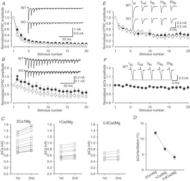
Figure 5. Activity-dependent changes in IpCa and EPSCs in KO and WT and mice.
A, EPSCs evoked by a train of 20 stimuli at 100 Hz in normal artificial cerebrospinal fluid (aCSF) in WT (upper sample trace, •) and KO (lower sample trace, ○) mice. B, EPSCs evoked by a train of 100 Hz stimulation in 1 mm [Ca2+]o (2 mm [Mg2+]o) solution in WT (•) and KO (○) mice. The asterisk indicates the point at which the EPSC amplitude in WT mice was significantly larger than that in KO mice (P < 0.05). C, the first and second amplitude of IpCa in WT mice evoked at 100 Hz (10 ms interstimulus interval) in normal aCSF (left panel), 1 mm [Ca2+]o (2 mm [Mg2+]o) solution (middle panel) and 0.6 mm [Ca2+]o (2 mm [Mg2+]o) solution (right panel). The second IpCa amplitudes were significantly larger than the first one in all solutions (P < 0.01 in paired t test). D, the mean magnitude of IpCa facilitation (%) in WT mice in three different solutions. E, EPSCs evoked by a train of 20 stimuli at 10 Hz in 1 mm [Ca2+]o (2-mm [Mg2+]o) solution in WT (•) and KO (○) mice. F, normalized amplitudes of IpCa (n = 5) in WT mice evoked by a train of 20 stimuli at 10 Hz in 1 mm [Ca2+]o (2 mm [Mg2+]o) solution. No IpCa facilitation was observed. Error bars indicate s.e.m.
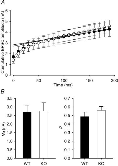
Figure 6. Release probability in WT and KO mice.
A, cumulative amplitudes of EPSCs during a 100 Hz train in WT (•) and KO (○) mice (data taken from Fig. 5A). Data points in the range of the 16–20th EPSC amplitudes were fitted by linear regression and extrapolated to time zero to estimate the readily releasable pool size of synaptic vesicles (Nq). B, left panel, Nq estimated in individual cells; right panel, release probability (p) estimated from the first EPSC amplitude divided by Nq. In WT and KO mice, Nq was 2.71 ± 0.40 nA (n = 12) and 2.76 ± 0.49 nA (n = 12), respectively, whereas p was 0.49 ± 0.05 (n = 12) and 0.56 ± 0.05 (n = 12), respectively. Neither p nor Nq was significantly different between WT and KO mice. Error bars indicate s.e.m.
Results
Pharmacological identification of presynaptic Ca2+ currents
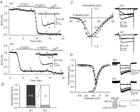
Ca2+ channel subtypes expressed at the calyx of Held terminal were pharmacologically identified using type-specific blockers. In WT mice, as previously reported in rats of a similar age (Iwasaki & Takahashi, 1998), the N-type Ca2+ channel blocker ω-CgTx (2 μm) only slightly (4.9 ± 2.0%, n = 8) attenuated the amplitude of IpCa, whereas the P/Q-type Ca2+ channel blocker ω-AgaTx (200 nm) blocked most of the remaining IpCa (89.8 ± 2.7% of total currents, n = 8) (Fig. 1A and B). Small currents remaining after application of ω-CgTx and ω-AgaTx (5.3 ± 0.9%) were abolished by Cd2+ (100 μm). In contrast, in KO mice, ω-CgTx blocked most of the IpCa (88.3 ± 2.3%, n = 8). The remaining currents (11.7 ± 2.3%) were not attenuated by ω-AgaTx, but they were abolished by Cd2+, suggesting that these were R-type currents (Wu et al. 1998). Thus IpCa was mainly (88%) N-type in KO mice, whereas it was mainly P/Q-type (90%) in WT mice (Fig. 1B). In KO mice, the total IpCa amplitude (500 ± 55 pA, n = 8) was 52% of that in WT mice (967 ± 169 pA, n = 8), whereas the amplitude of N-type currents in KO mice was 9.6 times larger than that in WT mice (see Fig. 1B legend for values), indicating that N-type channels were overexpressed in compensation for P/Q-type channels at the calyx of Held. Similar results have recently been reported at the calyx of Held for the KO and WT mice (Inchauspe et al. 2004).
Figure 1. Presynaptic Ca2+ current recorded from the calyx of Held presynaptic terminal in wild-type and knockout mice.
A and B, pharmacological dissection of presynaptic Ca2+ current (IpCa). A, ω-conotoxin GVIA (ω-CgTx; 2 μm), ω-agatoxin IVA (ω-AgaTx; 200 nm) and Cd2+ (100 μm) were applied in this sequence to block IpCa (evoked by a 10 ms depolarizing pulse) in wild-type (WT) and knockout (KO) mice. Sample records are IpCa (average of 6 records) before (a) and after (b) application of ω-CgTx, after ω-AgaTx application (c), and after Cd2+ application (d) (superimposed). B, percentage of P/Q-type (filled bar) and N-type (open bar) current components in WT and KO mice. Data are derived from 8 calyces each. The N-type current in KO mice was 443 ± 52 pA, whereas that in WT was 46 ± 21 pA. C, current–voltage relationships of IpCa in WT (•, n = 9) and KO (○, n = 11) mice. Sample records show IpCa evoked by depolarizing steps to −40, −20, 0 and +20 mV (superimposed). D, activation and inactivation curves. Activation curves (circles) were obtained from tail currents evoked by a 10 ms depolarizing pulse stepping to various membrane potentials. Data obtained from WT (•, n = 9) and KO (○, n = 11) mice were normalized to the maximal value, plotted against voltage, and fitted by the Boltzmann function I= 1/(Imax+ exp(V½−V)/k), where V½ and k represent the half-activation voltage and slope factor, respectively. Five data points between −5 and +15 mV from WT mice were excluded from the activation curve fit because inactivation, proceeding during 10 ms pulse, attenuated the tail current amplitude. Resting inactivation of IpCa was assessed using a protocol (right bottom) comprising conditioning prepulses of various amplitudes (500 ms in duration) followed by a test pulse (10 ms, to −10 mV). To formulate inactivation curves, amplitudes of IpCa evoked by the test pulse were normalized to that evoked after a −130 mV conditioning pulse, and plotted against the conditioning voltage, both in WT (▪, n = 6) and KO (□, n = 8) mice. Sample records show IpCa evoked by the test pulse after conditioning prepulses (10 mV incremental steps from −130 to −20 mV, 12 records superimposed for both WT and KO mice). Lines in A and C were eye-fitted. Error bars indicate s.e.m.
Activation properties of IpCa
Taking advantage of predominant expression of N- and P/Q-type channels, respectively, in the calyceal terminals of KO and WT mice, we first compared the activation properties of N- and P/Q-type IpCa. In the current–voltage relationship, IpCa in KO mice peaked at a membrane potential more positive than that in WT mice, with no difference in their reversal potentials (Fig. 1C). Consistently, the activation curve obtained from tail currents (Fig. 1D) indicated that the half-activation voltage was more positive, by 7.1 mV, in KO mice (−9.9 mV) than WT mice (−17.0 mV). The slope factor was similar between KO (9.4 mV) and WT (8.4 mV) mice. These results are similar to those reported by Inchauspe et al. (2004).
Inactivation properties of IpCa
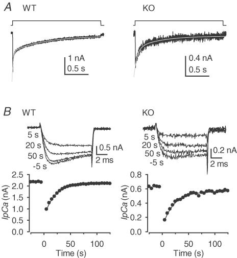
We next compared the inactivation properties of Ca2+ currents in KO and WT mice. As previously reported in rats (Forsythe et al. 1998), inactivation was observed only in association with activation, indicating no resting inactivation both in KO and WT mice (Fig. 1D). During a sustained depolarization, IpCa decayed with a double exponential time course (Fig. 2A). The fast and slow time constants of inactivation were 77 ± 22 ms (71 ± 5%) and 775 ± 187 ms, respectively, in KO mice (n = 6), and 28 ± 4 ms (41 ± 6%) and 609 ± 88 ms, respectively, in WT mice (n = 5). There was no significant difference in the weighted mean decay time constant between KO (282 ± 86 ms) and WT (364 ± 50 ms) mice. The recovery time course of IpCa from inactivation largely determines the late phase (>5 s) of recovery of EPSCs from post-tetanic depression (Forsythe et al. 1998). We compared the recovery time course of IpCa in KO and WT mice by monitoring IpCa elicited by a 10 ms depolarizing test pulse every 5 s, after inactivating IpCa with a 1.9 s depolarizing conditioning pulse (Fig. 2B). During the first 5 s after the conditioning pulse, the amplitude of IpCa rapidly recovered to 45 ± 6% (n = 5) in WT mice and 39 ± 4% (n = 5) in KO mice (no significant difference). Thereafter, the IpCa amplitude recovered more slowly with mono-exponential time course (Fig. 2B), with a similar time constant in WT mice (14.7 ± 1.7 s, n = 5) and KO mice (16.2 ± 2.3 s, n = 5). As IpCa recovered from inactivation, its rise time tended to become faster in WT mice, but not in KO mice (Fig. 2B).
Figure 2. Inactivation and recovery time courses of IpCa.
A, inactivation of IpCa during a 1.9 s depolarizing pulse in WT and KO mice. The inactivation time courses were fitted with double exponential curves (superimposed on records). B, the time course of recovery from inactivation. Ordinates indicate the amplitude of IpCa elicited by a 10 ms test pulse every 5 s before and after application (at time 0) of a 1.9 s conditioning pulse. Sample records show IpCa evoked at 5 s before, and 5, 20 and 50 s after the conditioning pulse (superimposed).
Activity-dependent facilitation of IpCa
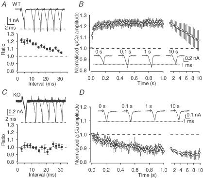
At the calyx of Held presynaptic terminal in WT mice, a pair of brief depolarizing pulses, given at a short interval, facilitated the second IpCa (Fig. 3A) as in rats (Borst & Sakmann, 1998; Cuttle et al. 1998; Forsythe et al. 1998; Tsujimoto et al. 2002). The magnitude of facilitation at 5 ms interstimulus interval (ISI) was 19 ± 4% (n = 10), similar to that in rats (Cuttle et al. 1998). When activated repetitively at high frequency (100 Hz), IpCa in WT mice underwent a marked facilitation (Fig. 3B), as in rats (Cuttle et al. 1998; Forsythe et al. 1998). These IpCa facilitations result from a Ca2+-dependent increase in activation kinetics (Borst & Sakmann, 1998; Cuttle et al. 1998), which is mediated by the Ca2+-binding protein neuronal calcium sensor 1 (NCS-1; Tsujimoto et al. 2002). In contrast, in KO mice, IpCa showed no facilitation in the paired-pulse protocol (Fig. 3C) (Inchauspe et al. 2004) or during high-frequency activation (Fig. 3D). These results indicate that IpCa facilitation is a unique property of P/Q-type Ca2+ channels (Ishikawa et al. 2003; Inchauspe et al. 2004).
Figure 3. Activity-dependent facilitation of IpCa.
A and C, IpCa evoked by a pair of 1 ms depolarizing pulses at different interpulse intervals (superimposed in sample records, dashed lines indicate the first IpCa amplitude). The paired-pulse ratio (the second amplitude relative to the first one) is plotted against interpulse intervals in WT (A, n = 10) and KO (C, n = 7) mice. Paired-pulse facilitation of IpCa was observed only in WT mice with interpulse intervals shorter than 30 ms. The results were essentially the same when the depolarizing pulse was stepped to 0 mV instead of −10 mV (n = 12, data not shown). B and D, IpCa evoked by a 100 Hz train of 1 ms depolarizing pulses (10 s). Ordinates indicate the relative IpCa amplitude normalized to the first amplitude (dashed line) in WT (B, n = 7) and KO (D, n = 6) mice. The time scale (abscissa) is squeezed between 2 and 10 s.
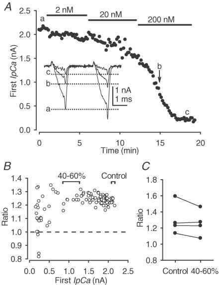
Given that IpCa facilitation is Ca2+ dependent (Cuttle et al. 1998) and that the mean IpCa amplitude in KO mice was 52% of that in WT mice, it might be argued that the lack of IpCa facilitation in KO mice resulted from a lower Ca2+ channel density in KO mice. We examined this possibility in WT mice by partially blocking Ca2+ channels with incremental concentrations of ω-AgaTx (2–200 nm, Fig. 4A). When IpCa was reduced to 40–60%, the IpCa facilitation ratio (3 ms ISI, 1.26 ± 0.08, n = 4) remained similar to control before ω-AgaTx application (1.30 ± 0.10, P > 0.2, paired t test, Fig. 4C), although further reduction of IpCa decreased the facilitation ratio (Fig. 4B). These results suggest that the lack of IpCa facilitation in KO mice did not result from their low Ca2+ channel density, but from the absence of activity-dependent facilitation in N-type Ca2+ channels.
Figure 4. The effect of Ca2+ influx on IpCa facilitation in WT mice.
A, the first IpCa amplitude of a pair during application of incremental concentrations of ω-AgaTx (2–200 nm). Ca2+ currents were evoked by a pair (3 ms interval) of 1 ms depolarizing pulses (from −80 to 0 mV) applied every 10 s. Sample records show pairs of IpCa evoked at different epochs (a–c, superimposed). The first IpCa amplitudes are indicated by dashed lines. B, the paired-pulse ratio of IpCa (ordinate) plotted against the first IpCa amplitude (abscissa). C, the paired-pulse ratio of IpCa when ω-AgaTx blocked IpCa to 40–60% was not significantly different (paired t test) from that before ω-AgaTx application (control).
Short-term plasticity of synaptic transmission in WT and KO mice
We next compared EPSCs mediated by P/Q-type Ca2+ channels in WT mice with those mediated by N-type Ca2+ channels in KO mice. Despite significantly smaller IpCa amplitude in KO mice, the EPSC amplitudes in WT mice (6.52 ± 0.86 nA, n = 12) and KO mice (6.87 ± 1.01 nA, n = 12) were surprisingly similar. This did not result from the postsynaptic receptor saturation, because the EPSC amplitude in the presence of the low affinity glutamate antagonist kynurenate (1 mm) was also similar between WT (1.37 ± 0.26 nA, n = 12) and KO (1.70 ± 0.45 nA, n = 12) mice. The rise and decay kinetics of EPSCs were also similar between WT and KO mice, with 10–90% rise time being 0.33 ± 0.01 ms (n = 12) in WT mice, and 0.32 ± 0.02 ms (n = 12) in KO mice, with the weighted mean decay time constant being 1.54 ± 0.08 ms (n = 12) in WT mice, and 1.75 ± 0.26 ms (n = 12) in KO mice.
Might the differential occurrence of IpCa facilitation between WT and KO mice be reflected in the short-term plasticity of EPSCs? During 100 Hz stimulation, EPSCs in both WT and KO mice underwent a depression (Fig. 5A), as previously reported in rats (von Gersdorff et al. 1997; Iwasaki & Takahashi, 2001) and mice (Wang & Kaczmarek, 1998). This depression is caused mainly by depletion of synaptic vesicles (Wang & Kaczmarek, 1998), although other factors such as inactivation of IpCa (Forsythe et al. 1998; Xu & Wu, 2005), desensitization of postsynaptic AMPA receptors (Wong et al. 2003) and inhibition of transmitter release mediated by presynaptic G-protein-coupled autoreceptors (von Gersdorff et al. 1997; Kimura et al. 2003) are also involved. The magnitude of depression was less in WT mice than KO mice, although the difference was statistically insignificant. At the calyx of Held, like other synapses many, synaptic facilitation is masked by stronger synaptic depression in normal aCSF, whereas lowering [Ca2+]o reduces vesicle depletion and postsynaptic receptor desensitization, thereby unmasking synaptic facilitation. In 1 mm [Ca2+]o (2 mm[Mg2+]o) solution, during a train of 100 Hz stimulation, EPSCs in WT mice underwent a transient facilitation (Fig. 5B). In the same condition, however, no significant facilitation was observed for EPSCs in KO mice. Lowering [Ca2+]o also reduces IpCa facilitation because of its Ca2+ dependence (Cuttle et al. 1998). In WT mice, in 1 mm[Ca2+]o (2 mm[Mg2+]o) solution, where IpCa was reduced to 73%± 2% (n = 9), IpCa facilitation (ISI, 10 ms) was still significant (6.7 ± 0.5%, P < 0.001, n = 10; Fig. 5C and D), although it was less than that in a normal solution (12 ± 0.6%, n = 14). We also tested 0.6 mm[Ca2+]o (2 mm[Mg2+]o) solution (Inchauspe et al. 2004). In this solution IpCa was reduced to 57 ± 8% (n = 6), and IpCa facilitation was 4.1 ± 0.7% (n = 6; Fig. 5C and D).
At synapses with higher release probability, synaptic responses generally undergo stronger depression during repetitive stimulation (Otsuka et al. 1962; Iwasaki & Takahashi, 2001). We examined whether different release probabilities underlie the different synaptic strength during repetitive stimulation (Fig. 5B) between KO and WT mice. During 10 Hz stimulation in 1 mm[Ca2+]o (2 mm[Mg2+]o) solution, EPSCs underwent a similar magnitude of depression in both WT and KO mice (Fig. 5E), implying that the release probability may be similar between WT and KO mice. In the same condition, in WT mice, IpCa did not show facilitation (Fig. 5F) as expected from the short-lasting effect of a conditional pulse on IpCa facilitation in WT mice (Fig. 3A) and rats (Cuttle et al. 1998) in normal aCSF.
We further compared release probability at the calyx of Held between WT and KO mice using a high-frequency stimulation protocol (Schneggenburger et al. 1999). In this method (Fig. 6A), the zero time intercept of the slope, fitted to a cumulative amplitude histogram of EPSCs evoked at 100 Hz, provides an estimate of the size of the readily releasable pool of synaptic vesicles (N) multiplied by the mean quantal amplitude (q). The release probability (p) can then be estimated by dividing the first EPSC amplitude by Nq. These estimations revealed no significant difference between KO and WT mice in both Nq and p (Fig. 6B), suggesting further that the short-term facilitation of EPSCs observed predominantly in WT mice (Fig. 5B) did not arise from a difference in release probability. Therefore, these results strongly suggest that the activity-dependent facilitation of P/Q-type Ca2+ currents underlies synaptic facilitation in WT mice in 1 mm[Ca2+]o (2 mm[Mg2+]o) solution.
Presynaptic inhibition mediated by GTP-binding-protein-coupled receptor in WT and KO mice
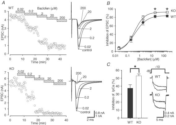
In neuronal and secretory cells, the inhibitory effect of GTP-binding (G) proteins on N-type Ca2+ channels is stronger than that on P/Q-type Ca2+ channels (Bourinet et al. 1996; Zhang et al. 1996; Currie & Fox, 1997). At central synapses, the G-protein-coupled receptor (GPCR)-mediated presynaptic inhibitions are selectively blocked by the N-type-channel-specific blocker ω-CgTX (Umemiya & Berger, 1994; Momiyama & Koga, 2001; Liang et al. 2003). The calyces of Held in WT and KO mice provide an ideal preparation to test the subtype selectivity in the coupling between GPCRs and Ca2+ channels in the nerve terminal. At the calyx of Held, the GABAB receptor agonist baclofen inhibits IpCa (Takahashi et al. 1998) via G protein βγ subunits, thereby presynaptically inhibiting EPSCs (Kajikawa et al. 2001). As illustrated in Fig. 7A, bath applications of baclofen in incremental concentrations attenuated EPSCs in a concentration-dependent manner in both WT and KO mice. The concentration–response relationship of baclofen (Fig. 7B) indicated that the maximal inhibition of EPSCs by baclofen (200 μm) in KO mice (92 ± 2%, n = 4) was significantly higher (P < 0.05) than that in WT mice (83 ± 3%, n = 5), whereas the IC50 was similar between WT (0.41 μm) and in KO (0.38 μm) mice. Consistently, inhibition of IpCa by 20-μM baclofen in KO mice (53 ± 2%, n = 6) was significantly higher (P < 0.05) than that in WT mice (37 ± 4%, n = 3; Fig. 7C). These results suggest that, at the calyx of Held, presynaptic GABAB receptors couple with N-type Ca2+ channels more strongly than with P/Q-type Ca2+ channels.
Figure 7. Inhibitory effects of baclofen on EPSCs and IpCa in WT and KO mice.
A, cumulative inhibitory effects of baclofen in WT (upper panel) and KO (lower panel) mice with sample traces of EPSCs (right panels, superimposed). B, concentration–response curves of inhibitory effect of baclofen on EPSCs in WT (•) and KO (○) mice. The magnitudes of inhibition (ordinate) at 20 and 200 μm were significantly larger in KO than in WT mice (*P < 0.05). Data were derived from 5 and 4 cells in WT and KO mice, respectively. A curve fit to the data points represents the equation: inhibition (%) =[maximal inhibition]/[1 + (IC50/baclofen concentration)n], where maximal inhibition was 84% in WT and 93% in KO mice, IC50 was 0.42 μm in WT and 0.39 μm in KO mice, and n values were 0.86 and 1.04 in WT and KO mice, respectively. C, inhibition of IpCa by baclofen (20 μm) in KO mice was significantly stronger (P < 0.05) than that in WT mice. Error bars indicate s.e.m.
Discussion
Common and different properties of N-type and P/Q-type Ca2+ currents recorded from calyceal nerve terminals
Using α1A-deficient mice and their WT littermates, we compared characteristics of N-type and P/Q-type Ca2+ currents directly recorded from the calyx of Held presynaptic terminals. Presynaptic N-type and P/Q-type Ca2+ currents shared common properties, with both lacking resting inactivation and both having similar kinetics in inactivation and recovery from inactivation. A small difference was found for the voltage range of activation, which was more negative in the P/Q-type than in N-type channels. The most striking difference was found for the activity-dependent facilitation, which was predominantly observed for P/Q-type Ca2+ currents. This Ca2+ current facilitation is dependent upon Ca2+ influx (Cuttle et al. 1998) and mediated by the Ca2+-binding protein NCS-1 (Tsujimoto et al. 2002), but independent of G proteins. It is therefore distinct from the depolarization-induced disinhibition of G proteins (Ikeda, 1991; Kasai, 1992; Brody & Yue, 2000), and also from the calmodulin-dependent Ca2+ current facilitation (Lee et al. 2000; DeMaria et al. 2001) (for a review, see Takahashi, 2005). Another difference between N- and P/Q-type Ca2+ currents in the calyceal nerve terminal was found for the heterotrimeric G-protein-mediated inhibition. The magnitude of inhibition of N-type IpCa by maximal concentration of baclofen was significantly greater than that of P/Q-type IpCa, as reported for Ca2+ currents in neuronal and secretory cells (Bourinet et al. 1996; Zhang et al. 1996; Currie & Fox, 1997).
Synaptic transmission mediated by P/Q-type or N-type Ca2+ channels
EPSCs mediated by P/Q-type and those mediated by N-type Ca2+ channels showed different short-term synaptic plasticity in low Ca2+ aCSF (1 mm[Ca2+]o 2 mm[Mg2+]o), with the former undergoing a transient facilitation during a 100 Hz train of stimulation, whereas the latter showed a depression throughout the train. Such a difference can, in principle, be caused by different release probability; however, this was not the case between WT and KO mice. Given that the activity-dependent IpCa facilitation was observed only for P/Q-type channels in WT mice, this is the most likely cause for the difference in short-term plasticity between KO and WT mice. In the low Ca2+ aCSF solution, the second IpCa following the first one at the 10 ms interval, was facilitated by 6.7%. Assuming fourth power for the IpCa–EPSC relationship, an increase of the EPSC amplitude caused by the IpCa facilitation is estimated to be 30%. This magnitude is close to the difference in the magnitude of the second EPSCs between WT and KO mice during 100 Hz stimulation in 1 mm[Ca2+]o/2 mm[Mg2+]o solution (42%, Fig. 5B), supporting further that the activity-dependent facilitation of P/Q-type channels significantly contributes to the facilitation of P/Q-type channel-mediated synaptic transmission. Inchauspe et al. (2004) reported that EPSCs at the calyx of Held evoked by the paired-pulse stimulation protocol in 0.6 mm[Ca2+]o/2–3 mm[Mg2+]o solution show facilitation in WT mice, but not in KO mice, and they assumed that this difference was caused by the difference in IpCa facilitation, although they did not measure IpCa facilitation in their study conditions. We tested the IpCa facilitation in their study conditions (0.6 mm[Ca2+]o/2 mm[Mg2+]o) and our results (Fig. 5D) generally support their conclusion. At the calyx of Held in rats, before postnatal day 10, synaptic transmission is mediated by N-type, P/Q-type and R-type Ca2+ channels (Iwasaki & Takahashi, 1998; Wu et al. 1998). After this period, however, P/Q-type Ca2+ channels become predominant in mediating synaptic transmission (Iwasaki & Takahashi, 1998; Iwasaki et al. 2000). Such a developmental switch of presynaptic Ca2+ channel subtypes is also observed at thalamic and cerebellar inhibitory synapses (Iwasaki et al. 2000). Our finding of P/Q-type-specific Ca2+ current facilitation suggests that the switch of Ca2+ channel subtypes from N- to P/Q-type in developing nerve terminals may strengthen reliability of transmission by raising synaptic efficacy during high-frequency transmission. At synapses with low release probability, synaptic facilitation is prominent, and it plays an important role in the generation of postsynaptic action potentials (Henze et al. 2002). At such synapses, P/Q-type Ca2+ current facilitation may play a critical role in the signal flow through neuronal circuits.
G-protein-coupled receptor-mediated inhibition of transmitter release triggered by P/Q-type or N-type Ca2+ channels
It is generally believed that N-type Ca2+ channels are more tightly linked to heterotrimeric G proteins. N–type Ca2+currents are selectively inhibited by adenosine A1 receptor agonists in brainstem neurones (Umemiya & Berger, 1994), and by ATP (Currie & Fox, 1996) or GTPγS (Currie & Fox, 1997) in chromaffin cells. Compared with recombinant α1A Ca2+ currents, α1B Ca2+currents are more strongly inhibited by opioids (Bourinet et al. 1996), somatostatin (Zhang et al. 1996) and ATP (Currie & Fox, 2002). Consistently A1 receptor agonist selectively blocks glycinergic transmission mediated by N-type Ca2+ channels in brainstem slices (Umemiya & Berger, 1994). However, at the calyx of Held, an A1 receptor agonist inhibited EPSCs mediated by N-type or P/Q-type Ca2+ channels to a similar extent (Kimura et al. 2003), suggesting that the selective coupling of N-type Ca2+ channels with heterotrimeric G proteins may not be general. In the present study, we re-examined this issue at the calyx of Held for the presynaptic inhibitory effect of baclofen on IpCa and EPSCs mediated by P/Q-type Ca2+ channels in WT mice and N-type Ca2+ channels in KO mice. Our results indicate that the maximal inhibition by baclofen of N-type channel-mediated EPSCs is by 8% greater than that of P/Q-type channel-mediated EPSCs, whereas the IC50 values of the baclofen effect on EPSCs were similar between those mediated by N-type and P/Q-type Ca2+ channels. Thus, compared with P/Q-type Ca2+ channels, N-type Ca2+ channels are more strongly coupled with GABAB receptors at the calyceal nerve terminal, although the difference is smaller than those previously reported (Umemiya & Berger, 1994; Currie & Fox, 1996, 1997, 2002; Bourinet et al. 1996; Zhang et al. 1996).
At the calyx of Held, EPSCs were not affected by a GABAB-receptor-selective antagonist (Yamauchi et al. 2000), suggesting that presynaptic GABAB receptors are not normally activated by endogenous GABA, at least in slices. Metabotropic glutamate receptors (von Gersdorff et al. 1997) and A1 adenosine receptors (Kimura et al. 2003) in the calyceal terminals are activated during repetitive transmission at the immature calyx of Held, but their contribution to synaptic depression is small. Nevertheless, given that less tight coupling of GPCRs with P/Q-type channels is widely reported at many synapses (Umemiya & Berger, 1994; Momiyama & Koga, 2001; Liang et al. 2003), the N-to-P/Q-type switch of presynaptic Ca2+ channels during postnatal development (Iwasaki et al. 2000) might potentially attenuate the GPCR-mediated presynaptic inhibition at various CNS synapses.
Compensatory changes associated with the α1A-subunit knockout
Despite significantly smaller IpCa in KO mice compared with WT mice, mean amplitude of EPSCs was surprisingly similar between KO and WT mice. This was not secondary to saturation of postsynaptic AMPA receptors because mean amplitudes of EPSCs in KO and WT mice were still similar after reducing the AMPA receptor occupancy with kynurenate. Analysis using the tetanic stimulation protocol indicated that neither the release probability nor the number of readily releasable synaptic vesicles is different between WT and KO mice. Inchauspe et al. (2004), however, reported that EPSCs in KO mice are on average by 33% smaller than those in WT mice. The main difference between their results and ours lies in the smaller EPSC amplitude in their KO mice (3.9 nA, n = 7) compared with ours (6.9 nA, n = 12). Although the reason for the difference is not entirely clear, it might arise from general conditions of KO mice, because their KO mice (11–15 days old) are older than ours (9–12 days old), and the α1A-subunit KO mice become increasingly weak after 12 postnatal days until they eventually die 3 weeks after birth (Jun et al. 1999).
In KO mice, a compensatory mechanism(s) seems to maintain release probability, despite reduced presynaptic Ca2+ influx. A possible mechanism may be that N-type Ca2+ channels overexpressed in KO mice are more efficiently coupled to the release machinery than P/Q-type channels in WT mice. This, however, is unexpected from previous reports. At the normal calyx of Held of rats, N- and R-type Ca2+ channels are distantly located from release sites, and Ca2+ influx through them is less effective in triggering transmitter release compared with that through P/Q-type Ca2+ channels (Wu et al. 1999). Furthermore, at the neuromuscular junction of α1A-KO mice, overexpressed N-type Ca2+ channels are distantly located from release sites (Urbano et al. 2003). Another possible mechanism for the compensatory increase in release probability would be that the release machinery might acquire higher Ca2+ sensitivity in KO mice. Activation of presynaptic protein kinase C directly facilitates transmitter release (Hori et al. 1999) by increasing Ca2+ sensitivity of the release machinery (Lou et al. 2005). Also, an increase in cAMP in the nerve terminal facilitates transmitter release without affecting IpCa (Sakaba & Neher, 2001; Kaneko & Takahashi, 2004). It remains to be seen whether these second messengers are involved in this compensatory mechanism.
Whilst the amplitude of IpCa in KO mice was about one half of that in WT mice, blocking P/Q-type Ca2+ currents in WT mice by 50% with ω-AgaTx had no significant effect on the magnitude of IpCa facilitation. In contrast, a reduction of IpCa to 57% by lowering external [Ca2+]o to 0.6 mm reduced IpCa facilitation from 12 to 4.1%. These results suggest that the magnitude of P/Q-type Ca2+ current facilitation is highly dependent upon the amount of Ca2+ influx through individual Ca2+ channels, but it is relatively independent of the Ca2+ channel density. Presumably the Ca2+-dependent IpCa facilitation occurs immediately around the site of Ca2+ entry.
Given that native presynaptic N-type Ca2+ currents in WT animals are too small for detailed analysis, our study cannot entirely exclude a possibility that the difference in IpCa observed between WT and KO mice might arise from an unexpected secondary effect induced by gene knockout. It should also be noted that, despite a clear correlation between IpCa facilitation and synaptic facilitation shown here, a causal relationship between them still remains to be established. Despite these reservations, results obtained here will provide a new step toward understanding the meaning behind different Ca2+ channel subtypes triggering transmitter release at CNS synapses.
Acknowledgments
We thank Kyoko Matsuyama for her technical assistance. We also thank Beverley Clark, Ian Forsythe and Tetsuhiro Tsujimoto for comments. This study was supported by Grant-in-Aid for Specially Promoted Research from the Ministry of Education, Culture, Sports, Science and Technology.
References
- Borst JGG, Sakmann B. Facilitation of presynaptic calcium currents in the rat brainstem. J Physiol. 1998;513:149–155. doi: 10.1111/j.1469-7793.1998.149by.x. [DOI] [PMC free article] [PubMed] [Google Scholar]
- Bourinet E, Soong TW, Stea A, Snutch TP. Determinants of the G protein-dependent opioid modulation of neuronal calcium channels. Proc Natl Acad Sci U S A. 1996;93:1486–1491. doi: 10.1073/pnas.93.4.1486. [DOI] [PMC free article] [PubMed] [Google Scholar]
- Brody DL, Yue DT. Relief of G-protein inhibition of calcium channels and short-term synaptic facilitation in cultured hippocampal neurons. J Neurosci. 2000;20:889–898. doi: 10.1523/JNEUROSCI.20-03-00889.2000. [DOI] [PMC free article] [PubMed] [Google Scholar]
- Currie KPM, Fox AP. ATP serves as a negative feedback inhibitor of voltage-gated Ca2+ channel currents in cultured bovine adrenal chromaffin cells. Neuron. 1996;16:1027–1036. doi: 10.1016/s0896-6273(00)80126-9. [DOI] [PubMed] [Google Scholar]
- Currie KPM, Fox AP. Comparison of N- and P/Q-type voltage-gated calcium channel current inhibition. J Neurosci. 1997;17:4570–4579. doi: 10.1523/JNEUROSCI.17-12-04570.1997. [DOI] [PMC free article] [PubMed] [Google Scholar]
- Currie KPM, Fox AP. Differential facilitation of N- and P/Q-type calcium channels during trains of action potential-like waveforms. J Physiol. 2002;539:419–431. doi: 10.1113/jphysiol.2001.013206. [DOI] [PMC free article] [PubMed] [Google Scholar]
- Cuttle MF, Tsujimoto T, Forsythe ID, Takahashi T. Facilitation of the presynaptic calcium current at an auditory synapse in rat brainstem. J Physiol. 1998;512:723–729. doi: 10.1111/j.1469-7793.1998.723bd.x. [DOI] [PMC free article] [PubMed] [Google Scholar]
- DeMaria CD, Soong TW, Alseikhan BA, Alvania RS, Yue DT. Calmodulin bifurcates the local Ca2+ signal that modulates P/Q-type Ca2+ channels. Nature. 2001;411:484–489. doi: 10.1038/35078091. [DOI] [PubMed] [Google Scholar]
- Forsythe ID, Barnes-Davies M. The binaural auditory pathway: membrane currents limiting multiple action potential generation in the rat medial nucleus of the trapezoid body. Proc R Soc Lond B Biol Sci. 1993;251:143–150. doi: 10.1098/rspb.1993.0021. [DOI] [PubMed] [Google Scholar]
- Forsythe ID, Tsujimoto T, Barnes-Davies M, Cuttle MF, Takahashi T. Inactivation of presynaptic calcium current contributes to synaptic depression at a fast central synapse. Neuron. 1998;20:797–807. doi: 10.1016/s0896-6273(00)81017-x. [DOI] [PubMed] [Google Scholar]
- Henze DA, Wittner L, Buzsaki G. Single granule cells reliably discharge targets in the hippocampal CA3 network in vivo. Nature Neurosci. 2002;5:790–795. doi: 10.1038/nn887. [DOI] [PubMed] [Google Scholar]
- Hori T, Takai Y, Takahashi T. Presynaptic mechanism for phorbol ester-induced synaptic potentiation. J Neurosci. 1999;19:7262–7267. doi: 10.1523/JNEUROSCI.19-17-07262.1999. [DOI] [PMC free article] [PubMed] [Google Scholar]
- Ikeda S. Double-pulse calcium channel current facilitation in adult sympathetic neurones. J Physiol. 1991;439:181–214. doi: 10.1113/jphysiol.1991.sp018663. [DOI] [PMC free article] [PubMed] [Google Scholar]
- Inchauspe CG, Martini FJ, Forsythe ID, Uchitel OD. Functional compensation of P/Q by N-type channels blocks short-term plasticity at the calyx of Held presynaptic terminal. J Neurosci. 2004;24:10379–10383. doi: 10.1523/JNEUROSCI.2104-04.2004. [DOI] [PMC free article] [PubMed] [Google Scholar]
- Ishikawa T, Shin H-S, Takahashi T. Distinct and common properties of N-type and P/Q-type calcium currents at the calyx of Held presynaptic terminal. J Physiol. 2003;551.P:C28. [Google Scholar]
- Iwasaki S, Momiyama A, Uchitel OD, Takahashi T. Developmental changes in calcium channel types mediating central synaptic transmission. J Neurosci. 2000;20:59–65. doi: 10.1523/JNEUROSCI.20-01-00059.2000. [DOI] [PMC free article] [PubMed] [Google Scholar]
- Iwasaki S, Takahashi T. Developmental changes in calcium channel types mediating synaptic transmission in rat auditory brainstem. J Physiol. 1998;509:419–423. doi: 10.1111/j.1469-7793.1998.419bn.x. [DOI] [PMC free article] [PubMed] [Google Scholar]
- Iwasaki S, Takahashi T. Developmental regulation of transmitter release at the calyx of Held in rat auditory brainstem. J Physiol. 2001;534:861–871. doi: 10.1111/j.1469-7793.2001.00861.x. [DOI] [PMC free article] [PubMed] [Google Scholar]
- Jun K, et al. Ablation of P/Q-type Ca2+ channel currents, altered synaptic transmission, and progressive ataxia in mice lacking the α1A-subunit. Proc Natl Acad Sci U S A. 1999;96:15245–15250. doi: 10.1073/pnas.96.26.15245. [DOI] [PMC free article] [PubMed] [Google Scholar]
- Kajikawa Y, Saitoh N, Takahashi T. GTP-binding protein βγ subunits mediate presynaptic calcium current inhibition by GABAB receptor. Proc Natl Acad Sci U S A. 2001;98:8054–8058. doi: 10.1073/pnas.141031298. [DOI] [PMC free article] [PubMed] [Google Scholar]
- Kaneko M, Takahashi T. Presynaptic mechanism underlying cAMP-dependent synaptic potentiation. J Neurosci. 2004;24:5202–5208. doi: 10.1523/JNEUROSCI.0999-04.2004. [DOI] [PMC free article] [PubMed] [Google Scholar]
- Kasai H. Voltage- and time-dependent inhibition of neuronal calcium channels by a GTP-binding protein in a mammalian cell line. J Physiol. 1992;448:189–209. doi: 10.1113/jphysiol.1992.sp019036. [DOI] [PMC free article] [PubMed] [Google Scholar]
- Katz B. The Release of Neural Transmitter Substances. Liverpool: Liverpool University Press; 1969. [Google Scholar]
- Kimura M, Saitoh N, Takahashi T. Adenosine A1 receptor-mediated presynaptic inhibition at the calyx of Held of immature rats. J Physiol. 2003;553:415–426. doi: 10.1113/jphysiol.2003.048371. [DOI] [PMC free article] [PubMed] [Google Scholar]
- Lee A, Scheuer T, Catterall WA. Ca2+/calmodulin-dependent facilitation and inactivation of P/Q-type Ca2+ channels. J Neurosci. 2000;20:6830–6838. doi: 10.1523/JNEUROSCI.20-18-06830.2000. [DOI] [PMC free article] [PubMed] [Google Scholar]
- Liang Y-C, Huang C-C, Hsu K-S, Takahashi T. Cannabinoid-induced presynaptic inhibition at the primary afferent trigeminal synapse of juvenile rat brainstem slices. J Physiol. 2003;555:85–96. doi: 10.1113/jphysiol.2003.056986. [DOI] [PMC free article] [PubMed] [Google Scholar]
- Lou X, Scheuss V, Schneggenburger R. Allosteric modulation of the presynaptic Ca2+ sensor for vesicle fusion. Nature. 2005;435:497–501. doi: 10.1038/nature03568. [DOI] [PubMed] [Google Scholar]
- Luebke JI, Dunlap K, Turner TJ. Multiple calcium channel types control glutamatergic synaptic transmission in the hippocampus. Neuron. 1993;11:895–902. doi: 10.1016/0896-6273(93)90119-c. [DOI] [PubMed] [Google Scholar]
- Momiyama T, Koga E. Dopamine D2-like receptors selectively block N-type Ca2+ channels to reduce GABA release onto rat striatal cholinergic interneurones. J Physiol. 2001;533:479–492. doi: 10.1111/j.1469-7793.2001.0479a.x. [DOI] [PMC free article] [PubMed] [Google Scholar]
- Otsuka M, Endo M, Nonomura Y. Presynaptic nature of neuromuscular depression. Jpn J Physiol. 1962;12:573–584. doi: 10.2170/jjphysiol.12.573. [DOI] [PubMed] [Google Scholar]
- Sakaba T, Neher E. Preferential potentiation of fast-releasing synaptic vesicles by cAMP at the calyx of Held. Proc Natl Acad Sci U S A. 2001;98:331–336. doi: 10.1073/pnas.021541098. [DOI] [PMC free article] [PubMed] [Google Scholar]
- Schneggenburger R, Meyer AC, Neher E. Released fraction and total size of a pool of immediately available transmitter quanta at a calyx synapse. Neuron. 1999;23:399–409. doi: 10.1016/s0896-6273(00)80789-8. [DOI] [PubMed] [Google Scholar]
- Takahashi T. Dynamic aspects of presynaptic calcium currents mediating synaptic transmission. Cell Calcium. 2005;37:507–511. doi: 10.1016/j.ceca.2005.01.018. [DOI] [PubMed] [Google Scholar]
- Takahashi T, Kajikawa Y, Tsujimoto T. G-protein-coupled modulation of presynaptic calcium currents and transmitter release by a GABAB receptor. J Neurosci. 1998;18:3138–3146. doi: 10.1523/JNEUROSCI.18-09-03138.1998. [DOI] [PMC free article] [PubMed] [Google Scholar]
- Takahashi T, Momiyama A. Different types of calcium channels mediate central synaptic transmission. Nature. 1993;366:156–158. doi: 10.1038/366156a0. [DOI] [PubMed] [Google Scholar]
- Tsujimoto T, Jeromin A, Saitoh N, Roder JC, Takahashi T. Neuronal calcium sensor 1 and activity-dependent facilitation of P/Q-type calcium currents at presynaptic nerve terminals. Science. 2002;295:2276–2279. doi: 10.1126/science.1068278. [DOI] [PubMed] [Google Scholar]
- Umemiya M, Berger AJ. Activation of adenosine A1 and A2 receptors differentially modulates calcium channels and glycinergic synaptic transmission in rat brainstem. Neuron. 1994;13:1439–1446. doi: 10.1016/0896-6273(94)90429-4. [DOI] [PubMed] [Google Scholar]
- Urbano FJ, Piedras-Renteria ES, Jun K, Shin H-S, Uchitel OD. Altered properties of quantal neurotransmitter release at endplates of mice lacking P/Q-type Ca2+ channels. Proc Natl Acad Sci U S A. 2003;100:3491–3496. doi: 10.1073/pnas.0437991100. [DOI] [PMC free article] [PubMed] [Google Scholar]
- von Gersdorff H, Schneggenburger R, Weis S, Neher E. Presynaptic depression at a calyx synapse: The small contribution of metabotropic glutamate receptors. J Neurosci. 1997;17:8137–8146. doi: 10.1523/JNEUROSCI.17-21-08137.1997. [DOI] [PMC free article] [PubMed] [Google Scholar]
- Wang L-Y, Kaczmarek LK. High-frequency firing helps replenish the readily releasable pool of synaptic vesicles. Nature. 1998;394:384–388. doi: 10.1038/28645. [DOI] [PubMed] [Google Scholar]
- Wong AYC, Graharm BP, Billups B, Forsythe ID. Distinguishing between presynaptic and postsynaptic mechanisms of short-term depression during action potential trains. J Neurosci. 2003;23:4868–4877. doi: 10.1523/JNEUROSCI.23-12-04868.2003. [DOI] [PMC free article] [PubMed] [Google Scholar]
- Wu L-G, Borst JGG, Sakmann B. R-type Ca2+ currents evoke transmitter release at a rat central synapse. Proc Natl Acad Sci U S A. 1998;95:4720–4725. doi: 10.1073/pnas.95.8.4720. [DOI] [PMC free article] [PubMed] [Google Scholar]
- Wu L-G, Westenbroek RE, Borst JGG, Catterall WA, Sakmann B. Calcium channel types with distinct presynaptic localization couple differentially to transmitter release in single calyx-type synapses. J Neurosci. 1999;19:726–736. doi: 10.1523/JNEUROSCI.19-02-00726.1999. [DOI] [PMC free article] [PubMed] [Google Scholar]
- Xu J, Wu L-G. The decrease in the presynaptic calcium current is a major cause of short-term depression at a calyx-type synapse. Neuron. 2005;46:633–645. doi: 10.1016/j.neuron.2005.03.024. [DOI] [PubMed] [Google Scholar]
- Yamauchi T, Hori T, Takahashi T. Presynaptic inhibition by mucimol through GABAB receptors. Eur J Neursci. 2000;12:3433–3436. doi: 10.1046/j.1460-9568.2000.00248.x. [DOI] [PubMed] [Google Scholar]
- Zhang J-F, Ellinor PT, Aldrich RW, Tsien RW. Multiple structural elements in voltage-dependent Ca2+ channels support their inhibition by G proteins. Neuron. 1996;17:991–1003. doi: 10.1016/s0896-6273(00)80229-9. [DOI] [PubMed] [Google Scholar]


