Abstract
Laparoscopic urologic procedures have become increasingly popular, but their widespread use has been limited by training issues. The use of 3-dimensional (3D) vision might aid in training and performance of laparoscopic tasks. The purpose of this study was to evaluate a 3D visual system used by novice laparoscopists. In this prospective, randomized study, 24 novice laparos-copists were evaluated on a validated and standardized laparoscopic task using both 2-dimensional (2D) and 3D visualization systems. The task was performed more rapidly with 3D visualization (108 vs. 127 seconds, P < .05). On subjective evaluations, participants believed the task was easier with the 3D system, and participants preferred the 3D system to the 2D system by a 2:1 margin. 3D visualization improves the learning curve for laparoscopic surgery. Surgeons should consider 3D systems when learning complex laparoscopic surgeries. Further evaluation of operative times and complications is needed in clinical studies.
Key words: Laparoscopic surgery, 3-Dimensional visualization, EndoSite 3Di, da Vinci System
Since the early 1990s, laparoscopic urologic surgeries have evolved from experimental techniques to commonly accepted procedures.1,2 Early pioneers in the field used instrumentation and optics that are primitive by today’s standards, but technological advances have made the operations more efficacious and easier to perform. Various devices, including energy sources, vascular staplers, 3-chip cameras, and robotic assistance, have allowed surgeons to perform complex operations with improved confidence.
Despite the advances with technology, training remains a major issue in the field.3 Most urologists today completed their formal training before the wide dissemination of laparoscopic techniques, and even in today’s academic climate there remain some teaching institutions that lack a dedicated laparoscopic or minimally invasive surgical program. Although laparoscopic courses and preceptorships are widely available, the urologist is often faced with a major reconstructive or ablative procedure as his or her first operation; such a situation is daunting and clearly not ideal.
One of the largest challenges in laparoscopic surgical training is adaptation to a 2-dimensional (2D) flat view of the surgical field. The lack of depth perception is a significant sensory loss for the surgeon. For example, the perception of depth during a laparoscopic radical prostatectomy is critical to proper hemostasis and effective reconstruction of the sutured anastomosis. Although the challenges of lost depth perception can be overcome with a long experience of cases, most urologists cannot depend on such a high volume over a short period.
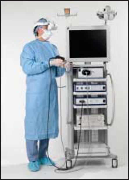
Three-dimensional (3D) laparoscopic visual systems have been developed to augment laparoscopic skills. The da Vinci® Surgical System (Intuitive Surgical, Sunnyvale, CA) is one such device, though its extraordinary price limits its dissemination.4 Another device, the EndoSite 3Di Digital Vision System, developed by Viking Systems (La Jolla, CA), couples a 3D view with an ergonomic head-mounted display, allowing improved spectral depth perception with the use of traditional laparoscopic instrumentation (Figure 1). Such a system, at less than one-tenth the cost of the da Vinci System, might offer similar benefits. In the study described here, inexperienced urologic laparoscopists were tested in a routine task with both a traditional 2D laparoscopic view and the 3D head-mounted display to determine whether there was any advantage in using the 3D system to acquire laparoscopic skills.
Figure 1.

The EndoSite 3Di Digital Vision System (Viking Systems, La Jolla, CA) couples a 3D view with an ergonomic head-mounted display.
Materials and Methods
During a laparoscopic training course at the Washington University School of Medicine, 24 participants were timed while performing a basic laparoscopic task. The task, a standardized validated laparoscopic skills test, was to transfer 10 beads from one container to another container. A standard laparoscopic trainer was used to house the testing items. The first container, holding the beads, had no lid or top. The second container had a modified top, which was cut so as to accept only 1 bead at a time. The total time needed to place all 10 beads in the second container, using either 2D or 3D video and identical laparoscopic instruments, was recorded for each subject.
The 2D system (Karl Storz, Culver City, CA) consisted of a standard laparoscopic video tower, with a 3-chip charge-coupled device (3CCD) digital system and 10 mm/30° scope, attached to a 23-in cathode ray tube monitor. The 3D system (EndoSite 3Di) included a stereo digital scope (dual 3CCD optical channel) attached to a 3D data-processing unit, which conveyed information to a head-mounted display. The head-mounted display consisted of dual 800 × 600 miniature liquid crystal display (LCD) screens attached to a padded ergonomic headset, allowing a stereoscopic 3D view. The optics were optimized in both systems before performance of the task, and lighting was adjusted to similar levels for both systems. In both tasks, the participants used the same laparoscopic instrumentation to complete the task.
Results
Demographic information was obtained for all participants. The mean age of the participants was 46 years (range, 33–61 years), and mean years in practice was 12. All participants had performed fewer than 25 laparoscopic procedures. There were 22 men and 2 women in the study group.
The order in which the participants completed the task was randomized between 2D and 3D. Thirteen subjects performed the task with 2D first, and 11 performed with 3D first. The mean time to complete the task with 2D vision was 127 seconds (range, 83–208 seconds), and the mean time to complete the task with 3D vision was 108 seconds (range, 75–147 seconds). The difference was statistically significant (P < .05, t test).
The participants were asked to rate the difficulty of performing the task on a subjective basis, before they learned their times to complete the tasks. The tasks were rated on a scale of 1 (easy) to 10 (difficult). The mean rating for the task with 2D vision was 3.6, compared with 2.9 for 3D. When subjects were asked to rate subjectively which system they preferred (before learning their performance results), 14 preferred the 3D system, 3 had no preference, and 7 preferred the 2D system.
Discussion
One of the challenges of complex reconstructive and ablative laparoscopic surgery is training and dissemination of operative technique. Although there is little doubt that minimally invasive surgical options benefit the patient, little has been written regarding methods to train surgeons in these complex skills. Residencies and fellowships might allow adequate skills development, but such education is not practical for the majority of certified and practicing physicians. Weekend courses and preceptorships might aid in bridging the educational gap, but without close tutelage, surgeons still might be intimidated by complex minimally invasive procedures. Educational challenges likely will continue to be an issue in laparoscopic skill acquisition.
Besides education, instrumentation can shorten the learning curve and operative times. A modern example is the gastrointestinal anastomosis stapler, which is a common alternative to hand-sewn bowel anastomosis and allows excellent efficacy with a short learning curve. Similarly, surgeons would be wise to consider improvements in laparoscopic technology to aid in reconstructive cases. Much of the focus of surgeons has been on operative instrumentation rather than on video or audio systems. Therefore, despite allocation of resources to disposable instruments or energy sources, analogue laparoscopic and cystoscopic towers remain the norm, and their 2D view might limit the efficiency of the operations.
The goal of this study was to evaluate a 3D system used by neophyte laparoscopists, to assess ease of performing a complex laparoscopic task. In this study, there was a marked and significant improvement in operative times with a stereoscopic 3D view of the operative field. Because the task was performed with identical mechanical instrumentation (graspers, needle drivers), the difference can be attributable only to the changes in depth perception obtained with the 3D system. This skill acquisition ultimately might prove to be beneficial when laparoscopic radical prostatectomy, laparoscopic pyeloplasty, or other complex reconstructive techniques are performed.
Much has been written about the improved learning curve associated with the da Vinci Surgical System.5 It is unclear whether the robotic system offers any advantages over a smaller and less expensive 3D head-mounted system. Although wristed instruments might seem to be a technological advance, with an existing 3D view it might be possible for surgeons to become proficient over an equally short learning curve. The cost savings for such proficiency, however, are overwhelmingly in favor of a 3D non-robotic tower, which costs less than one tenth the price of a surgical robot. In addition, the non-robotic tower is more mobile and might be used for any laparoscopic procedure.
Other benefits of an advanced visual system are obvious but less tangible. Surgeon fatigue might be minimized by the use of a head-mounted display instead of a statically positioned LCD screen. Surgeons also might be less prone to being distracted by movement in the operating room, because their visual field is largely filled with the operative view. The perception of depth might aid in more meticulous dissection, which might limit complications.
It is unclear from this study whether experienced laparoscopists could benefit from a 3D system. It also is unclear whether the improvement in operative times can translate into real clinical improvements. Nevertheless, for other than laparoscopic experts, a 3D system seems to be an excellent tool for performing complex laparoscopic tasks and undoubtedly should be considered by every minimally invasive surgeon. The benefits might translate into improved operative times, shortened learning curves, and greater surgeon comfort. These benefits might allow the beginning laparoscopic surgeon to become an expert quickly and without limitations.
Main Points.
Despite advances in the technology, training remains a major issue in the field of laparoscopic urologic surgery.
One of the largest challenges in laparoscopic surgical training is adaptation to a 2-dimensional (2D) flat view of the surgical field; 3-dimensional (3D) laparoscopic visual systems have been developed to augment laparoscopic skills.
The EndoSite 3Di (Viking Systems, La Jolla, CA) couples a 3D view with an ergonomic head-mounted display, allowing improved spectral depth perception with the use of traditional laparoscopic instrumentation.
In a prospective, randomized study in which 24 novice laparoscopists were evaluated on a laparoscopic task using both 2D and the EndoSite 3Di visualization systems, mean operative time was significantly faster with the 3D system; participants also rated the 3D system as easier to use.
The non-robotic EndoSite 3Di system is approximately one-tenth as expensive as a robotic 3D surgical system (da Vinci; Intuitive Surgical, Sunnyvale, CA); in addition, the non-robotic tower is more mobile and might be used for any laparoscopic procedure.
References
- 1.Clayman RV, Kavoussi LR, Soper NJ, et al. Laparoscopic nephrectomy: initial case report. J Urol. 1991;146:278–282. doi: 10.1016/s0022-5347(17)37770-4. [DOI] [PubMed] [Google Scholar]
- 2.Bhayani SB, Clayman RV, Sundaram CP, et al. Surgical treatment of renal neoplasia: evolving toward a laparoscopic standard of care. Urology. 2003;62:821–826. doi: 10.1016/s0090-4295(03)00670-8. [DOI] [PubMed] [Google Scholar]
- 3.Abdelshehid CS, Eichel L, Lee D, et al. Current trends in urologic laparoscopic surgery. J Endourol. 2005;19:15–20. doi: 10.1089/end.2005.19.15. [DOI] [PubMed] [Google Scholar]
- 4.Bhayani SB, Link RE, Varkarakis JM, Kavoussi LR. Complete da Vinci versus laparoscopic pyeloplasty: cost analysis. J Endourol. 2005;19:327–332. doi: 10.1089/end.2005.19.327. [DOI] [PubMed] [Google Scholar]
- 5.Binder J, Brautigam R, Jonas D, Bentas W. Robotic surgery in urology: fact or fantasy? BJU Int. 2004;94:1183–1187. doi: 10.1046/j.1464-410x.2004.05130.x. [DOI] [PubMed] [Google Scholar]


