Abstract
Recent work has demonstrated that Salmonella enterica serovar Typhimurium forms biofilms on HEp-2 tissue culture cells in a type 1 fimbria-dependent manner. To investigate how biofilm growth of HEp-2 tissue culture cells affects gene expression in Salmonella, we compared global gene expression during planktonic growth and biofilm growth. Microarray results indicated that the transcription of ∼100 genes was substantially altered by growth in a biofilm. These genes encode proteins with a wide range of functions, including antibiotic resistance, central metabolism, conjugation, intracellular survival, membrane transport, regulation, and fimbrial biosynthesis. The identification of five fimbrial gene clusters was of particular interest, as we have demonstrated that type 1 fimbriae are required for biofilm formation on HEp-2 cells and murine intestinal epithelium. Mutations in each of these fimbriae were constructed in S. enterica serovar Typhimurium strain BJ2710, and the mutants were found to have various biofilm phenotypes on plastic, HEp-2 cells, and chicken intestinal tissue. The pef and csg mutants were defective for biofilm formation on each of the three surfaces tested, while the lpf mutant exhibited a complete loss of the ability to form a biofilm on chicken intestinal tissue but only an intermediate loss of the ability to form a biofilm on tissue culture cells and plastic surfaces. The bcf mutant displayed increased biofilm formation on both HEp-2 cells and chicken intestinal epithelium, while the sth mutant had no detectable biofilm defects. In all instances, the mutants could be restored to a wild-type phenotype by a plasmid carrying the functional genes. This is the first work to identify the genomic responses of Salmonella to biofilm formation on host cells, and this work highlights the importance of fimbriae in adhering to and adapting to a eukaryotic cell surface. An understanding of these interactions is likely to provide new insights for intervention strategies in Salmonella colonization and infection.
Pathogenic Salmonella strains cause disease in a range of mammalian hosts (6, 62). Some Salmonella strains have a narrow host range, such as Salmonella enterica serovar Typhi, that causes disease in humans only, while other strains, such as Salmonella enterica serovar Typhimurium, cause infections in a wide range of animal species including mice, poultry, pigs, sheep, cattle, horses, and humans (20, 62). S. enterica serovar Typhimurium human gastroenteritis is initiated by the colonization of the intestinal epithelium followed by invasion and destruction of M cells and enterocytes, which disrupts the integrity of the mucosal surface and allows access to the underlying tissue (33, 34). While colonization of host intestinal tissue is considered an essential early step of infection, several studies have provided specific evidence that fimbriae mediate Salmonella adherence to and growth on the intestinal mucosa (7, 11, 32, 38, 62).
Fimbriae are rigid, filamentous structures present on the surface of a bacterium that mediate attachment to a receptor. Many fimbriae are assembled using a chaperone-usher system of assembly, and the filamentous structure is usually composed of several subunits (31). The shaft of the fimbria is typically comprised of a repeating major subunit as well as minor subunits that are integrated into the shaft of the fimbria and have unknown functions (12, 49). The published S. enterica serovar Typhimurium genome sequence contains a total of 13 fimbrial gene clusters (32, 39, 62), including eight gene clusters that have been identified by sequence homology alone.
The best characterized of the Salmonella fimbriae is type 1 fimbriae. This fimbrial type is encoded by the fim gene cluster and is assembled by the chaperone-usher system (31). These fimbriae are termed mannose sensitive because exogenous mannose inhibits binding by the fimbriae (10). The fimA gene encodes the major structural subunit, while the fimH gene encodes the adhesin protein that is located at the tip of the assembled fimbrial structure and mediates binding to the receptor (22, 46). These fimbriae are expressed in vitro after static growth for 48 h at 37°C. Our research group has recently demonstrated that these fimbriae are involved in biofilm formation on HEp-2 tissue culture cells, murine intestinal epithelium, and chicken intestinal epithelium (7, 37).
Several other Salmonella fimbriae have been partially characterized. The long polar fimbriae (Lpf) are encoded by the lpfABCDE genes and have been implicated in the colonization of the murine intestinal mucosa (3, 5, 36). Recent evidence indicates that expression of the lpf genes occurs by a phase variation mechanism that helps to evade the host immune system (36, 42, 43). Studies of phase variation in Escherichia coli and Proteus mirabilis indicate that in vivo selection for fimbriate (on) or nonfimbriate (off) bacteria depends on the organ colonized and the requirement for fimbriae in the organ (42). Plasmid-encoded fimbriae (Pef) are encoded on the 90-kb Salmonella virulence plasmid and are encoded by two divergently transcribed operons (23). The Pef fimbriae mediate adhesion of Salmonella to murine intestinal epithelium, which results in fluid accumulation (4). Regulation of these fimbriae also occurs via a mechanism of phase variation that is analogous to the E. coli Pap system (41). Thin aggregative fimbriae (Tafi) are atypical appendages that possess highly adhesive properties. The components of this adherence factor are encoded by the divergently transcribed operons csgBAC and csgDEFG (39). Tafi mediate adherence to extracellular matrix proteins, contact phase proteins, and major histocompatibility complex class I molecules (8, 30, 48). Tafi have also been implicated in biofilm formation (58) and may contribute to mouse virulence (63). In vitro expression of Tafi is optimal at low temperatures during in vitro growth (57). Environmental signals regulating Tafi biosynthesis proceed through the csgD gene product that directly regulates expression of the major subunit CsgA (8, 25, 26, 57). In addition to regulating expression of Tafi, CsgD also regulates cellulose biosynthesis (8, 24-26), a component of exopolysaccharides in Salmonella biofilms formed on some surfaces (24, 37, 53, 54, 60).
Recent evidence from our group as well as others indicates that following adherence to a solid surface (i.e., glass, plastic, or eukaryotic cells), many serotypes of S. enterica are able to form complex, three-dimensional biofilms (7, 12, 24, 37). In natural settings, biofilms are often composed of multiple bacterial species and are believed to be a prevalent mode of growth for many bacterial populations (15). Biofilms can also be a persistent source of infections (15). For instance, Pseudomonas aeruginosa biofilms contribute significantly to the persistence of the bacteria in cystic fibrosis lung infections, as the organisms in the biofilm are more resistant to antibiotic treatment, and the presence of the biofilm provides a constant source of infecting bacteria (67). The ability of Salmonella to form biofilms is likely to be important for intestinal carriage in domestic animals such as poultry, swine, and cattle (1, 40, 56). Salmonella biofilm formation on nonbiological surfaces may also be an important consideration for the food processing industry (35, 54).
As the ability of Salmonella to form biofilms appears to be important in a variety of environments, characterization of the molecular mechanisms that control biofilm formation may yield new insights for the efforts to control Salmonella. In the work described in this paper, we have performed Salmonella genome microarray hybridization experiments to identify genes that are upregulated during biofilm formation on eukaryotic cell surfaces. We found that biofilm formation on HEp-2 cells alters the expression of several classes of S. enterica serovar Typhimurium genes, including genes involved in fimbrial production. Growth of Salmonella biofilms on the HEp-2 cells resulted in significant upregulation of five fimbrial operons. To more carefully define the role of these fimbriae in biofilm formation on eukaryotic cells, we constructed defined mutants in fimbrial genes and assayed the mutants for their ability to form a biofilm on HEp-2 cells, tissue culture plastic, and chicken intestinal tissue. This work reveals that biofilm conditions may act as inducing signals for the expression of various Salmonella fimbriae and that these fimbriae contribute to various stages of biofilm formation in a process that appears to be important for intestinal colonization.
MATERIALS AND METHODS
Growth conditions, bacterial strains, and plasmids.
The bacterial strains and plasmids used in this study are listed in Table 1. For biofilm experiments, strains were grown statically at 37°C in Lennox broth (0.5% NaCl) (Gibco-Invitrogen, Carlsbad, CA) supplemented with the appropriate antibiotics as necessary. Antibiotics were added at the following concentrations: ampicillin, 100 μg ml−1; kanamycin, 25 μg ml−1; chloramphenicol, 25 μg ml−1; tetracycline, 100 μg ml−1; and spectinomycin, 100 μg ml−1. When inoculated into a biofilm chamber, the bacteria were grown in RPMI 1640 tissue culture medium (Gibco-Invitrogen, Carlsbad, CA), which is also used to culture the HEp-2 tissue culture cells. Plasmid pNAL105, carrying the csgBAC operon, was constructed by PCR amplification of the csgBAC genes (∼2 kb) from genomic DNA with primers csgB5 (5′-TAGATAATTTTCGCTATGTA-3′) and csgC3 (5′-CCAATCCATTTCCGCCACCA-3′). The amplified PCR fragment was ligated into pCR2.1 (Invitrogen, Carlsbad, CA), and a plasmid clone in which expression of the csgBAC genes was driven by the lac promoter was selected.
TABLE 1.
Strains and plasmids
| Strain or plasmid | Genotype or phenotypea | Source or reference |
|---|---|---|
| Strains | ||
| Escherichia coli | ||
| DH12S | mcrA Δ(mrr-hsdRMS-mcrBC) F′ lacIqΔM15 | Invitrogen |
| Top10 | F−mcrA Δ(mrr-hsdRMS-mcrBC) φ80lacZΔM15 | Invitrogen |
| S. enterica serovar Typhimurium | ||
| χ8307 | UK-1 derivative with disruptions in csgA::spc, fimA::cat, lpfABCDE::kan, and pefC::tet | R. J. Curtiss III |
| ATCC 14028 | Virulent wild-type strain | 21 |
| SL1344 | Virulent wild-type strain | 68 |
| LB5010 | S. enterica serovar Typhimurium LT2 strain containing a complete fim gene cluster | 68 |
| BJ2508 | BJ2710 fimH::kan | 7 |
| BJ2710 | SL1344 derivative containing the LB5010 fimH gene | 37 |
| BJ3514 | BJ2710 bcfF::cat | This work |
| BJ3539 | BJ2710 pefC::tet | This work |
| BJ3571 | BJ2710 lpfABCDE::kan | This work |
| BJ3577 | BJ2710 sthD::cat | This work |
| BJ3660 | BJ2710 csgA::spc | This work |
| UK-1 | Virulent wild-type strain | 70 |
| Plasmids | ||
| p30 | Plasmid encoding all of the Salmonella pef genes | 23 |
| pNAL105 | pCR2.1 derivative carrying Salmonella csgBAC genes | This work |
| pJTN350 | bcfABCDEFGH genes; Tetr | A. J. Baumler |
| pKD3 | pANTSγ vector containing the cat template gene cloned from pSC140; Ampr | 17 |
| pKD4 | pANTSγ vector containing the kan template gene cloned from pCP15; Ampr | 17 |
| pKD46 | Temperature-sensitive red helper plasmid expressing araC-ParaB and γβexo from λ phage; Ampr | 17 |
| pMRP9-1 | Plasmid encoding GFP; Ampr | 50 |
| pMS1000 | Plasmid carrying the lpfABCDE genes; Tetr | 3 |
| pISF204 | Plasmid pBR322 carrying functional fimH and fimF; Ampr | 28 |
| pBBRMCS-1 | Plasmid encoding GFP; Catr | 50 |
Ampr, ampicillin resistant; Kanr, kanamycin resistant; Catr, chloramphenicol acetyltransferase resistant.
Construction of S. enterica serovar Typhimurium fimbrial mutants using a linear transformation protocol.
S. enterica serovar Typhimurium strains mutated in specific fimbrial gene clusters were constructed using the λ red recombinase system described previously by Datsenko and Wanner (17). Briefly, primers were designed so that the entire open reading frame of the gene to be deleted was replaced by the chloramphenicol resistance gene. Mutants were complemented with the appropriate genes to ensure that the mutations constructed were responsible for the phenotypes observed. PCR analysis demonstrated that transformants had the chloramphenicol resistance cassette inserted at the proper site. This technique was used to construct S. enterica serovar Typhimurium strain BJ3577 (BJ2710 sthD::cat), S. enterica serovar Typhimurium strain BJ3571 (BJ2710 lpfABCDE::cat), and S. enterica serovar Typhimurium strain BJ3514 (BJ2710 bcfF::cat). All primers were purchased from IDT (Coralville, IA), and sequences will be provided upon request.
Strains BJ3539 (BJ2710 pefC::tet) and BJ3660 (BJ2710 csgA::spc) were created using P22 transduction. A P22 lysate was prepared using S. enterica serovar Typhimurium strain χ8307, a derivative of strain UK-1 (52), and S. enterica serovar Typhimurium strain BJ2710 was transduced with either tetracycline or spectinomycin resistance to create strains BJ3539 (pefC::tet) and BJ3660 (csgA::spc).
Growth curves in Lennox broth and RPMI 1640 tissue culture medium were performed for the parent strain and each mutant strain. No differences in growth rates were detected for any of the strains (data not shown).
Expression and detection of type 1 fimbriae.
To initiate biofilm formation, bacterial strains were grown to express type 1 fimbriae. Strains were cultured in 10 ml of LB broth and incubated without shaking for 48 h at 37°C. Bacterial suspensions for use in mannose-sensitive hemagglutination assays were prepared as previously described (61). The presence of fimbrial antigens on the surface of bacteria was detected using monospecific fimbrial antiserum as described previously by Hancox et al. (28).
Growth conditions for planktonically grown and biofilm-grown S. enterica serovar Typhimurium BJ2710.
Planktonic (nonbiofilm) populations of S. enterica serovar Typhimurium BJ2710 were obtained by inoculating 10 ml of a statically grown starter culture into 60 ml of Dulbecco's modified Eagle's medium tissue culture medium (Invitrogen) with 10% newborn calf serum (BioWhittaker) and supplemented with the appropriate antibiotics. The cultures were grown for 3 h at 37°C in the presence of 5% CO2.
To obtain large quantities of bacteria grown in biofilm conditions on HEp-2 cells, the tissue culture cells were cultivated in a modified T-75 tissue culture flask. Two ports were aseptically drilled into opposite ends of one side of a T-75 tissue culture flask. The flasks were seeded with HEp-2 cells using standard procedures (51) and incubated with the modified sides down, with the ports plugged, overnight at 37°C in the presence of 5% CO2 to allow the cells to form a monolayer. Cultures of S. enterica serovar Typhimurium BJ2710 grown statically for 48 h were inoculated into a flask of HEp-2 cells and allowed to adhere for 30 min before the ports were connected to sterile tubing. Dulbecco's modified Eagle's medium supplemented with 10% newborn calf serum was washed across the cell monolayer at a flow rate of 130 μl min−1 to create conditions favorable for biofilm formation. All incubations were performed at 37°C in the presence of 5% CO2, and the biofilm was harvested after 24 h. The bacterial biofilms were removed from the chambers and collected in 50-ml centrifuge tubes that contained 0.75% acidic phenol, 10% ethanol, and 1% Triton X-100. Following 10 min of centrifugation at 4,500 × g, the bacterial pellet was immediately frozen at −80°C for future use.
RNA isolation.
RNA was isolated from planktonically grown or biofilm-grown cultures of BJ2710 for use in microarray hybridization experiments. Frozen bacterial pellets were thawed on ice and resuspended in 1 ml of cold resuspension buffer (9). One hundred sixty microliters of Tris-buffered phenol (Invitrogen) and 2 ml of hot phenol buffer (9) were added to the bacterial suspension. The cell suspensions were heated to 95°C for 1 min and centrifuged at 6,000 rpm for 15 min. The nucleic acids were subsequently purified by phenol-chloroform extraction and precipitated with isopropanol. Following DNase I (Promega) digestion, samples were phenol-chloroform extracted and precipitated with a 1/10 volume of 3 M sodium acetate and 3 volumes of cold ethanol. The RNA yield ranged from 10 to 20 μg per extraction, as determined by the optical density at 260 nm (OD260). RNA purity was determined by OD260/OD280 ratios.
cDNA synthesis and aa-dUTP incorporation.
Purified bacterial RNA was converted to cDNA in two steps. First, 15 μg of random hexamers (Roche) was annealed to 10 μg of total RNA in a 15-μl reaction mixture by heating the mixture to 65°C for 10 min, followed by a 10-min incubation on ice. Next, 0.5 μl RNasin (Promega), 3 μl of 0.1 M dithiothreitol (Invitrogen), 6 μl of 5× first-strand buffer (Invitrogen), 2.9 μl RNase-free water (Invitrogen), 0.6 μl deoxynucleoside triphosphate-aminoallyl-dUTP mix (25 mM dATP, 25 mM dCTP, 25 mM dGTP, 10 mM dTTP, and 15 mM aa-dUTP) (Sigma) were added to each reaction mixture. The reaction mixtures were preincubated to 42°C for 2 min, followed by the addition of 2 μl of SuperScript II reverse transcriptase (RT) (Invitrogen). cDNA synthesis was allowed to proceed at 42°C for 3 h. Following cDNA synthesis, the RNA template was degraded by adding 10 μl of 1 M NaOH and 10 μl of 0.5 M EDTA and incubating the reaction mixture at 70°C for 15 min (29). The reaction mixture was then neutralized by the addition of 10 μl of 1 M HCl. Unincorporated nucleotides were removed with a QiaQuick PCR purification column (QIAGEN), and the eluted samples were vacuum dried in a Speed Vac.
cDNA labeling.
cDNA samples were resuspended in 4.5 μl of 0.1 M NaCO3 buffer and incubated with either Cy5 monoreactive dye or Cy3 monoreactive dye (Amersham) for 1 h at room temperature in the dark. Unincorporated dyes were removed with a QiaQuick PCR purification column (QIAGEN), and the eluted samples were dried in a Speed Vac. Prior to hybridization, the probes were resuspended and combined in 45 μl of a solution containing 5× SSC (1× SSC is 0.15 M NaCl plus 0.015 M sodium citrate), 25% formamide (Fisher), and 1% sodium dodecyl sulfate (Sigma). Twenty micrograms of salmon sperm carrier DNA was added to the probe mixtures and heated to 95°C for 5 min to denature the probes. The entire cDNA probe sample was then hybridized to a Salmonella genomic microarray and incubated at 42°C overnight (29).
Microarray quantitation and analysis.
Microarrays hybridized to labeled probe pools were scanned and quantified using a Packard Scientific 4000XL spotted array scanner and the accompanying ScanArray Express software. The median signal intensity, median background, and median signal corrected for local background were imported into Microsoft Excel from ScanArray Express. The background-corrected median signal intensity for each gene was normalized to the total signal for each dye. The normalized signal for each gene was then averaged over a total of 12 arrays.
Quantitative RT-PCR.
Total RNA was isolated from bacterial cultures grown in liquid medium or as biofilm and purified as described above for the microarray experiments. Random hexamers were added to 2 μg of total RNA, and the hexamers were allowed to anneal for 5 min at 65°C. cDNA synthesis was carried out using the SuperScript III reverse transcriptase kit (Invitrogen, Carlsbad, CA) according to the instructions of the manufacturer. To compare expression levels of fimbrial genes in planktonically grown cultures to those of fimbrial genes in biofilm-grown cultures, primers and probes (IDT, Coralville, IA) (Table 2) were designed to specifically detect the amplification of a small segment (∼60 bp) of each of the five fimbrial gene clusters that were upregulated in the microarray analysis. To ensure that contaminating DNA was not present in the RNA preparation, a control reaction in which the template for the reaction was RNA that had not been treated with reverse transcriptase was performed. The PCR amplifications were performed with the TaqMan Universal Master Mix (Applied Biosystems, Branchburg, NJ) and monitored in real time with the ABI Prism 7000 sequence detector (Perkin Elmer, Boston, MA) and ABI Prism 1.1 software at the University of Iowa DNA Facility.
TABLE 2.
Primers and probes for real-time RT-PCR analysisa
| Probe or primer | Gene probed | Sequence | 5′ reporter dye | 3′ quencher |
|---|---|---|---|---|
| Probes | ||||
| agfA-180T | agfA | TGCTCTGCAAAGCGATGCCCG | FAM | BlackHole Quencher 1 |
| bcfF-290T | bcfF | TGCCAGCGGCGTAACGGTCA | FAM | BlackHole Quencher 1 |
| fimA-85T | fimA | TGGCGCAGCGGTTGCGG | FAM | BlackHole Quencher 1 |
| lpfE-257T | lpfE | ACCGTCCATCGTAACGCTGGCCT | FAM | BlackHole Quencher 1 |
| pefA-232T | pefA | CGATCAGTATGGTCACGCCGCG | FAM | BlackHole Quencher 1 |
| sthD-383T | sthD | CCACCGGCCACGGCGATATC | FAM | BlackHole Quencher 1 |
| rpoD-1679T | rpoD | TGCTGCGTATGCGTTTCGGTATCG | TET | TAMRA |
| Primers | ||||
| agfA-163F | agfA | TCCGCTAACGCTGCGC | ||
| agfA-224R | agfA | TGGGTAATGGTCGTTTCAGATTT | ||
| bcfF-269F | bcfF | TTTACAAAACTGCGGCAGCA | ||
| bcfF-326R | bcfF | CGCCGCACCGCTAAAA | ||
| fimA-63F | fimA | TTGCGAGTCTGATGTTTGTCG | ||
| fimA-124R | fimA | CACGCTCACCGGAGTAGGAT | ||
| lpfE-238F | lpfE | GGTCAGTCGGGTCCGGA | ||
| lpfE-298R | lpfE | GATTGCGCGTATGCCACA | ||
| pefA-214F | pefA | GCAAAAACGCGCAGCCT | ||
| pefA-273R | pefA | CCCATACCGCCTTGTTCAA | ||
| sthD-360F(a) | sthD | ATGGTGAAAGGAATGGTGGC | ||
| sthD-420R(a) | sthD | ACAATATGCCCGGGCGGATAT |
FAM, 6-carboxyfluorescein; TAMRA, 6-carboxytetramethylrhodamine; TET, 6-carboxy-2′,4,7,7′-tetrachlorofluorescein.
Formation of biofilm on tissue culture plastic, HEp-2 cells, and chicken intestinal epithelium.
The ability of Salmonella strains to form biofilms on the surface of HEp-2 cells was investigated by using a modification of the flowthrough continuous culture system described previously by Parsek and Greenberg (50). Flow chambers were seeded with HEp-2 cells grown in RPMI 1640 medium (Gibco-Invitrogen, Carlsbad, CA) with 10% fetal calf serum (Gibco-Invitrogen, Carlsbad, CA), and the biofilm was cultivated in a manner identical to that described previously by Ledeboer and Jones (37). All bacterial strains used in the assay were transformed with either plasmid pMRP9-1 or pBBRMCS-1, which both express green fluorescent protein (GFP) (50). For biofilm assays, HEp-2 cells were stained with 1 μM cell tracker orange (CMTMR) (Molecular Probes, Eugene, OR) according to the instructions of the manufacturer. Imaging of the biofilm was performed using either a Bio-Rad MRC600 or a Zeiss LSM510 confocal scanning laser microscope and appropriate software to analyze the images. Biofilm development was monitored at 24 h, and all experiments were repeated at least three times. The data presented are representative of the general appearance of each biofilm in multiple microscopic fields. Confocal images of bacterial growth on the HEp-2 cells are presented as composite images of the x-y plane.
For biofilm experiments using tissue culture plastic as the solid surface, the flow cell was assembled as described previously by Parsek and Greenberg (50), with the modification that a plastic tissue culture slide (Thermanox; Nunc) was used instead of glass. Chambers were inoculated with 1 × 10⁹ CFU of bacteria and allowed to adhere to the plastic surface for 30 min prior to initiating the flow of the RPMI 1640 plus 10% newborn calf serum tissue culture medium. All other parameters were the same as those for the HEp-2 biofilm experiments.
For chicken intestinal biofilm experiments, intestinal tissue was removed from euthanized 1- to 2-week-old Black Australorpe chickens. The distal one-third of the small intestine was removed and washed with phosphate-buffered saline (PBS). Peristaltic tubing (1/16-in. diameter) was inserted into each end of a 10-cm length of excised intestine, and the tubing was secured with 6-0 silk sutures (Ethicon, Cincinnati, OH). The intestinal loops were inoculated with 1 × 10⁵ bacteria and incubated, without flow, for 30 min at 37°C in 5% CO2 to allow initial bacterial adherence. Subsequently, the loops were infused with RPMI 1640 tissue culture medium supplemented with 7% bovine calf serum and 3% chicken serum (Invitrogen, Carlsbad, CA) at a rate of 130 μl/min and incubated for 24 h in a CO2 incubator. Biofilm formation on the mucosal surfaces was examined over the entire length of the intestinal segment using a Zeiss LSM510 confocal scanning laser microscope under low power, and representative images were generated and analyzed. Topographical maps were generated using Zeiss confocal software.
Quantitation of Salmonella biofilm formed on HEp-2 cells by direct agar plating.
Bacterial strains and biofilm chambers were prepared as described above, except that HEp-2 cells were seeded into the biofilm chamber without first labeling the cells with CMTMR dye. Biofilm development was monitored at 24 h, and all experiments were repeated on at least three separate occasions. The bacterial HEp-2 cell biofilms were removed from the coverslips using a cell scraper (Corning, Corning, NY), resuspended in 10 ml PBS, and homogenized by vortexing for 3 min. The biofilm suspensions were then diluted, plated onto Lennox agar (Difco, Sparks, MD), and counted after incubation overnight.
RESULTS
Differential gene expression in S. enterica serovar Typhimurium biofilms.
To identify genes whose expression levels are increased when S. enterica serovar Typhimurium grows as a biofilm on HEp-2 cells, total RNA was isolated from both biofilm-grown bacteria and planktonically grown bacteria and analyzed by hybridization to an S. enterica serovar Typhimurium DNA microarray. Expression of each gene in the genome during growth in biofilm conditions was compared to expression of each gene during growth in liquid medium.
The median hybridization results of 12 arrays revealed that ∼100 genes were upregulated more than sevenfold (Table 3), which represents 2.2% of the Salmonella genome. Analysis of the microarray data revealed differential regulation of genes from several different functional groups that included genes encoding fimbriae, antibiotic resistance genes, genes encoding exopolysaccharide biosynthesis, etc. In addition, several other open reading frames containing putative or unknown proteins were also substantially upregulated. Of particular interest was the upregulation of five fimbrial gene clusters (Bcf, Lpf, Pef, Tafi, and Sth fimbriae), since fimbriae are involved in several biofilm-associated processes, including attachment to the substratum (7), intercellular adhesion (65, 66), and biosynthesis of the extracellular matrix (54, 60).
TABLE 3.
Identification of selected S. enterica serovar Typhimurium genes induced by biofilm growth by microarray analysis
| Gene | Function | Ratio of expression in biofilm/expression in planktonic conditions | q value (%)a |
|---|---|---|---|
| Genes significantly induced by biofilm conditions | |||
| sthD | Putative fimbrial subunit | 11.8 | 0.9419504 |
| traI | Conjugative transfer | 10.6 | 1.1587486 |
| ssaM | Secretion system apparatus | 10.4 | 1.2559339 |
| traR | Conjugative transfer | 9.9 | 2.5028969 |
| pefA | Plasmid-encoded fimbriae | 9.4 | 3.4318071 |
| marB | Multiple antibiotic resistance | 8.9 | 0.9419504 |
| ssaI | Secretion system apparatus | 8.4 | 1.2559339 |
| pagD | PhoP-regulated gene | 7.4 | 4.4211969 |
| msgA | Macrophage survival gene | 7.2 | 4.4211969 |
| sopD | Secreted protein (Sop family) | 6.8 | 0.9954703 |
| pipB | SPI-3-encoded protein | 6.7 | 6.2146759 |
| spvR | Plasmid virulence regulator | 6.7 | 3.0513243 |
| ssaS | Secretion system apparatus | 6.5 | 2.8107398 |
| trbE | Conjugative transfer | 6.2 | 0.9954703 |
| sifA | Macrophage replication | 6.0 | 0.9419504 |
| traO | Conjugative transfer | 6.0 | 2.5028969 |
| wza | Polysaccharide transport | 6.0 | 3.0513243 |
| pipD | SPI-3-encoded protein | 5.9 | 3.0513243 |
| ssaL | Secretion system apparatus | 5.7 | 2.4221583 |
| traK | Conjugative transfer | 5.2 | 0.9419504 |
| traD | Conjugative transfer | 5.2 | 0.7684332 |
| traQ | Conjugative transfer | 5.2 | 2.5028969 |
| traJ | Conjugative transfer | 5.2 | 0.7684332 |
| traA | Conjugative transfer | 5.1 | 2.5028969 |
| csgA | Curlin major subunit | 5.1 | 2.5028969 |
| bcfF | Bovine colonization factor | 5.1 | 4.4211969 |
| traM | Conjugative transfer | 5.0 | 3.0513243 |
| pefI | Plasmid-encoded fimbriae | 4.7 | 3.4318071 |
| lpfE | Long polar fimbriae | 4.2 | 1.2559339 |
| bcfG | Bovine colonization factor | 3.8 | 4.4211969 |
| marA | Multiple antibiotic resistance | 3.2 | 0.9419504 |
| Control gene unaffected by biofilm conditions | |||
| rpoD | Sigma 70 factor | 0.9 | 1.2559339 |
The q value is the percent chance that the gene expression change observed is random.
Measurement of the induction of fimbrial genes after growth in biofilm using quantitative RT-PCR.
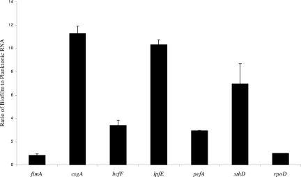
Quantitative RT-PCR was employed to independently determine if the expression of the bcf, lpf, pef, csg (encoding Tafi), and sth genes were significantly upregulated during HEp-2 cell biofilm growth. To establish uninduced baseline controls, we examined the rpoD gene, which previous work in our laboratory has found is expressed at almost constant levels throughout the growth curve (our unpublished data). mRNA from biofilm-grown bacteria and from broth-grown bacteria was converted to a cDNA template and used in quantitative RT-PCR experiments. As expected, expression of the rpoD gene did not vary (biofilm expression/planktonic expression ratio, 1.0), while expression of the fimA gene dropped slightly in biofilm conditions (biofilm expression/planktonic expression ratio, 0.85) (Fig. 1). Each of the five genes assayed (csgA, bcfF, lpfE, pefA, and sthD) was induced by various amounts in biofilm conditions, as measured with the quantitative RT-PCR technique. csgA, lpfE, and sthD were each induced ≥7.0-fold (11.3-fold, 10.3-fold, and 7.0-fold, respectively), as is shown in Fig. 1. While induction of the bcfF gene (3.4-fold) and the pefA gene (3.0-fold) was less than that observed for csgA, lpfE, and sthD, the level of upregulation was still significant. These quantitative RT-PCR findings confirmed the microarray results.
FIG. 1.
Quantitative RT-PCR comparing fimbrial gene expression in S. enterica serovar Typhimurium strain BJ2710 grown in a 24-h biofilm to that grown planktonically. Total RNA was isolated from either biofilm-grown or planktonically grown BJ2710, converted into cDNA, and used as a template in real-time PCRs. Individual fimbrial genes were quantitated in real time using a TaqMan probe as a transcriptional reporter for each gene examined. The ratio of each fimbrial gene transcript was determined by dividing the biofilm growth expression level by the planktonic growth expression level. The ratio for the control rpoD gene transcript was set at 1 since work in our laboratory has shown that this gene is expressed at constant levels under the two conditions (our unpublished data). The results were averaged and plotted as shown. The P values for the ratios shown for fimA, csgA, bcfF, lpfE, pefA, and sthD are 0.25, 0.01, 0.04, 0.04, 0.07, and 0.02, respectively.
Analysis of biofilm formation by S. enterica serovar Typhimurium Bcf, Lpf, Pef, Tafi, and Sth fimbrial mutants.
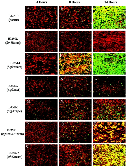
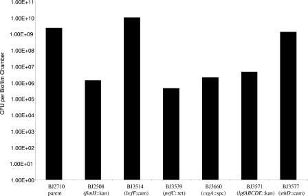
Based on the results from the microarray and quantitative RT-PCR experiments, expression of the Bcf, Lpf, Pef, Tafi, and Sth fimbrial gene clusters are induced by biofilm growth. We constructed a mutation in a gene(s) required for fimbria production in each of the five operons and compared the ability of each S. enterica serovar Typhimurium mutant to form biofilms on HEp-2 cells with that of the parent strain, BJ2710, which has recently been described (37). Each strain was transformed with a plasmid that carries the GFP gene to allow visualization of the strains by confocal microscopy (37). We examined growth on HEp-2 cells at 4 h, 8 h, and 24 h so that any differences in the kinetics of biofilm formation by the Salmonella strains could be uncovered. Representative areas of biofilm formed by each strain are shown as composite images of the x-y plane (Fig. 2). Four hours after infection of HEp-2 cells with the parent strain, BJ2710, the bacteria had attached to numerous locations on the tissue culture cell monolayer that were visible as randomly distributed foci of low numbers of fluorescent bacteria (Fig. 2A). At 8 h, these areas of adherence had increased in size, presumably due to bacterial replication (Fig. 2B), and by 24 h, virtually the entire surface of the HEp-2 cells was covered with an extensive biofilm which also extended outward (∼40 μm) from the tissue culture cell surfaces (Fig. 2C). This general sequence of biofilm development has been well characterized in several bacterial systems, including S. enterica serovar Typhimurium (64). A negative control strain, S. enterica serovar Typhimurium BJ2508 (fimH::kan), lacked the ability to attach to and form biofilm on HEp-2 cells over the course of the experiment (Fig. 2D to F). Similar to the parent strain, BJ2710, organisms of Bcf mutant strain BJ3514 were adherent and had begun to form microcolonies at 4 h (Fig. 2G). Surprisingly, at 8 h, the Bcf mutant already displayed significant biofilm development compared to the parent strain (Fig. 2B and H). At the 8-h time point, the biofilm height of the Bcf mutant was 10 to 20 μm taller, as determined by confocal microscope measurements, than any of the microcolonies produced by the parent strain. While not visibly noticeable, this trend continued to the 24-h time point, where the bcfF mutant produced a biofilm that was ∼10 μm taller and thicker than the wild-type biofilm. To quantitate the number of bacteria in the biofilms, the biofilm was removed from the chamber surface with a cell scraper and vortexed to homogenize the suspension, and dilutions were plated. Biofilms produced by strain BJ2710 contained an average of 2 × 10⁹ CFU per biofilm chamber, while the biofilm from the bcfF mutant contained an average of 1.1 × 10¹⁰ CFU per biofilm chamber (Fig. 3). These results indicate that the Bcf mutant is approximately fivefold more efficient at biofilm formation than strain BJ2710.
FIG.2.
Comparison of biofilm formation by S. enterica serovar Typhimurium strains BJ2710 (parent), BJ2508 (BJ2710 fimH::kan), BJ3514 (BJ2710 bcfF::cat), BJ3539 (BJ2710 pefC::tet), BJ3660 (BJ2710 csgA::spc), BJ3571 (BJ2710 lpfABCDE::kan), and BJ3577 (BJ2710 sthD::cat) on cultured HEp-2 cells over a 24-h time course. Shown are composite images of the biofilm formed by adherent S. enterica serovar Typhimurium strain BJ2710 at 4 h (A), 8 h (B), and 24 h (C). Biofilms formed by the indicated Salmonella fimbrial mutants at 4 h (D, G, J, M, P, and S), 8 h (E, H, K, N, Q, and T), and 24 h (F, I, L, O, R, and U) are shown. The bacteria carried pMRP9-1, which encodes GFP, and appeared green under the confocal microscope. The HEp-2 cells were stained with CMTMR and appeared red under the confocal microscope. The level of HEp-2 cell staining for each of the samples was comparable to what is observed in D, which is almost exclusively HEp-2 cell staining.
FIG. 3.
Enumeration of bacteria in biofilms formed by S. enterica serovar Typhimurium strains BJ2710 (parent), BJ2508 (BJ2710 fimH::kan), BJ3514 (BJ2710 bcfF::cat), BJ3539 (BJ2710 pefC::tet), BJ3660 (BJ2710 csgA::spc), BJ3571 (BJ2710 lpfABCDE::kan), and BJ3577 (BJ2710 sthD::cat) on HEp-2 cells over 24 h. Biofilms containing bacteria and HEp-2 cells were scraped from the biofilm chambers with a cell scraper, diluted in PBS, and plated onto Lennox agar to determine the number of bacteria in each biofilm. Data collected from three separate biofilm experiments on separate days were averaged and plotted. The data were subjected to statistical analysis using the two-tailed Student's t test. The numbers of organisms isolated from biofilms formed by the fimH, bcfF, pefC, or csgA mutant were found to be significantly different from the numbers of bacteria isolated from the BJ2710 biofilm (P value of >0.05). The difference in the number of organisms isolated from the biofilm formed by BJ2710 and the number of organisms isolated from the biofilm formed by the lpfABCDE mutant was not statistically different, nor was the difference between BJ2710 and the sthD mutant statistically different.
Strain BJ3539 (BJ2710 pefC::tet) was severely reduced in its ability to form a biofilm on HEp-2 cells. Similar to the parent strain, the pefC mutant attached to the HEp-2 cells and formed microcolonies at 4 h and 8 h postinoculation (Fig. 2A and J and B and K, respectively). However, at 24 h, the biofilm formed by the pefC mutant still had the appearance of an 8-h biofilm formed by S. enterica serovar Typhimurium BJ2710 (Fig. 2B, C, and L), indicating that the pefC mutation caused an arrest in biofilm development that occurs after the microcolony formation stage (8 h). These visual observations correlated with colony counts from the 24-h biofilm chamber that revealed a 500-fold decrease in CFU per biofilm compared to BJ2710 (Fig. 3). We also determined the effect of a csgA mutation on biofilm formation. Since previous studies have found that csgA is not expressed during early stages of growth but is maximally expressed during stationary phase (47, 69), we expected, and observed, no differences in the abilities of the csgA mutant and the parent strain, BJ2710, to attach to and initiate biofilm formation on the HEp-2 cells at 4 h and 8 h. However, at the 24-h time point, the csgA knockout displayed a significant inability to form a mature biofilm (Fig. 2O). The numbers of bacteria in the csgA mutant biofilm were quantitated and found to be reduced 100-fold compared to that in the parent strain (Fig. 3). Using the same approach, we examined biofilm formation in strain BJ3571, which is deleted for the lpfABCDE genes. The Lpf mutant produced microbial growth on HEp-2 cells at 4 h and 8 h postinfection that appeared similar to that produced by BJ2710 (Fig. 2A and P and B and Q, respectively). However, at 24 h postinoculation, the biofilm formed by BJ3571 (lpfABCDE) covered significantly less of the HEp-2 cell apical surface than BJ2710 (Fig. 2R), although the height of the growth was as tall as biofilm columns formed by the parent strain (∼40 μm). Quantitation of the number of organisms in the Lpf mutant biofilm confirmed that slightly fewer lpf mutant organisms were able to grow on HEp-2 cells after 24 h (Fig. 3). Thus, S. enterica serovar Typhimurium mutants defective in the production of Pef, Tafi, and Lpf fimbriae were substantially decreased in their ability to form biofilms on HEp-2 tissue culture cells compared to the parent strain, BJ2710.
Finally, we analyzed biofilm formation by an S. enterica serovar Typhimurium sthD mutant, since the sthD gene was upregulated by both microarray and quantitative RT-PCR analyses (Table 3 and Fig. 1). Analysis of biofilm formation by the sthD mutant revealed no significant differences from parent strain, BJ2710, in the ability to attach to, form microcolonies on, or form profuse growth on HEp-2 cells at 4, 8, and 24 h (Fig. 2A to C and S to U, respectively). In addition, there was no significant difference in the number of CFU recovered from biofilms formed by strains BJ3577 (sthD) and BJ2710 (Fig. 3). These results suggest that while expression of sth genes is induced by biofilm conditions, the genes are not required for biofilm formation.
To ensure that the phenotypes associated with each mutant were due to the defined mutation, each mutant was complemented with a plasmid carrying the intact gene(s). In all cases, the complemented strains formed a biofilm that was indistinguishable from that formed by the parent strain, BJ2710 (data not shown).
Growth phenotypes of S. enterica serovar Typhimurium fimbrial mutants.
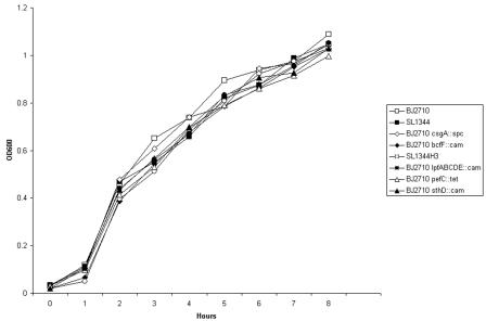
One mechanism by which organisms would display a reduced ability to form biofilms would be if the strain has a reduced growth rate. Since some of the fimbrial mutants displayed an altered biofilm phenotype, we compared the growth rate of each of the fimbrial mutants with the parent strain, BJ2710. As observed in Fig. 4, each of the S. enterica serovar Typhimurium strains displayed essentially the same growth rate throughout an 8-h growth experiment using Lennox agar. The same experiment, performed with RPMI 1640 tissue culture medium, also revealed no differences in growth rates (our unpublished data).
FIG. 4.
Growth curves of S. enterica serovar Typhimurium strains BJ2710 (parent), BJ2508 (BJ2710 fimH::kan), BJ3514 (BJ2710 bcfF::cat), BJ3539 (BJ2710 pefC::tet), BJ3660 (BJ2710 csgA::spc), BJ3571 (BJ2710 lpfABCDE::kan), and BJ3577 (BJ2710 sthD::cat) in Lennox broth. Growth curves were performed to ensure that the biofilm phenotypes observed for each fimbrial mutant were not due to a growth defect resulting from the mutation.
Characterization of biofilm formation by S. enterica serovar Typhimurium fimbrial mutants on chicken intestinal tissue.
Previous studies have demonstrated that Salmonella biofilms formed on various substrates (i.e., gallstones, glass, and eukaryotic cells) require different genes for proper development (37, 53, 54). Based on these observations, we examined biofilm development by the Salmonella fimbrial mutants on another, and perhaps more relevant, eukaryotic cell surface, chicken intestinal epithelia.
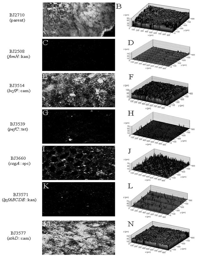
Biofilm formation by BJ2710 or the fimbrial mutants on excised chicken intestinal sections was monitored and compared after a 24-h incubation period. Representative areas of biofilm formed by each strain after 24 h are shown as composite images of the x-y plane and as x-y-z three-dimensional topographical maps (Fig. 5). S. enterica serovar Typhimurium strain BJ2710 formed an extensive biofilm on the chicken intestine during the 24-h incubation period (Fig. 5A and B). Figure 5A is representative of the biofilm that completely covered the surface of the intestinal tissue, and the average height of the bacterial mass across the section was 40 to 50 μm (Fig. 5B). The fimH mutant that is defective for biofilm formation on HEp-2 cells displayed virtually no ability to attach to and form biofilm on the chicken intestinal tissue (Fig. 5C and D). Similar to the results from the HEp-2 cell biofilm assay, strain BJ3514 (BJ2710 bcfF::cat) demonstrated a biofilm that was as dense as (Fig. 5E) but 10 to 20 μm taller than the biofilm produced by BJ2710 (Fig. 5B and F). Strain BJ3539 (BJ2710 pefC::tet) attached to and formed microcolonies on the chicken intestinal tissue but was unable to form a mature biofilm (Fig. 5G and H). Strain BJ3660 (BJ2710 csgA::spc) also formed sparse biofilm in what appeared to be areas protected from the medium flow by intestinal cells (Fig. 5I and J), but the immature biofilm appeared to be unable to extend into the luminal space of the intestine. Strain BJ3577 (BJ2710 sthD::cat) displayed no significant differences from the parent (Fig. 5A and B and M and N, respectively). In comparison, the biofilm phenotypes of strains BJ3539, BJ3660, and BJ3577 on chicken intestinal epithelium were essentially the same as those observed on HEp-2 cell monolayers.
FIG. 5.
Biofilm formation by S. enterica serovar Typhimurium strains BJ2710 (parent), BJ2508 (BJ2710 fimH::kan), BJ3514 (BJ2710 bcfF::cat), BJ3539 (BJ2710 pefC::tet), BJ3660 (BJ2710 csgA::spc), BJ3571 (BJ2710 lpfABCDE::kan), and BJ3577 (BJ2710 sthD::cat) on chicken intestinal epithelium. Panels A, C, E, G, I, K, and M show x-y-z composite sections generated by confocal microscopy of BJ2710 and its respective fimbrial mutants that were able to adhere to and grow as a biofilm on the chicken intestinal epithelial surface. The organisms were originally labeled with GFP and appear white in the image. Panels B, D, F, H, J, L, and N are topographical maps and correspond to BJ2710 and each of the respective fimbrial mutants and show a dramatic difference in the height and extent of the biofilm formed. The height of biofilm formed varied from 50 to 60 μm (BJ2710, BJ3514, and BJ3577) to less than 10 μm (BJ2508, BJ3539, and BJ3571).
We also examined the ability of the lpfABCDE mutant strain to form biofilm on the chicken intestinal tissue. In contrast to biofilm formation on HEp-2 cells, which was only partially attenuated, the lpfABCDE mutant had a phenotype similar to that of the pef mutant, which was able to adhere to and form microcolonies on the chicken epithelium but was blocked in its ability to form mature biofilm on the intestine epithelium (Fig. 5K and L). In conclusion, the defects in biofilms formed by the Bcf, Pef, Tafi, and Sth S. enterica serovar Typhimurium fimbrial mutants were similar on HEp-2 cells and chicken intestinal epithelium, while the Lpf mutant had a more severe biofilm defect on chicken intestinal epithelium than on HEp-2 cells.
Since degradation of the excised chicken intestinal tissue was of some concern, each of the strains was also orally inoculated into chickens. Following a 10-day incubation period to allow the Salmonella strains to adhere to and colonize the intestine, the intestines were excised and examined for biofilm formation by confocal microscopy. The appearances of the in vivo biofilms formed by each of the Salmonella mutant strains were nearly identical to the ex vivo results (data not shown).
Biofilm formation by S. enterica serovar Typhimurium on tissue culture-coated plastic.
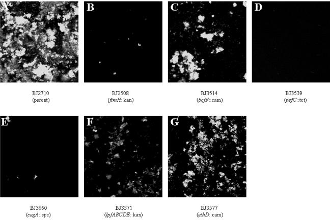
To determine if the defect in biofilm formation by the fimbrial mutants extended to nonbiological surfaces, we examined biofilm formation by each of the mutants on Thermanox (tissue culture-treated plastic) coverslips. Biofilm formation was monitored over a 24-h incubation period. Representative areas of biofilm formed by each strain after 24 h are shown as composite images of the x-y plane (Fig. 6). S. enterica serovar Typhimurium strains BJ2710 and BJ3577 (BJ2710 sthD::cat) each grew as extensive biofilms during the 24-h incubation period (Fig. 6A and G, respectively). The biofilms were on average ∼60 μm in height and were characterized by towers of biofilm bacteria. The fimH::kan mutant BJ2508 was completely unable to form a biofilm (Fig. 6B). Strain BJ3514 (bcfF::cat mutant) demonstrated a slight reduction in biomass but produced towers of biofilm that were similar in height to those of the wild type (Fig. 6C). Neither the Pef mutant (BJ3539) nor the CsgA mutant (BJ3660) formed any detectable biofilm on the plastic surface (Fig. 6D and E, respectively). Strain BJ3571 (BJ2710 lpfABCDE) adhered to and formed biofilm, but the resulting biofilm was reduced in height and thickness compared to that of the parent strain (Fig. 6A and F).
FIG. 6.
Biofilm formation by S. enterica serovar Typhimurium strains BJ2710 (wild type), BJ2508 (BJ2710 fimH::kan), BJ3514 (BJ2710 bcfF::cat), BJ3539 (BJ2710 pefC::tet), BJ3660 (BJ2710 csgA::spc), BJ3571 (BJ2710 lpfABCDE::kan), and BJ3577 (BJ2710 sthD::cat) on plastic slides. Images shown in panels A and B depict GFP-labeled BJ2710 and BJ3402, respectively, after growth for 24 h as a biofilm on the plastic slides. Panels C, D, E, F, and G are x-y composite images of strains BJ3514, BJ3539, BJ3660, BJ3571, and BJ3577. Each strain adheres incompletely to the plastic surface; however, each of the fimbrial mutants displayed various levels of adherence to the plastic surface.
DISCUSSION
Bacterial biofilms are now recognized as an important strategy of bacterial survival and growth in nature (13-15). Biofilms allow bacteria to stick to solid surfaces submerged in liquid environments and to bind to various host surfaces as part of pathogenic strategies in a host. Adherence to a solid surface is the initial step in biofilm formation, followed by the production and expression of an exopolysaccharide matrix that is involved in the shaping and maintaining of the structural integrity and three-dimensional architecture of a developing bacterial community (16, 18). Within this environment, bacteria multiply and undergo changes in gene expression to establish a biofilm community (59).
Previous work from our research group has shown that type 1 fimbriae are required to initiate biofilm formation on HEp-2 cells, murine intestinal tissue, and chicken intestinal tissue (7, 37). In this work, microarray hybridization results indicated that five additional fimbrial operons (encoding Bcf, Lpf, Pef, Sth, and Tafi) were upregulated in biofilm conditions and might therefore be involved in forming the bacterial community on the surface of cells. A subsequent analysis of mutants in genes encoding these five fimbriae revealed several different biofilm phenotypes. The observation that this subset of genes identified by microarray analysis was important for biofilm formation is worth noting. The analysis of strains with mutations in these genes confirmed their important role in the biofilm. However, other genes identified by microarray analysis as upregulated in conditions of biofilm growth could also play a role in biofilm formation but may not be required.
A mutant in the bcfF gene displayed an increased ability to form biofilms on both HEp-2 cells and chicken intestinal epithelium. The reason for this phenomenon is unknown, but one possibility is that the absence of Bcf fimbriae may allow an increased expression of other fimbriae that contribute to biofilm formation. A study using E. coli found that a bacterial culture can be divided into subpopulations that express only one fimbrial type at a time (P, type 1C, type 1, or S fimbriae), and turning off the expression of one fimbrial type (by mutation) allowed the expression of a new fimbrial type (44, 45). A mutant in the sthD gene that is part of a putative fimbrial operon had no detectable phenotype in any of the biofilm assays that we used.
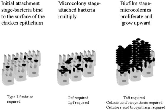
Three of the fimbrial mutants (pefC, csgA, and lpfABCDE) that we examined did have reduced biofilm production. The pefC mutant was the most defective for biofilm formation on HEp-2 cells when viewed by confocal microscopy, although both the csgA and lpfABCDE mutants also displayed reduced abilities to form a biofilm on the tissue culture cells (Fig. 2J to R). Quantitative plating of organisms in the biofilm revealed that the pefC mutant had 5,000-fold fewer organisms than BJ2710, while the csgA and lpfABCDE mutants had 1,000-fold and 500-fold fewer CFU than BJ2710, respectively (Fig. 3). However, on chicken intestinal epithelium, the alterations in the biofilm were qualitatively different. The pefC and lpfABCDE mutants were profoundly defective for biofilm production on chicken intestinal tissue (Fig. 5G and K), while the csgA mutant appeared to be able to bind around the circumference of the intestinal villus structures but could not form biofilm extensions into the lumen of the gut (Fig. 5I). This phenotype is similar to that observed for colanic acid and cellulose biosynthetic S. enterica serovar Typhimurium mutants (37) and supports the hypothesis that Tafi may be part of the extracellular matrix, along with secreted polysaccharides, on glass surfaces (2, 66). These results indicate that at least four types of fimbriae, Lpf, Pef Tafi, and type 1 fimbriae, are important for Salmonella biofilm formation on eukaryotic cells. Furthermore, our data suggest that each fimbria may have a separate function that is critical to biofilm formation, since a mutation in each of the fimbrial gene clusters significantly reduces biomass accumulation. The model presented in Fig. 7 summarizes these experimental observations into a working model.
FIG. 7.
Model of biofilm formation on chicken intestinal epithelium. The first stage in the formation of biofilm is initial attachment. S. enterica serovar Typhimurium strains lacking functional type 1 fimbriae are severely defective in the ability to establish initial colonization of the chicken intestinal epithelium (Fig. 5C). Following initial attachment events, the adherent organisms begin to multiply and form microcolonies on the epithelium. In some cases, the microcolonies can grow to the point that the silhouette of microvilli are visible, as for strain BJ3660, which carries a mutation in the csgA gene (Fig. 5I). A similar phenotype was observed for colanic acid and cellulose biosynthetic genes (37). Mutants in Pef and Lpf are unable to express this biofilm phenotype on chicken intestinal cells and therefore appear to have an early defect in biofilm maturation. Finally, mature biofilm is characterized by almost complete coverage of the epithelial surface and significant height of bacterial growth on top of the mucosa. The S. enterica serovar Typhimurium CsgA mutant as well as colanic acid and cellulose biosynthetic mutants are unable to form mature biofilm, which indicates that those genes are required for the final stages of biofilm formation.
This work did not address the role of seven additional Salmonella fimbrial operons in establishing a biofilm. While these operons were not upregulated to the same extent during growth on tissue culture cells, it may be worthwhile to investigate their role in biofilm formation since their maintenance in the Salmonella genome suggests that they provide a selective advantage. Also, it is possible that the narrow window of gene expression used for the microarray analysis may have overlooked conditions in which some of these genes may be upregulated.
Much research has focused on attachment, and several groups have been instrumental in identifying and characterizing fimbriae of S. enterica serovar Typhimurium. The csgBAC genes encode Tafi (12), and the fimbriae are expressed well from 18°C to 30°C, although wild-type strains were found to express Tafi at 37°C as well (19). Other work established a role for these fimbriae in forming a biofilm on a stainless steel surface (2). The Pef fimbriae are encoded by genes on the Salmonella 90-kb virulence plasmid (23) and have been shown to be important for adhesion to the small intestine of the mouse and for subsequent fluid accumulation (4). The genes encoding the Lpf fimbriae were identified because of their presence in S. enterica serovar Typhimurium and their absence in related strains (3). The fimbrial structures were observed by electron microscopy, and subsequent work indicated that they mediated adhesion to murine Peyer's patch tissue (5). Another interesting study examined the expression of 11 different S. enterica serovar Typhimurium fimbrial operons (32). Antibodies were raised against purified subunits of each of the 11 fimbrial types and were used to monitor expression of the various fimbriae under different growth conditions. Static growth at pH 5.1, at 32°C, or within bovine ligated intestinal loops increased expression of all fimbriae, except the type 1 fimbriae, in a relatively uniform fashion. In contrast, our work suggests that expression of the various S. enterica serovar Typhimurium fimbriae is not quite so uniform since, as mentioned above, seven of the fimbrial gene operons were not substantially upregulated, as determined in the microarray experiments. While this work was not directed at identifying the signals that induce biofilm expression, it is tantalizing to speculate that there is a variety of signals within the developing, complex environment of biofilm that orchestrates the proper expression of each factor when appropriate. Because of this complexity, efforts to understand these interactions in the laboratory setting using broth cultures may be unsuitable because the required biofilm environment is not properly established.
In addition to the identification of genes involved in fimbrial biosynthesis, we have also found that conjugative transfer genes (traI, traR, trbE, traO, traK, traD, traQ, traJ, traA, and traM), multiple antibiotic resistance genes (marA and marB), genes that play a role in Salmonella intracellular survival (ssaM, ssaI, pagD, msgA, pipB, spvR, ssaS, sifA, pipD, and ssaL), and many genes with no known functions are induced by biofilm growth. While many of these genes may not be required for biofilm formation, it does seem possible that growth on intestinal cells is an activating signal for bacterial processes such as conjugation and induction of antibiotic resistance genes and/or intracellular survival mechanisms. Genes induced in mature biofilms, such as the conjugative transfer mechanisms, may be involved in biofilm formation as adherence factors or in biofilm maturation (27, 55). Consequently, biofilm growth may be an effective way to identify and study factors that regulate and sense environmental conditions that control the expression of these phenotypes. Future work will be aimed at exploring these possibilities.
Our data indicate that fimbriae play a critical role in S. enterica serovar Typhimurium colonization or biofilm formation on eukaryotic cell surfaces. Fimbriae have previously been shown to be involved in adherence to the substratum (7), intracellular adhesion (65), and stabilization of the extracellular matrix (66). While this report identifies three fimbrial gene clusters essential for biofilm formation on eukaryotic cell surfaces, many questions about the role of these genes in the biofilm remain. Areas of interest include identifying the environmental regulators that induce changes in gene expression, characterizing the regulatory cascade, identifying the receptors of the non-type 1 fimbriae, and examining expression of the non-type 1 fimbriae on the bacterial surface.
Acknowledgments
We thank Andreas Baumler, Roy J. Curtiss III, and Robert Kadner for their generous gifts of strains and plasmids; Jenny Jagnow and Steve Clegg for their helpful discussions and assistance in strain construction; Mark Simmons for his expert assistance in the development and analysis of the real-time RT-PCR assays; Steffen Porwollik for his role in the construction of the Salmonella microarray; Jessica Linton and the staff of the University of Iowa DNA Facility for their expert assistance in microarrays and real-time RT-PCR; and Tom Moninger and Randy Nessler of the University of Iowa Central Microscopy facility for their expert technical assistance with the confocal microscopy.
This research was funded by NIH grants AI23829 (M.M.), AI38268, and AI059541 (B.D.J.) and Sidney Kimmel. N.A.L. was a fellow on the Mechanisms of Parasitology Training Grant, supported by NIH grant AI007511.
Editor: A. D. O'Brien
REFERENCES
- 1.Althouse, C., S. Patterson, P. Fedorka-Cray, and R. E. Isaacson. 2003. Type 1 fimbriae of Salmonella enterica serovar Typhimurium bind to enterocytes and contribute to colonization of swine in vivo. Infect. Immun. 71:6446-6452. [DOI] [PMC free article] [PubMed] [Google Scholar]
- 2.Austin, J. W., G. Sanders, W. W. Kay, and S. K. Collinson. 1998. Thin aggregative fimbriae enhance Salmonella enteritidis biofilm formation. FEMS Microbiol. Lett. 162:295-301. [DOI] [PubMed] [Google Scholar]
- 3.Baumler, A. J., and F. Heffron. 1995. Identification and sequence analysis of lpfABCDE, a putative fimbrial operon of Salmonella typhimurium. J. Bacteriol. 177:2087-2097. [DOI] [PMC free article] [PubMed] [Google Scholar]
- 4.Baumler, A. J., R. M. Tsolis, F. A. Bowe, J. G. Kusters, S. Hoffmann, and F. Heffron. 1996. The pef fimbrial operon of Salmonella typhimurium mediates adhesion to murine small intestine and is necessary for fluid accumulation in the infant mouse. Infect. Immun. 64:61-68. [DOI] [PMC free article] [PubMed] [Google Scholar]
- 5.Baumler, A. J., R. M. Tsolis, and F. Heffron. 1996. The lpf fimbrial operon mediates adhesion of Salmonella typhimurium to murine Peyer's patches. Proc. Natl. Acad. Sci. USA 93:279-283. [DOI] [PMC free article] [PubMed] [Google Scholar]
- 6.Baumler, A. J., R. M. Tsolis, T. A. Ficht, and L. G. Adams. 1998. Evolution of host adaptation in Salmonella enterica. Infect. Immun. 66:4579-4587. [DOI] [PMC free article] [PubMed] [Google Scholar]
- 7.Boddicker, J. D., N. A. Ledeboer, J. Jagnow, B. D. Jones, and S. Clegg. 2002. Differential binding to and biofilm formation on, HEp-2 cells by Salmonella enterica serovar Typhimurium is dependent upon allelic variation in the fimH gene of the fim gene cluster. Mol. Microbiol. 45:1255-1265. [DOI] [PubMed] [Google Scholar]
- 8.Brown, P. K., C. M. Dozois, C. A. Nickerson, A. Zuppardo, J. Terlonge, and R. Curtiss III. 2001. MlrA, a novel regulator of curli (AgF) and extracellular matrix synthesis by Escherichia coli and Salmonella enterica serovar Typhimurium. Mol. Microbiol. 41:349-363. [DOI] [PubMed] [Google Scholar]
- 9.Chuang, S. E., D. L. Daniels, and F. R. Blattner. 1993. Global regulation of gene expression in Escherichia coli. J. Bacteriol. 175:2026-2036. [DOI] [PMC free article] [PubMed] [Google Scholar]
- 10.Clegg, S., and D. C. Old. 1979. Fimbriae of Escherichia coli K-12 strain AW405 and related bacteria. J. Bacteriol. 137:1008-1012. [DOI] [PMC free article] [PubMed] [Google Scholar]
- 11.Clegg, S., B. K. Purcell, and J. Pruckler. 1987. Characterization of genes encoding type 1 fimbriae of Klebsiella pneumoniae, Salmonella typhimurium, and Serratia marcescens. Infect. Immun. 55:281-287. [DOI] [PMC free article] [PubMed] [Google Scholar]
- 12.Collinson, S., S. Clouthier, J. Doran, P. Banser, and W. Kay. 1996. Salmonella enteritidis agfBAC operon encoding thin, aggregative fimbriae. J. Bacteriol. 178:662-667. [DOI] [PMC free article] [PubMed] [Google Scholar]
- 13.Costerton, J. W., K. J. Cheng, G. G. Geesey, T. I. Ladd, J. C. Nickel, M. Dasgupta, and T. J. Marrie. 1987. Bacterial biofilms in nature and disease. Annu. Rev. Microbiol. 41:435-464. [DOI] [PubMed] [Google Scholar]
- 14.Costerton, J. W., Z. Lewandowski, D. E. Caldwell, D. R. Korber, and H. M. Lappin-Scott. 1995. Microbial biofilms. Annu. Rev. Microbiol. 49:711-745. [DOI] [PubMed] [Google Scholar]
- 15.Costerton, J. W., P. S. Stewart, and E. P. Greenberg. 1999. Bacterial biofilms: a common cause of persistent infections. Science 284:1318-1322. [DOI] [PubMed] [Google Scholar]
- 16.Danese, P. N., L. A. Pratt, and R. Kolter. 2000. Exopolysaccharide production is required for development of Escherichia coli K-12 biofilm architecture. J. Bacteriol. 182:3593-3596. [DOI] [PMC free article] [PubMed] [Google Scholar]
- 17.Datsenko, K. A., and B. L. Wanner. 2000. One-step inactivation of chromosomal genes in Escherichia coli K-12 using PCR products. Proc. Natl. Acad. Sci. USA 97:6640-6645. [DOI] [PMC free article] [PubMed] [Google Scholar]
- 18.Davies, D. G., A. M. Chakrabarty, and G. G. Geesey. 1993. Exopolysaccharide production in biofilms: substratum activation of alginate gene expression by Pseudomonas aeruginosa. Appl. Environ. Microbiol. 59:1181-1186. [DOI] [PMC free article] [PubMed] [Google Scholar]
- 19.Dibb-Fuller, M., E. Allen-Vercoe, M. J. Woodward, and C. J. Thorns. 1997. Expression of SEF17 fimbriae by Salmonella enteritidis. Lett. Appl. Microbiol. 25:447-452. [DOI] [PubMed] [Google Scholar]
- 20.Edwards, P. R., and D. W. Bruner. 1943. The occurrence and distribution of Salmonella types in the United States. J. Infect. Dis. 72:58-67. [DOI] [PubMed] [Google Scholar]
- 21.Fields, P. I., R. V. Swanson, C. G. Haidaris, and F. Heffron. 1986. Mutants of Salmonella typhimurium that cannot survive within the macrophage are avirulent. Proc. Natl. Acad. Sci. USA 83:5189-5193. [DOI] [PMC free article] [PubMed] [Google Scholar]
- 22.Firon, N., I. Ofek, and N. Sharon. 1984. Carbohydrate-binding sites of the mannose-specific fimbrial lectins of enterobacteria. Infect. Immun. 43:1088-1090. [DOI] [PMC free article] [PubMed] [Google Scholar]
- 23.Friedrich, M. J., N. E. Kinsey, J. Vila, and R. J. Kadner. 1993. Nucleotide sequence of a 13.9 kb segment of the 90 kb virulence plasmid of Salmonella typhimurium: the presence of fimbrial biosynthetic genes. Mol. Microbiol. 8:543-558. [DOI] [PubMed] [Google Scholar]
- 24.Garcia, B., C. Latasa, C. Solano, F. Garcia-del Portillo, C. Gamazo, and I. Lasa. 2004. Role of the GGDEF protein family in Salmonella cellulose biosynthesis and biofilm formation. Mol. Microbiol. 54:264-277. [DOI] [PubMed] [Google Scholar]
- 25.Gerstel, U., C. Park, and U. Romling. 2003. Complex regulation of csgD promoter activity by global regulatory proteins. Mol. Microbiol. 49:639-654. [DOI] [PubMed] [Google Scholar]
- 26.Gerstel, U., and U. Romling. 2003. The csgD promoter, a control unit for biofilm formation in Salmonella typhimurium. Res. Microbiol. 154:659-667. [DOI] [PubMed] [Google Scholar]
- 27.Ghigo, J. M. 2001. Natural conjugative plasmids induce bacterial biofilm development. Nature 412:442-445. [DOI] [PubMed] [Google Scholar]
- 28.Hancox, L. S., K. S. Yeh, and S. Clegg. 1997. Construction and characterization of type 1 non-fimbriate and non-adhesive mutants of Salmonella typhimurium. FEMS Immunol. Med. Microbiol. 19:289-296. [DOI] [PubMed] [Google Scholar]
- 29.Hegde, P., R. Qi, K. Abernathy, C. Gay, S. Dharap, R. Gaspard, J. E. Hughes, E. Snesrud, N. Lee, and J. Quackenbush. 2000. A concise guide to cDNA microarray analysis. BioTechniques 29:548-556. [DOI] [PubMed] [Google Scholar]
- 30.Herwald, H., M. Morgelin, A. Olsen, M. Rhen, B. Dahlback, W. Muller-Esterl, and L. Bjorck. 1998. Activation of the contact-phase system on bacterial surfaces—a clue to serious complications in infectious diseases. Nat. Med. 4:298-302. [DOI] [PubMed] [Google Scholar]
- 31.Hultgren, S. J., S. Normark, and S. N. Abraham. 1991. Chaperone-assisted assembly and molecular architecture of adhesive pili. Annu. Rev. Microbiol. 45:383-415. [DOI] [PubMed] [Google Scholar]
- 32.Humphries, A. D., M. Raffatellu, S. Winter, E. H. Weening, R. A. Kingsley, R. Droleskey, S. Zhang, J. Figueiredo, S. Khare, J. Nunes, L. G. Adams, R. M. Tsolis, and A. J. Baumler. 2003. The use of flow cytometry to detect expression of subunits encoded by 11 Salmonella enterica serotype Typhimurium fimbrial operons. Mol. Microbiol. 48:1357-1376. [DOI] [PubMed] [Google Scholar]
- 33.Jones, B. D., and S. Falkow. 1993. Phenotypic and genetic aspects of Salmonella invasion, p. 4-16. In C. Kado and J. Crosa (ed.), Molecular mechanisms of bacterial virulence. Kluwer Academic Publishers, Hingham, Mass.
- 34.Jones, B. D., N. Ghori, and S. Falkow. 1994. Salmonella typhimurium initiates murine infection by penetrating and destroying the specialized epithelial M cells of the Peyer's patches. J. Exp. Med. 180:15-23. [DOI] [PMC free article] [PubMed] [Google Scholar]
- 35.Joseph, B., S. K. Otta, and I. Karunasagar. 2001. Biofilm formation by Salmonella spp. on food contact surfaces and their sensitivity to sanitizers. Int. J. Food Microbiol. 64:367-372. [DOI] [PubMed] [Google Scholar]
- 36.Kingsley, R. A., E. H. Weening, A. M. Keestra, and A. J. Baumler. 2002. Population heterogeneity of Salmonella enterica serotype Typhimurium resulting from phase variation of the lpf operon in vitro and in vivo. J. Bacteriol. 184:2352-2359. [DOI] [PMC free article] [PubMed] [Google Scholar]
- 37.Ledeboer, N. A., and B. D. Jones. 2005. Exopolysaccharide sugars contribute to biofilm formation by Salmonella enterica serovar Typhimurium on HEp-2 cells and chicken intestinal epithelium. J. Bacteriol. 187:3214-3226. [DOI] [PMC free article] [PubMed] [Google Scholar]
- 38.Lockman, H. A., and R. Curtiss III. 1992. Virulence of non-type 1-fimbriated and nonfimbriated nonflagellated Salmonella typhimurium mutants in murine typhoid fever. Infect. Immun. 60:491-496. [DOI] [PMC free article] [PubMed] [Google Scholar]
- 39.McClelland, M., K. E. Sanderson, J. Spieth, S. W. Clifton, P. Latreille, L. Courtney, S. Porwollik, J. Ali, M. Dante, F. Du, S. Hou, D. Layman, S. Leonard, C. Nguyen, K. Scott, A. Holmes, N. Grewal, E. Mulvaney, E. Ryan, H. Sun, L. Florea, W. Miller, T. Stoneking, M. Nhan, R. Waterston, and R. K. Wilson. 2001. Complete genome sequence of Salmonella enterica serovar Typhimurium LT2. Nature 413:852-856. [DOI] [PubMed] [Google Scholar]
- 40.Morgan, E., J. D. Campbell, S. C. Rowe, J. Bispham, M. P. Stevens, A. J. Bowen, P. A. Barrow, D. J. Maskell, and T. S. Wallis. 2004. Identification of host-specific colonization factors of Salmonella enterica serovar Typhimurium. Mol. Microbiol. 54:994-1010. [DOI] [PubMed] [Google Scholar]
- 41.Nicholson, B., and D. Low. 2000. DNA methylation-dependent regulation of pef expression in Salmonella typhimurium. Mol. Microbiol. 35:728-742. [DOI] [PubMed] [Google Scholar]
- 42.Norris, T. L., R. A. Kingsley, and A. J. Bumler. 1998. Expression and transcriptional control of the Salmonella typhimurium Ipf fimbrial operon by phase variation. Mol. Microbiol. 29:311-320. [DOI] [PubMed] [Google Scholar]
- 43.Norris, T. L., and A. J. Baumler. 1999. Phase variation of the lpf operon is a mechanism to evade cross-immunity between Salmonella serotypes. Proc. Natl. Acad. Sci. USA 96:13393-13398. [DOI] [PMC free article] [PubMed] [Google Scholar]
- 44.Nowicki, B., M. Rhen, V. Vaisanen-Rhen, A. Pere, and T. K. Korhonen. 1984. Immunofluorescence study of fimbrial phase variation in Escherichia coli KS71. J. Bacteriol. 160:691-695. [DOI] [PMC free article] [PubMed] [Google Scholar]
- 45.Nowicki, B., M. Rhen, V. Vaisanen-Rhen, A. Pere, and T. K. Korhonen. 1985. Organization of fimbriate cells in colonies of Escherichia coli strain 3040. J. Gen. Microbiol. 131:1263-1266. [DOI] [PubMed] [Google Scholar]
- 46.Old, D. C., and J. P. Duguid. 1970. Selective outgrowth of fimbriate bacteria in static liquid medium. J. Bacteriol. 103:447-456. [DOI] [PMC free article] [PubMed] [Google Scholar]
- 47.Olsen, A., A. Arnqvist, M. Hammar, and S. Normark. 1993. The RpoS sigma factor relieves H-NS mediated transcriptional repression of csgA, the subunit gene of fibronectin-binding curli in Escherichia coli. Mol. Microbiol. 7:523-536. [DOI] [PubMed] [Google Scholar]
- 48.Olsen, A., M. Wick, M. Morgelin, and L. Bjorck. 1998. Curli, fibrous surface proteins of Escherichia coli, interact with major histocompatibility complex class I molecules. Infect. Immun. 66:944-949. [DOI] [PMC free article] [PubMed] [Google Scholar]
- 49.Paranchych, W., and L. S. Frost. 1988. The physiology and biochemistry of pili. Adv. Microb. Physiol. 29:53-114. [DOI] [PubMed] [Google Scholar]
- 50.Parsek, M. R., and E. P. Greenberg. 1999. Quorum sensing signals in development of Pseudomonas aeruginosa biofilms. Methods Enzymol. 310:43-55. [DOI] [PubMed] [Google Scholar]
- 51.Penheiter, K. L., N. Mathur, D. Giles, T. Fahlen, and B. D. Jones. 1997. Non-invasive Salmonella typhimurium mutants are avirulent because of an inability to enter and destroy M cells of ileal Peyer's patches. Mol. Microbiol. 24:697-709. [DOI] [PubMed] [Google Scholar]
- 52.Porter, S. B., and R. Curtiss III. 1997. Effect of inv mutations on Salmonella virulence and colonization in 1-day-old White Leghorn chicks. Avian Dis. 41:45-57. [PubMed] [Google Scholar]
- 53.Prouty, A. M., W. H. Schwesinger, and J. S. Gunn. 2002. Biofilm formation and interaction with the surfaces of gallstones by Salmonella spp. Infect. Immun. 70:2640-2649. [DOI] [PMC free article] [PubMed] [Google Scholar]
- 54.Prouty, A. M., and J. S. Gunn. 2003. Comparative analysis of Salmonella enterica serovar Typhimurium biofilm formation on gallstones and on glass. Infect. Immun. 71:7154-7158. [DOI] [PMC free article] [PubMed] [Google Scholar]
- 55.Reisner, A., J. A. Haagensen, M. A. Schembri, E. L. Zechner, and S. Molin. 2003. Development and maturation of Escherichia coli K-12 biofilms. Mol. Microbiol. 48:933-946. [DOI] [PubMed] [Google Scholar]
- 56.Ricke, S. C. 2003. The gastrointestinal tract ecology of Salmonella enteritidis colonization in molting hens. Poult. Sci. 82:1003-1007. [DOI] [PubMed] [Google Scholar]
- 57.Romling, U., Z. Bian, M. Hammar, W. D. Sierralta, and S. Normark. 1998. Curli fibers are highly conserved between Salmonella typhimurium and Escherichia coli with respect to operon structure and regulation. J. Bacteriol. 180:722-731. [DOI] [PMC free article] [PubMed] [Google Scholar]
- 58.Romling, U., W. D. Sierralta, K. Eriksson, and S. Normark. 1998. Multicellular and aggregative behaviour of Salmonella typhimurium strains is controlled by mutations in the agfD promoter. Mol. Microbiol. 28:249-264. [DOI] [PubMed] [Google Scholar]
- 59.Schembri, M. A., K. Kjaergaard, and P. Klemm. 2003. Global gene expression in Escherichia coli biofilms. Mol. Microbiol. 48:253-267. [DOI] [PubMed] [Google Scholar]
- 60.Solano, C., B. Garcia, J. Valle, C. Berasain, J. M. Ghigo, C. Gamazo, and I. Lasa. 2002. Genetic analysis of Salmonella enteritidis biofilm formation: critical role of cellulose. Mol. Microbiol. 43:793-808. [DOI] [PubMed] [Google Scholar]
- 61.Tinker, J. K., L. S. Hancox, and S. Clegg. 2001. FimW is a negative regulator affecting type 1 fimbrial expression in Salmonella enterica serovar Typhimurium. J. Bacteriol. 183:435-442. [DOI] [PMC free article] [PubMed] [Google Scholar]
- 62.Townsend, S. M., N. E. Kramer, R. Edwards, S. Baker, N. Hamlin, M. Simmonds, K. Stevens, S. Maloy, J. Parkhill, G. Dougan, and A. J. Baumler. 2001. Salmonella enterica serovar Typhi possesses a unique repertoire of fimbrial gene sequences. Infect. Immun. 69:2894-2901. [DOI] [PMC free article] [PubMed] [Google Scholar]
- 63.van der Velden, A. W., A. J. Baumler, R. M. Tsolis, and F. Heffron. 1998. Multiple fimbrial adhesins are required for full virulence of Salmonella typhimurium in mice. Infect. Immun. 66:2803-2808. [DOI] [PMC free article] [PubMed] [Google Scholar]
- 64.Webb, J. S., M. Givskov, and S. Kjelleberg. 2003. Bacterial biofilms: prokaryotic adventures in multicellularity. Curr. Opin. Microbiol. 6:578-585. [DOI] [PubMed] [Google Scholar]
- 65.White, A. P., S. K. Collinson, P. A. Banser, D. L. Gibson, M. Paetzel, N. C. J. Strynadka, and W. W. Kay. 2001. Structure and characterization of AgfB from Salmonella enteritidis thin aggregative fimbriae. J. Mol. Biol. 311:735-749. [DOI] [PubMed] [Google Scholar]
- 66.White, A. P., D. L. Gibson, S. K. Collinson, P. A. Banser, and W. W. Kay. 2003. Extracellular polysaccharides associated with thin aggregative fimbriae of Salmonella enterica serovar Enteritidis. J. Bacteriol. 185:5398-5407. [DOI] [PMC free article] [PubMed] [Google Scholar]
- 67.Whiteley, M., M. G. Bangera, R. E. Bumgarner, M. R. Parsek, G. M. Teitzel, S. Lory, and E. P. Greenberg. 2001. Gene expression in Pseudomonas aeruginosa biofilms. Nature 413:860-864. [DOI] [PubMed] [Google Scholar]
- 68.Wray, C., and W. J. Sojka. 1978. Experimental Salmonella typhimurium infection in calves. Res. Vet. Sci. 25:139-143. [PubMed] [Google Scholar]
- 69.Wu, H., and P. M. Fives-Taylor. 2001. Molecular strategies for fimbrial expression and assembly. Crit. Rev. Oral Biol. Med. 12:101-115. [DOI] [PubMed] [Google Scholar]
- 70.Zhang, X., S. M. Kelly, W. S. Bollen, and R. Curtiss III. 1997. Characterization and immunogenicity of Salmonella typhimurium SL1344 and UK-1 Δcrp and Δcdt deletion mutants. Infect. Immun. 65:5381-5387. [DOI] [PMC free article] [PubMed] [Google Scholar]


