Abstract
We describe the development of a Virtual Slide System for creating and viewing clinico-pathologic cases with embedded interactive digital microscopy. The system supports rich text-to-image annotation, including (1) hotlinks of text descriptions that move the student to the correct part of the slide, and (2) annotations such as arrows and circles that appear on the Virtual Slide on request. The interface can be configured by the student to alter the degree of guidance the system provides. The authoring layer provides a graphical user interface to authors for creating new case sets, cases, questions, and annotated virtual slides, which are saved to a database and automatically added to the Virtual Slide homepage. The system has been used in two pilot studies at the University of Pittsburgh.
BACKGROUND
Virtual Slides are whole slide images captured as multiple partial digital images at high magnification and then joined to create a single large (~1 GB) image file. Virtual Slides allow panning, zooming, and magnification without loss of resolution - creating an interface that reproduces the main functions of a microscope 1. Virtual slides are just starting to be used in web-based teaching 2. Our system represents the first virtual slide curriculum development system that we are aware of.
SYSTEM DESCRIPTION
The Virtual Slide Set (http://virtualslide.upmc.edu) consists of five-tiers: Client, Presentation, Business, Integration and Resource. The system includes a GUI for authoring which allows instructors to create their own virtual slide resources.
Client.
The client tier includes only a web browser with Java Plug-in. Students can view the slides from any location with an Internet connection.
Presentation.
This tier is responsible for handling authentication and authorization, managing client sessions, receiving requests from a client, interpreting them, forwarding them to the business tier for processing data, and presenting clients with the target slides. The technologies used in this tier include HTML, JavaScript, Java Servlets, JSP, and Java applets.
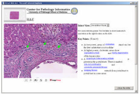
To avoid architectural complexity, we follow the Model Viewer Controller (MVC) design pattern. The design separates the presentation and authoring tools (View) from the content such as student data, course data or virtual slide data (Model). When the Controller (Java Servlet) receives requests from clients, it instantiates Views (JSP and Java applet) and associates them with the Model (JavaBean). The applet based virtual slide viewer provides three modes: Discover, Guide and Annotate. Students can change mode based on their preference for degree of annotation, to see the slides with maximum annotation or as unknowns (Figure 1).
Figure 1.
Student Interface showing Virtual Slide in “Annotate” mode. Text hyperlinks on right move the slide and show annotations.
Business.
The business tier manages: (a) student and instructor's behavior and (b) image manipulation. For example, if students go to a case page, the tier retrieve s case information, and it passes it to the presentation tier. Also, when students zoom or unzoom an image, this tier calculates the position of the cursor and provides the correct magnification. When instructors register information, the business tier manages the data transaction between authoring tool and database.
Integration.
The integration tier is responsible for connection between business tier and resource tier. We use JDBC for connecting with an Oracle 9i Server.
Resource.
This tier provides external resources that provide the actual data to the application. We have two external resources: (a) Oracle 9i database for student data, instructor data, course information and virtual slide information, and (b) image storage for the virtual slides.
Acknowledgments
The Virtual Slide Set is funded by an Innovations in Education Award grant from the Office of the Provost, University of Pittsburgh.
REFERENCES
- 1.Dick, F. Implementation of Virtual Microscopy in a Histology and Pathology Course. Sixth Annual Conference on Advancing Pathology Informatics, Imaging and the Internet. Pittsburgh, PA October 4–6, 2002.
- 2.Afework A, Benyon M, Bustamante F, et al. Digital Dynamic Telepathology –the Virtual Microscope. Proc AMIA Symp. 1998:912–916. [PMC free article] [PubMed] [Google Scholar]


