Abstract
A recombinant clone encoding enzymes for Klebsiella pneumoniae O12-antigen lipopolysaccharide (LPS) was found when we screened for serum resistance of a cosmid-based genomic library of K. pneumoniae KT776 (O12:K80) introduced into Escherichia coli DH5α. A total of eight open reading frames (ORFs) (wbO12 gene cluster) were necessary to produce K. pneumoniae O12-antigen LPS in E. coli K-12. A complete analysis of the K. pneumoniae wbO12 cluster revealed an interesting coincidence with the wbO4 cluster of Serratia marcescens from ORF5 to ORF8 (or WbbL to WbbA). This prompted us to generate mutants of K. pneumoniae strain KT776 (O12) and to study complementation between the two enterobacterial wb clusters using mutants of S. marcescens N28b (O4) obtained previously. Both wb gene clusters are examples of ABC 2 transporter-dependent pathways for O-antigen heteropolysaccharides. The wzm-wzt genes and the wbbA or wbbB genes were not interchangeable between the two gene clusters despite their high level of similarity. However, introduction of three cognate genes (wzm-wzt-wbbA or wzm-wzt-wbbB) into mutants unable to produce O antigen allowed production of the specific O antigen. The K. pneumoniae O12 WbbL protein performs the same function as WbbL from S. marcescens O4 in either the S. marcescens O4 or E. coli K-12 genetic background.
In gram-negative bacteria lipopolysaccharide (LPS) is one of the major structural and immunodominant molecules of the outer membrane (OM). LPS consists of three main regions: lipid A, the core oligosaccharide, and the O antigen. The O antigen is the most external component of LPS, and it consists of a polymer of oligosaccharide repeating units. An interesting feature is the high chemical variability shown by O antigens, which is reflected by the genetic variation in the genes involved in O-antigen biosynthesis, designated the wb cluster. The genetics of O-antigen biosynthesis have been intensively studied in the Enterobacteriaceae, and it has been shown that the wb clusters usually contain genes involved in biosynthesis of activated sugars, glycosyl transferases, O-antigen polymerases, and O-antigen export (20, 29).
Escherichia coli K-12 strains, including DH5α, are rough (O−), unable to produce O-antigen LPS, and serum sensitive. We and others have shown with different gram-negative bacteria (5, 13, 14) that the presence of O-antigen LPS (smooth phenotype) can confer serum resistance. We have used this phenotype to clone the O-antigen LPSs from different bacteria into E. coli DH5α.
In a recent study of the prevalence of the O serogroups among clinical Klebsiella isolates from different sources and countries, serogroup O12 accounted for 0.3% of the isolates (8). The chemical structure of the Klebsiella O12-antigen LPS was reported to be a heteropolymer of rhamnose and N-acetylglucosamine (4) and was recently determined to be →4)-β-d-GlcNAcp-(1→4)-α-l-Rhap-(1→ (26).
In this study we cloned and sequenced the wbO12 gene cluster of Klebsiella pneumoniae. As the K. pneumoniae wbO12 cluster showed a high level of similarity to the Serratia marcescens wbO4 cluster, complementation analysis was performed with both enterobacterial wb clusters by using the previously described O-antigen-deficient mutants of S. marcescens N28b (serotype O4) (17).
MATERIALS AND METHODS
Bacterial strains, plasmids, and growth conditions.
The bacterial strains, cosmids, and plasmids used are listed on Table 1. Bacteria were grown in Luria-Bertani (LB) broth and LB agar (15). LB medium was supplemented with ampicillin (100 μg/ml or 3 mg/ml), chloramphenicol (25 μg/ml), kanamycin (30 μg/ml), and tetracycline (20 μg/ml) when necessary.
TABLE 1.
Bacterial strains, cosmids, and plasmids used
| Strain, cosmid, or plasmid | Relevant characteristics | Source or reference |
|---|---|---|
| E. coli strains | ||
| DH5α | F−end A hsdR17 (rK− mK+) supE44 thi-1 recA1 gyr-A96 φ80lacZM15 | 7 |
| XL1-Blue | recA1 endA1 gyrA96 thi-1 hsdR17 supE44 relA1 lac (F′ proAB lacIqZΔM15 Tn10) | Stratagene |
| CLM4 | lacZ trp (sbcB-rfb) upp rel rpsL recA | 13 |
| K. pneumoniae strains | ||
| KT776 | Wild type, serotype O12:K80 | I. Ørskov |
| KT776-1 | O−wzm-wzr KT776 deletion mutant obtained with pKO3Wzm-t | This study |
| KT776-2 | O−wbbB KT776 deletion mutant obtained with pKO3WbbB | This study |
| S. marcescens strains | ||
| N28b | Wild-type O4 | 17 |
| N28b-2 | O−wbbL insertion mutant from N28b, Kmr | 17 |
| N28b-3 | O−wbbA insertion mutant from N28b, Kmr | 17 |
| N28b-4 | O−wzm-wzt N28b deletion mutant | 17 |
| Plasmids | ||
| pLA2917 | Tcr Kmr | 1 |
| pCosKT12 | pLA2917 with 20-kb chromosomal KT772 Sau3A insert | This study |
| pST-Blue | Kmr | Novagen |
| pST1 | pST-Blue with complete wzm-wzt genes of wbKpO12, Kmr | This study |
| pST1-m | pST1 plasmid with altered Walker box A in Wzt, Kmr | This study |
| pST5 | pST-Blue with complete wbbA gene of wbSmO4, Kmr | This study |
| pST6 | pST-Blue with complete wzm-wzt genes of wbSmO4, Kmr | This study |
| pST7 | pST-Blue with complete wzm-wzt-wbbA genes of wbSmO4, Kmr | This study |
| pBR328 | Apr Cmr Tcr | 21 |
| pJTO12 | pBR328 with complete wbKpO12, Cmr Tcr | This study |
| pBR1 | pBR328 with complete wbbL gene of wbKpO12, Cmr Tcr | This study |
| pBR2 | pBR328 with complete wbbB gene of wbKpO12, Cmr Tcr | This study |
| pBR3 | pBR328 with wzm-wzt-wbbB genes of wbKpO12, Cmr Tcr | This study |
| pKO3 | Cmr, temperature sensitive for replication, sacB | 12 |
| pKO3Wzm-t | pKO3 with internal fragment of wzm-wzt genes | This study |
| pKO3WbbB | pKO3 with internal fragment of wbbB gene | This study |
General DNA methods.
DNA manipulations were carried out essentially as previously described (18). DNA restriction endonucleases, T4 DNA ligase, E. coli DNA polymerase (Klenow fragment), and alkaline phosphatase were used as recommended by the suppliers. Recombinant clones were selected on LB agar plates containing the appropriate antibiotics.
Construction of a K. pneumoniae KT776 (O12:K80) genomic library.
K. pneumoniae KT776 genomic DNA was isolated and partially digested with Sau3A as described by Sambrook et al. (18). Cosmid pLA2917 (1) was digested with BglII, dephosphorylated, and ligated to Sau3A genomic DNA fragments. DNA packaging by using Gigapack Gold III (Stratagene) and infection of E. coli DH5α were carried out as previously described (6). Recombinant clones were selected on LB agar plates supplemented with tetracycline (20 μg/ml).
Cell surface isolation and analysis.
The OM was isolated as previously described (14). OM proteins were analyzed by sodium dodecyl sulfate-polyacrylamide gel electrophoresis (SDS-PAGE) by using the Laemmli procedure (11). Protein gels were fixed and stained with Coomassie blue. LPS was purified by the method of Westphal and Jann (28). For screening purposes LPS was obtained after proteinase K digestion of whole cells by the procedure of Darveau and Hancock (3). SDS-PAGE was performed, and LPS bands were detected by the silver staining method of Tsai and Frasch (25).
Serum killing.
The survival of exponential-phase bacteria in nonimmune human serum was measured as previously described (14) or by using a microtiter plate-based assay for screening (27).
Antisera.
Anti-K. pneumoniae O12 LPS serum was obtained and assayed as previously described for other LPSs (14, 23). Anti-S. marcescens O4 LPS serum was obtained previously (17).
Western immunoblotting.
After SDS-PAGE, immunoblotting was carried out by transferring preparations to polyvinylidene fluoride membranes (Millipore Corp., Bedford, Mass.) at 1.3 A for 1 h in the buffer of Towbin et al. (24). The membranes were then incubated sequentially with 1% bovine serum albumin, specific anti-O serum (1:500), alkaline phosphatase-labeled goat anti-rabbit immunoglobulin G, and disodium 5-bromo-4-chloro-indolylphosphate-nitroblue tetrazolium. Incubations were carried out for 1 h, and washing steps with 0.05% Tween 20 in phosphate-buffered saline were included after each incubation step. Colony blotting was performed by using K. pneumoniae O12 antiserum as indicated above.
ELISA.
Cytosol, whole membrane, inner membrane (IM), and OM fractions were analyzed by enzyme-linked immunosorbent assays (ELISAs). ELISAs were performed by dispensing standardized suspensions of each fraction in coating buffer (pH 9.6) into 96-well microtiter plates. The plates were left to stand overnight at 4°C. The wells were blocked with 1% bovine serum albumin in phosphate-buffered saline for 2 h at 37°C. Anti-O12 polyclonal serum (1:200) was added and incubated for 2 h at 37°C. Detection was performed by using peroxidase-labeled sheep anti-rabbit immunoglobulin G (1:1,000) and 2,2′-azino-di-(3-ethylbenzthiazoline sulfonate) as the substrate. Cytosol, whole membrane, IM, and OM were prepared as previously described (17).
DNA sequencing.
Double-stranded DNA sequencing was performed by the Sanger dideoxy chain termination method (19) with an ABI Prism dye terminator cycle sequencing kit (Perkin-Elmer). The primers used for DNA sequencing were purchased from Pharmacia LKB Biotechnology. Primers 5′-GACTGGGCGGTTTTATGG-3′ and 5′-CCATCTTGTTCAAT CATGCA-3′, designed from the sequence of cosmid pLA2917 (1), were used to sequence the inserts in the BglII restriction site on pLA2917.
DNA and protein sequence analysis.
The DNA sequence was translated in all six frames, and all open reading frames (ORFs) more than 100 bp long were inspected. Deduced amino acid sequences were compared with the deduced amino acid sequences encoded by DNA translated in all six frames from nonredundant GenBank and EMBL databases by using the BLAST network service at the National Center for Biotechnology Information (2). Multiple-sequence alignments were constructed by using the Clustal W program (22). Possible terminator sequences were deduced by using the Terminator program from the Genetics Computer Group package (Genetics Computer Group, Madison, Wis.) in a VAX 4300. Hydropath profiles were calculated by the method of Kyte and Doolittle (10).
K. pneumoniae KT776 (wzm-wzt) and wbbB mutant construction.
The method of Link et al. (12) was used to create chromosomal in-frame deletion mutants. Briefly, plasmid pJTO12 and primers A (5′-CGCGGATCCTGGTGGTTGTCAGTGGGC-3′) and B (5′-CCCATCCACTAAACATGATGAAAGTTTTTGATGAGG-3′) or primers C (5′-TGTTTAAGTTTAGTGGATGGGCCGTTAAAAATCATTACAATTG-3′) and D (5′-CGCGGATCCACAATCCCAGCAACTTGG-3′) were used in two sets of asymmetric PCRs to amplify a 785-bp DNA fragment (fragment AB) and a 733-bp DNA fragment (fragment CD) of the wzm-wzt region. DNA fragments AB and CD were annealed at the overlapping region and amplified by PCR as a single fragment by using primers A and D. The fusion product was purified, digested with BamHI, ligated to BamHI-digested vector pKO3, electroporated into DH5α, and selected by using Kmr to obtain plasmid pKO3Wzm-t.The same approach was used for the wbbB gene by using pJTO12 and primers A1 (5′-CGCGGATCCTTGAGTATGCTGATCGCTGC-3′) and B1 (5′-CCCATCCACTAAACTTAAACACCCTCTGAACGGATATGGAG-3′) or primers C1 (5′-TGTTTAAGTTTAGTGGATGGGGCGGAATTGGGGATTATCTC-3′) and D1 (5′-CGCGGATCCCGGAGCACTAAAAGAAAGCC-3′) amplifying a 734-bp fragment (fragment A1B1) and a 431-bp fragment (fragment C1D1) to obtain plasmid pKO3WbbB. Allelic replacement was performed by the method of Link et al. (12) by using plasmids pKO3Wzm-t and pKO3WbbB.
Plasmid construction.
Plasmids pST5, pST6, and pST7, harboring the S. marcescens wbO4 genes wbbA, wzm-wzt, and wzm-wzt-wbbA, respectively, were obtained from the pSUB6 plasmid (17) by PCR amplification. Primers Sm1 (5′-AACAATGGCGAGCGAGAAG-3′) and Sm2 (5′-GGTGAAAGCAAGTCGGAA AG-3′) amplified a 3,900-bp DNA fragment containing wbbA; primers Sm3 (5′-CTGCCGTGATCATACAGGG-3′) and Sm4 (5′-CTCTAAAGGGGTAAGCCGG-3′) amplified a 2,212-bp DNA fragment containing wzm-wzt; and primers Sm3 and Sm2 amplified a 5,950-bp DNA fragment containing wzm-wzt-wbbA. The amplified DNA fragments were independently ligated into the vector pST-Blue (Novagen) and introduced into E. coli by Kmr selection.
Plasmid pST1 was constructed by using the pJTO12 plasmid (complete K. pneumoniae wbO12 cluster) and PCR. Primers Kp1 (5′-AACAACTATTGTCCATGCC-3′) and Kp2 (5′-GGAATCGTCTTCTACCTG-3′) amplified a 3,043-bp DNA fragment containing the wzm-wzt genes from K. pneumoniae wbO12. The amplified DNA fragment was ligated into the vector pST-Blue to obtain plasmid pST1. Plasmid pST1-m was constructed by using pST1 (wzm-wzt from the K. pneumoniae wbO12 cluster) and PCR. Primers A2bis (5′-TGGTACCATATTCGCTCCG-3′) and B2 (5′-GCAGATAATCTGCAGCAAAGTCGACGCCCCGGC-3′) amplified a 1,220-bp DNA fragment, and primers C2 (5′-ACTTTGCTGCAGATTATCTGCGGCACACTAACC-3′) and D2bis (5′-AATAAGGGGCTTTCACCCC-3′) amplified a 1,149-bp DNA fragment. Using both amplified DNA fragments and primers A2bis and D2bis, we amplified a 2,369-bp DNA fragment that was ligated to vector pST-Blue to obtain plasmid pST1-m. Plasmid pST1-m contains the wzm gene and a mutant wzt gene from K. pneumoniae wbO12 with an AAG codon (lysine) substituted for a CGC codon (alanine) (underlined in the primer sequence). DNA sequencing of the pST1-m insert confirmed this change. Because some of the S. marcescens O4− mutants were Kmr and Apr is not a suitable marker, we used the following approach to generate plasmids pBR1, pBR2, and pBR3 using pJTO12 for PCR amplification. Primers Kp3 (5′-CGCCAGCTGGTGCT GTCTCTGTCGGAG-3′) and Kp4 (5′-CGCCAGCTGGGCAATACTGCGCACCAC-3′) amplified a 1,075-bp DNA fragment (wbbL); primers Kp5 (5′-CGCCAGCTGTCGGGCGATTACCTAATAC-3′) and Kp6 (5′-CGCCAGCTGGCCATTTAGCCTCAAGTAC-3′) amplified a 3,015-bp DNA fragment (wbbB); and primers Kp7 (5′-CGCCAGCTGTTGGTACCATATTCGCTCC-3′) and Kp6 amplified a 5,183-bp DNA fragment (wzm-wzt-wbbL). Each of the amplified DNA fragments was independently digested with PvuII (all the primers contained a PvuII restriction site) and ligated to PstI-digested and blunt-ended vector pBR328. This approach allowed us to maintain resistance to chloramphenicol and tetracycline as selective markers.
Nucleotide sequence accession number.
The nucleotide sequence of the genes described here has been deposited in the GenBank database under accession number AY130997.
RESULTS
Cloning of LPS O12-antigen genes of K. pneumoniae (O12:K80).
K. pneumoniae (O12:K80) strain KT776, like other encapsulated and smooth strains, is serum resistant (14), whereas E. coli K-12 strains like DH5α are serum sensitive. A cosmid-based genomic library of K. pneumoniae strain KT776 chromosomal DNA was constructed and introduced into E. coli DH5α, and recombinant clones were selected on LB agar containing tetracycline. Several serum-resistant clones were isolated by using a microtiter plate based assay (27), and pCosKT12 was representative of these clones. DH5α harboring pCosKT12 was characterized by analysis of the OM protein and LPS profile by SDS-PAGE. No major differences were found in the OM protein pattern, but cosmid pCosKT12 conferred K. pneumoniae O12-antigen LPS production to E. coli DH5α (Fig. 1). No O12 antigen was detected in strain DH5α with or without the cosmid vector pLA2917. pCosKT12 was cured from the recipient strain DH5α by serial growth without antibiotics, single-colony isolation, and testing for antibiotic sensitivity and lack of the plasmid DNA. The cured strain lacked the O12-antigen LPS and was serum sensitive.
FIG. 1.
Silver-stained polyacrylamide gel (A) and Western immunoblot of LPS reacted with K. pneumoniae O12-specific antiserum (B). Antiserum was obtained and assayed as described in Materials and Methods. LPS samples were prepared by the method of Darveau and Hancock (3). Lane 1, K. pneumoniae KT776; lane 2, E. coli DH5α; lane 3, DH5α harboring pCosKT12; lane 4, cured strain from lane 3; lane 5, DH5α harboring pJTO12; lane 6, E. coli CLM4 (with wb deleted [13]); lane 7, CLM4 harboring pJTO12.
Subcloning and sequencing of wbO12.
By using PstI partial digestion of pCosKT12 and ligation to PstI-digested plasmid vector pBR328, plasmid pJTO12 was obtained and introduced into DH5α. Plasmid pJTO12 contained a 13-kb insert from pCosKT12. As determined by LPS gel analysis and Western blotting with specific anti-O12 antibody, plasmid pJTO12 conferred production of K. pneumoniae O12 antigen to DH5α (Fig. 1). When pJTO12 was transferred to E. coli CLM4 (a strain with the wb cluster deleted [13]), it was still able to confer O12-antigen LPS production (Fig. 1). This result suggested that pJTO12 contains all the genetic information necessary for O12-antigen production. The DNA sequence of the insert was determined. Eight ORFs were deduced, and their characteristics are shown in Table 2. Upstream of each ORF putative ribosome binding sequences were found. No Rho-independent transcription termination sequences were found in the eight ORFs. This feature, in addition to the overlaps between the ORF2 stop codon and the ORF3 start codon and between the ORF6 stop codon and the ORF7 start codon and the short spaces between the ORFs, strongly suggests that these ORFs are part of an operon (wbO12).
TABLE 2.
K. pneumoniae KT776 (O12) wb gene cluster
| Locus | Base positions | G+C content (%) | Size of protein encoded (kDa) | pIa | GRAVYb |
|---|---|---|---|---|---|
| rmlB (ORF1) | 1-1065 | 57.8 | 39.4 | 6.1 | −0.287 |
| rmlA (ORF2) | 1083-1988 | 55.6 | 33.1 | 5.3 | −0.106 |
| rmlD (ORF3) | 1985-3875 | 61.3 | 31.7 | 5.8 | −0.065 |
| rmlC (ORF4) | 2890-3444 | 54.4 | 20.2 | 5.7 | −0.217 |
| wbbL (ORF5) | 3536-4366 | 34.5 | 32.1 | 8.7 | −0.187 |
| wzm (ORF6) | 4396-5229 | 37.5 | 31.2 | 9.7 | +0.978 |
| wzt (ORF7) | 5219-6541 | 43.8 | 48.2 | 6.1 | −0.084 |
| wbbB (ORF8) | 6545-9346 | 39.9 | 106.8 | 5.8 | −0.203 |
Isoelectric point of the protein calculated by using ProtParam at the Expassy server.
GRAVY, grand average hydropathicity of the protein calculated by using the Kyte-Doolitle method (10).
Analysis of the ORF deduced amino acid sequences.
Analysis of the deduced amino acid sequences showed that the ORF1 to ORF4 sequences are very similar to the sequences of the four enzymes involved in the biosynthesis of dTDP-rhamnose in other enterobacteria (Table 3). Accordingly, we suggest that these ORFs correspond to the genes rmlB, rmlA, rmlD, and rmlC, respectively. The ORF5 product showed homology with the WbbL protein from S. marcescens wbO4, which is a rhamnosyl transferase; it also exhibited less homology with other glycosyl transferases (Table 3). ORF5 was designated wbbL. The ORF6 and ORF7 products were similar to ABC 2-type transport system integral membrane and ATP-binding proteins, respectively (Table 3). Hydrophobicity analysis and identification of the putative transmembrane domains of the ORF6-encoded protein (amino acid residues 53 to 69, 84 to 100, 133 to 149, 158 to 174, 194 to 210, and 245 to 261), performed by using the method of Klein et al. (9), suggested that this protein is an integral membrane protein. Also, the ORF7-encoded protein contained the sequence GRNGAGKS (amino acid residues 70 to 77), which correspond to Walker box A, a motif present in ATP-binding proteins, as well as the ABC transporter family signature YSSGMYVRLAFAVQA (amino acid residues 158 to 172). Thus, ORF6 and ORF7 were designated wzm and wzt, respectively. Finally, the ORF8-encoded protein (933 amino acids) exhibited homology only with the WbbA protein (1,191 amino acids) encoded by the S. marcescens wbO4 cluster. However, homology between the two proteins was present only in the first 460 amino acid residues. Both WbbA and the ORF8 product contained a putative glycosyl transferase motif in the first half of the protein. Both proteins were predicted to be anchored to the membrane (by residues 334 to 350 in the ORF8-encoded protein), and no relevant similarities were found in the C-terminal halves of the proteins. Protein WbbA has been suggested to be bifunctional, and the glycosyl transferase function is also found in the ORF8-encoded protein; thus, we designated ORF8 wbbB.
TABLE 3.
Levels of identity and similarity of the amino acid sequences encoded by the K. pneumoniae KT776 gene cluster and the most significant other proteins
| Protein | No. of amino acids | % Similaritya | % Identitya | Accession no. |
|---|---|---|---|---|
| ORF1 (RmlB) of K. pneumoniae KT776 | 354 | AY130997 | ||
| RffG (RmlB) of Yersinia pestis | 355 | 81 | 72 | NP407310 |
| RmlB of E. coli of O157:H7 | 355 | 79 | 70 | NP312748 |
| ORF2 (RmlA) of K. pneumoniae KT776 | 301 | AY130997 | ||
| RmlA of Yersinia enterocolitica | 289 | 86 | 76 | P55257 |
| RffH (RmlA) of Yersinia pestis | 293 | 86 | 76 | NP407309 |
| ORF3 (RmlD) of K. pneumoniae KT776 | 296 | AY130997 | ||
| RmlD of Salmonella enterica | 299 | 58 | 47 | AAG09514 |
| RfbD (RmlD) of Salmonella enterica serovar Typhimurium | 299 | 58 | 47 | NP461041 |
| ORF4 (RmlC) of K. pneumoniae KT776 | 184 | AY130997 | ||
| RmlC of Shigella boydii | 189 | 86 | 72 | AAL27313 |
| RfbC (RmlC) of Shigella flexneri | 181 | 78 | 68 | P37780 |
| ORF5 (WbbL) of K. pneumoniae KT776 | 276 | AY130997 | ||
| WbbL of S. marcescens O4 | 282 | 69 | 55 | T31088 |
| ORF6 (Wzm) of K. pneumoniae KT776 | 277 | AY130997 | ||
| Wzm of S. marcescens O4 | 277 | 84 | 75 | T31089 |
| ORF7 (Wzt) of K. pneumoniae KT776 | 440 | AY130997 | ||
| Wzt of S. marcescens O4 | 441 | 83 | 71 | T31090 |
| ORF8 (WbbB) of K. pneumoniae KT776b | 933 | AY130997 | ||
| WbbA of S. marcescens O4 | 1,191 | 58 | 39 | T31091 |
The values were obtained from pairwise comparisions by using the Gap program. The Gap program settings, were as follows: gap weight, 12; and length weight, 12.
The similarity between WbbB (from residue 1 to residue 454) and WbbA (from residue 1 to residue 460) is limited to this region of the two proteins.
The complete sequence of the K. pneumoniae wbO12 cluster exhibited an interesting similarity with the sequence of the S. marcescens wbO4 cluster from ORF5 to ORF8 (or WbbL to WbbA), which prompted us to generate mutants of K. pneumoniae strain KT776 (O12) and to study complementation of the two wb clusters by using mutants previously created in S. marcescens N28b (O4).
Construction of mutant strains KT776-1 (wzm-wzt) and KT776-2 (wbbB).
Mutants KT776-1 (wzm-wzt) and KT776-2 (wbbB) were obtained by the method of Link et al. (12) to create in-frame deletions. Plasmids pKO3Wzm-t and pKO3WbbB were independently electroporated into K. pneumoniae KT776, and colonies were selected for Cmr at 42°C. Colonies were plated at 30°C in the presence of sucrose (5%). Candidate mutant colonies (chloramphenicol sensitive) were screened by PCR. Mutant KT776-1 (wzm-wzt) was characterized by producing an amplified 1,479-bp DNA fragment with primers A and D. Mutant KT776-2 (wbbB) was characterized by producing an amplified 1,125-bp DNA fragment with primers A1 and D1. The wild-type strain in similar PCR experiments produced 3,545- and 3,844-bp amplification fragments with primers A and D and primers A1 and D1, respectively. DNA sequencing of the amplified fragments confirmed the deletion of 2,066 bp for KT776-1 and the deletion of 2,718 bp for KT776-2.
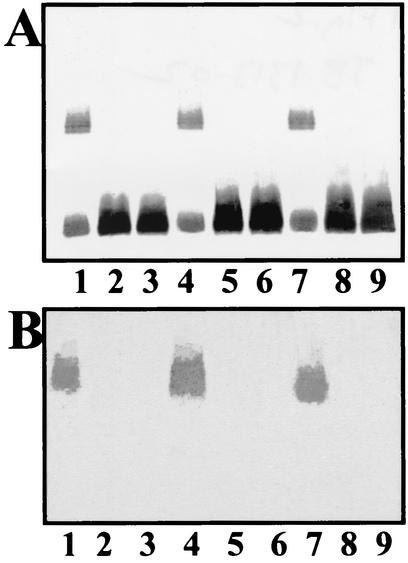
Both mutant KT776-1 and mutant KT776-2 were devoid of O12-antigen LPS as determined by using gels and Western blots (Fig. 2). No other major differences were observed in the OM molecules and the capsules of these mutants. Analysis of purified LPS from both mutants showed a complete lack of rhamnose, a characteristic sugar of the O12-antigen LPS. ELISAs (Table 4) with K. pneumoniae O12-antigen-specific antiserum were performed with different cellular fractions from KT776 (wild type) and mutants KT776-1 and KT776-2. As expected, the OM fraction of wild-type strain KT776 showed a high affinity for the specific antiserum. By contrast, the IM fraction obtained from mutant strain KT776-1 showed a high response to specific antiserum, while no response to the same antiserum was observed with the OM fraction of this mutant strain. Neither the OM fraction nor the IM fraction from mutant strain KT776-2 reacted with the specific antiserum. These results suggest that KT776-2 was unable to form O12-antigen LPS, while KT776-1 was able to form this antigen but was unable to transport it to the OM (i.e., it accumulated in the IM).
FIG. 2.
Silver-stained polyacrylamide gel (A) and Western immunblot of LPS reacted with K. pneumoniae O12-specific antiserum (B). Antiserum was obtained and assayed as described in Materials and Methods. LPS samples were prepared by the method of Darveau and Hancock (3) from K. pneumoniae strains. Lane 1, KT776 (wild type); lane 2, mutant KT776-1 (wzm-wzt); lane 3, mutant KT776-2 (wbbB); lane 4, KT776-1 harboring pST1 (wzm-wzt of wbKpO12); lane 5, KT776-1 harboring pBR1 (wbbL of wbKpO12); lane 6, KT776-1 harboring pBR2 (wbbB of wbKpO12); lane 7, KT776-2 harboring pBR2 (wbbB of wbKpO12); lane 8, KT776-2 harboring pBR1 (wbbL of wbKpO12); lane 9, KT776-2 harboring pST1 (wzm-wzt of wbKpO12).
TABLE 4.
K. pneumoniae O12-antigen export assaysa
| Strain |
A405 of fraction extract
|
|||
|---|---|---|---|---|
| Crude extract | Whole membrane | IM | OM | |
| KT776 (wild type) | 0.1 (0.01) | 0.5 (0.04) | 0.1 (0.03) | 0.7 (0.13) |
| KT776-1 (wzm-wzt) | 0.3 (0.05) | 0.6 (0.08) | 0.8 (0.12) | <0.1 |
| KT776-2 (wbbB) | <0.1 | <0.1 | <0.1 | <0.1 |
| KT776-1(pST1) (harboring wzm-wztKp) | 0.1 (0.03) | 0.4 (0.06) | 0.1 (0.02) | 0.8 (0.07) |
| KT776-1(pST1-m) (pST1 with altered Wzt) | 0.2 (0.06) | 0.5 (0.04) | 0.7 (0.10) | <0.1 |
| KT776-2(pBR2) (harboring wbbBKp) | 0.1 (0.03) | 0.6 (0.04) | 0.1 (0.05) | 0.6 (0.15) |
The (A405) values are means (standard deviations) calculated from three independent determinations (ELISAs with different cellular fractions and K. pneumoniae O12-specific antiserum).
Complementation studies with K. pneumoniae O12− mutants.
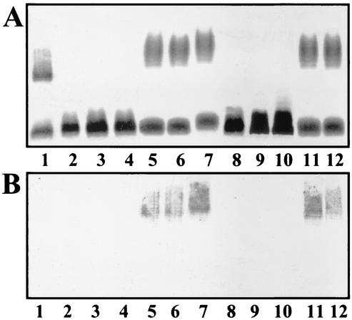
K. pneumoniae KT776-1 and KT776-2 were fully complemented for O12-antigen production by plasmid pJTO12 (wbKp) but not by the pBR328 vector, as determined by using LPS gels and Western blots with anti-O12-specific serum. Furthermore, mutant KT776-1 (wzm-wzt) was complemented by pST1 (wzm-wzt from K. pneumoniae wbO12) but not by plasmid pST-Blue, pBR1 (wbbLKp), or pBR2 (wbbBKp). Mutant KT776-2 (wbbB) was complemented by pBR2 (wbbB from K. pneumoniae wbO12) but not by the pBR328 vector, pBR1 (wbbLKp), or pST1 (wzm-wztKp) (Fig. 2). Furthermore, the OM fractions of mutants KT776-1 and KT776-2 transformed with pST1 (wzm-wztKp) and pBR2 (wbbBKp), respectively, again showed affinity for the O12-specific antiserum (Table 4). The results obtained so far suggest that an ABC 2-type dependent transporter is involved in the export of the K. pneumoniae O12-antigen LPS. In order to further examine this possibility, a Wzt mutant was obtained on plasmid pST1-m. The mutation resulted in substitution of the conserved lysine (amino acid residue 76) from the Walker box A of Wzt for an alanine. Mutant KT776-1 harboring pST1-m was unable to export the O12-antigen LPS, as determined by using LPS gels (data not shown) and the export assay with ELISA experiments (Table 4). Due to the similarities between the WbbA and WbbB proteins encoded by the S. marcescens wbO4 and K. pneumoniae wbO12 gene clusters, respectively, we decided to study the complementation of these mutants with plasmids harboring S. marcescens wbO4 genes. Mutants KT776-1 and KT776-2 were not complemented by plasmids pST5 (wbbASm) and pST6 (wzm-wztSm), as judged by using LPS gels or Western blots with specific anti-O12 serum. However, both mutants were able to produce O-antigen LPS when either pSUB6 (wbSm) or pST7 (wzm-wzt-wbbASm) plasmids were introduced by transformation (Fig. 3A). The O-antigen LPS produced was not able to react with specific K. pneumoniae anti-O12 serum (data not shown) but reacted with specific S. marcescens anti-O4 serum (Fig. 3B). No O-antigen LPS production was observed when these mutants were transformed with the plasmid vectors.
FIG. 3.
Silver-stained polyacrylamide gel (A) and Western immunblot of LPS reacted with S. marcescens O4-specific antiserum (B). LPS samples were prepared by the method of Darveau and Hancock (3). Lane 1, K. pneumoniae KT776 (wild type); lane 2, K. pneumoniae mutant KT776-1 (wzm-wzt); lane 3, KT776-1 harboring the pST-Blue vector; lane 4, KT776-1 harboring pST6 (wzm-wzt of wbSmO4); lane 5, KT776-1 harboring pST7 (wzm-wzt-wbbA of wbSmO4); lane 6, KT776-1 harboring pSUB6 (complete S. marcescens wbO4 cluster); lane 7, S. marcescens N28b (serotype O4, wild type); lane 8, K. pneumoniae mutant KT776-2 (wbbB); lane 9, KT776-2 harboring the pST-Blue vector; lane 10, KT776-1 harboring pST5 (wbbA of wbSmO4); lane 11, KT776-1 harboring pST7 (wzm-wzt-wbbA of wbSmO4); lane 12, KT776-2 harboring pSUB6 (complete S. marcescens wbO4 cluster).
Complementation studies with S. marcescens O4− mutants.
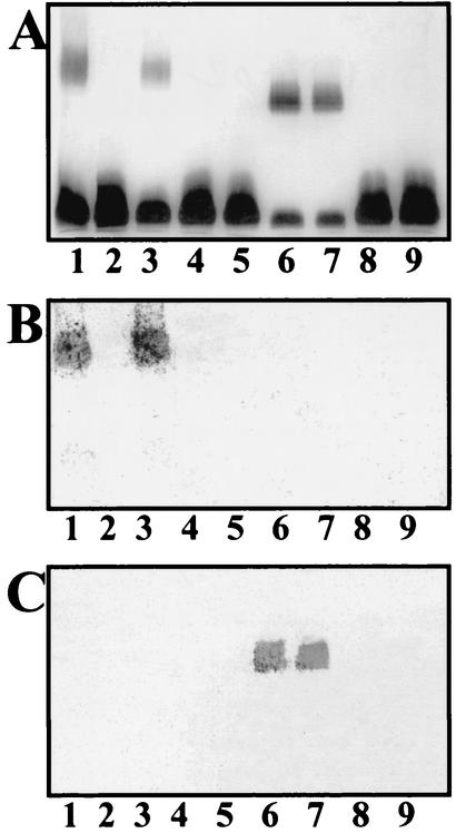
Mutant N28b-2 (wbbL) was complemented by plasmid pBR1 harboring the homologous wbbL gene from K. pneumoniae wbO12 (Fig. 4). Furthermore, as happens with the S. marcescens wbbL gene, plasmid pBR1 is able to produce O-antigen LPS in E. coli DH5α. This O-antigen LPS was unable to react with K. pneumoniae anti-O12 serum or S. marcescens anti-O4 serum, but it reacted with specific E. coli anti-O16 serum (data not shown). This feature is the same as that described for the S. marcescens wbbL gene introduced into E. coli (16).
FIG. 4.
Silver-stained polyacrylamide gel (A) and Western immunblots of LPS reacted with S. marcescens O4-specific antiserum (B) and K. pneumoniae O12-specific antiserum (C). LPS samples were prepared by the method of Darveau and Hancock (3) from S. marcescens strains. Lane 1, S. marcescens N28b (serotype O4, wild type); lane 2, S. marcescens mutant N28b-2 (wbbL); lane 3, N28b-2 harboring pBR1 (wbbL of wbKpO12); lane 4, S. marcescens mutant N28b-4 (wzm-wzt); lane 5, S. marcescens mutant N28b-4 harboring pST1 (wzm-wzt of wbKpO12); lane 6, S. marcescens mutant N28b-4 harboring pBR3 (wzm-wzt-wbbB of wbKpO12); lane 7, S. marcescens mutant N28b-3 harboring pBR3 (wzm-wzt-wbbB of wbKpO12); lane 8, S. marcescens mutant N28b-3 harboring pBR2 (wbbB of wbKpO12); lane 9, S. marcescens mutant N28b-3 (wbbA).
Introduction of plasmid pJTO12 (K. pneumoniae wbO12) into the S. marcescens N28b-3 and N28b-4 mutants allowed the mutants to produce O-antigen LPS, as judged by using LPS gels. This O-antigen LPS was unable to react with S. marcescens anti-O4 serum, but it reacted with K. pneumoniae anti-O12 serum. Identical results were obtained when both S. marcescens mutants were transformed with plasmid pBR3 (wzm-wzt-wbbB from K. pneumoniae wbO12) but not when they were transformed with plasmid vector pBR328 (Fig. 4). Mutant N28b-3 was unable to produce O-antigen LPS (as determined by using gels) when it was transformed with plasmid pBR2 (wbbBKp) (Fig. 4). Also, mutant N28b-4 was unable to produce O-antigen LPS when it was transformed with plasmid pST1 (wzm-wztKp). However, mutant N28b-4 was fully complemented when it was transformed with the pST6 plasmid (wzm-wztSm) but not when the pST-Blue vector was used (Fig. 4).
DISCUSSION
One recombinant clone encoding K. pneumoniae O12-antigen LPS was found when we screened for serum resistance of a cosmid-based genomic library of K. pneumoniae KT776 (O12:K80) introduced into E. coli DH5α. A total of eight ORFs (wbO12 gene cluster) were necessary to produce K. pneumoniae O12-antigen LPS in E. coli K-12, as judged by the fact that pJTO12 (wbO12) in E. coli CLM4 (a strain with a deletion in the wb cluster [13]) was able to confer O12-antigen LPS production. This result suggested that pJTO12 contains all the genetic information necessary for O12-antigen production.
Sugar nucleotides are the activated precursors for cell surface polysaccharides. To synthesize O antigens, monomers are assembled on a lipid carrier (undecaprenol phosphate) by enzymes encoded in the wb gene cluster before their incorporation into the LPS molecule. Two known pathways for O-antigen export have been established, the Wzx dependent pathway for heteropolysaccharide structures and the ABC 2 transporter-dependent pathway for homopolysaccharides (29). However, the similarities found between the ABC 2-type pathway and the group II capsular heteropolysaccharide export pathway suggest that not only homopolysaccharide O antigens could be exported by an ABC 2 pathway. The first example of a heteropolysaccharide exported through an ABC 2 export system that was described was the S. marcescens O4-antigen LPS (17). In this study we showed that the K. pneumoniae wbO12 cluster is also able to export a heteropolysaccharide (with two monosaccharides) by using an ABC 2 transport system. This conclusion was supported by the results of the KT776-1 complementation experiments. Plasmid pST1 (wzm-wztKpO12) was fully able to complement the KT776-1 mutant. Plasmid pST1-m (coding for a wild-type Wzm and a Walker box A Wzt mutant) was unable to restore export of O12-antigen LPS. These results allowed us to generalize that the ABC 2 transporter-dependent pathway can export not only O-antigen homopolysaccharides but also heteropolysaccharides (containing only two monosaccharides).
The high level of similarity between the S. marcescens wbO4 and K. pneumoniae wbO12 gene clusters and the similarity between the O4 and O12 chemical structures [in S. marcescens O4, →4)-β-d-Glcp-(1→4)-α-l-Rhap-(1→; and in K. pneumoniae O12, →4)-β-d-GlcNAcp-(1→4)-α-l-Rhap-(1→] gave us the opportunity to study the specificity of both systems by performing cross complementation experiments with genetically defined mutants.
The gene encoding the WbbL putative rhamnosyl transferase is interchangeable between the two wb gene clusters. Furthermore, when the K. pneumoniae wbO12 wbbL gene was introduced into E. coli K-12 strain DH5α, this strain was able to produce O16-antigen LPS, as previously described for S. marcescens wbO4 wbbL in E. coli K-12 (16). These results strongly suggest that the WbbL proteins can transfer l-Rha residues to the O4 position of d-Glc or d-GlcNAc, thus indicating that this enzyme is not highly specific.
Despite the high level of similarity between the ABC 2 export proteins for the two O-antigen gene clusters, neither the integral membrane protein (Wzm) nor the ATP-binding protein (Wzt) is interchangeable between the organisms. This fact indicates the high specificity for O-antigen export by the membrane components of the ABC 2 transporter-dependent pathway. The last proteins encoded by the two gene clusters (WbbA for S. marcescens wbO4 and WbbB for K. pneumoniae wbO12) exhibited similarity only in their first halves, and these two proteins were not interchangeable. This is not surprising, since WbbA should transfer a Glc residue by a β-1,4 linkage to α-l-Rha, whereas WbbB should transfer a GlcNAc residue by a β-1,4 linkage to α-l-Rha. It can be speculated that the N-terminal similarity between the WbbA and WbbB proteins could be related to the glycosyl transferase activity of both proteins. The overall results suggest that the genetic determinants responsible for the differences in the structures of these rather simple heteropolysaccharide O antigens are the last genes of the clusters (wbbA and wbbB). Despite the similarities in chemical O-antigen structure, the two heteropolysaccharides appear to use their own, noninterchangeable ABC 2 transport systems. The O antigen could be changed in both genetic backgrounds (Serratia and Klebsiella) when the three genes (wzm, wzt, and wbbA or wbbB) were introduced into a mutant unable to produce O antigen. Thus, both the ABC 2 export system and the putative d-GlcNAc transferase are specific for the K. pneumoniae O12 antigen.
To summarize, K. pneumoniae wbO12 contains four genes (rmlA, rmlB, rmlC, and rmlD) involved in the biosynthesis of dTDP-l-Rha, two genes involved in the sequential transfer of l-Rha (wbbL) and d-GlcNAc (wbbB), and two genes (wzm and wzt) coding for a specific O12 ABC 2 export system. These proposed enzymatic activities could explain the biosynthesis and export of K. pneumoniae O12-antigen LPS. It can be hypothesized that K. pneumoniae O12-antigen LPS biosynthesis begins with the transfer of N-acetylglucosamine to the acyl carrier protein, followed by sequential transfer of l-Rha and d-GlcNAc to obtain the complete O12-antigen. This O antigen is exported through the specific ABC 2 transport system and finally is ligated to the core LPS by the O-antigen ligase (WaaL) located in the K. pneumoniae waa cluster.
Acknowledgments
This work was supported by grants from the Plan Nacional de I+D (Ministerio de Ciencia y Tecnología, Spain) and Generalitat de Catalunya. L.I. and F.R were supported by FPI fellowships from the Ministerio de Ciencia y Tecnología (Spain) and Universidad de Barcelona, respectively.
We thank Maite Polo for her technical assistance.
REFERENCES
- 1.Allen, L. N., and R. S. Hanson. 1985. Construction of broad-host-range cosmid cloning vector: identification of genes necessary for growth of Methylobacterium organophilum on methanol. J. Bacteriol. 161:955-962. [DOI] [PMC free article] [PubMed] [Google Scholar]
- 2.Altschul, S. F., W. Gish, W. Miller, E. W. Myers, and D. Lipman. 1990. Basic local alignment search tool. J. Mol. Biol. 215:403-410. [DOI] [PubMed] [Google Scholar]
- 3.Darveau, R. P., and R. E. W. Hancock. 1983. Procedure for the isolation of bacterial lipopolysaccharides from both smooth and rough Pseudomonas aeruginosa and Salmonella typhimurium strains. J. Bacteriol. 155:831-838. [DOI] [PMC free article] [PubMed] [Google Scholar]
- 4.Erbing, C., B. Lindberg, and J. Lönngren. 1977. Structural studies on the Klebsiella O group 12 lipopolysaccharide. Carbohydr. Res. 59:831-838. [DOI] [PubMed] [Google Scholar]
- 5.Frank, M. M. 1990. Complement in host defense against bacterial infections, p. 305-317. In E. M. Ayoub, G. H. Cassell, W. C. Branche, Jr., and P. J. Henry (ed.), Microbial determinants of virulence and host response. American Society for Microbiology, Washington, D.C.
- 6.Guasch, J. F., N. Piqué, N. Climent, S. Ferrer, S. Merino, X. Rubires, J. M. Tomás, and M. Regué. 1996. Cloning and characterization of two Serratia marcescens genes involved in core lipopolysaccharide biosynthesis. J. Bacteriol. 178:5741-5747. [DOI] [PMC free article] [PubMed] [Google Scholar]
- 7.Hanahan, D. 1983. Studies on transformation of Escherichia coli with plasmids. J. Mol. Biol. 166:557-580. [DOI] [PubMed] [Google Scholar]
- 8.Hansen, D. S., F. Mestre, S. Albertí, S. Hernández-Allés, D. Alvarez, A. Domenech-Sanchez, J. Gil, S. Merino, J. M. Tomás, and V. J. Benedí. 1999. Klebsiella pneumoniae lipopolysaccharide O typing: revision of prototype strains and O-group distribution among clinical isolates from different sources and countries. J. Clin. Microbiol. 37:56-62. [DOI] [PMC free article] [PubMed] [Google Scholar]
- 9.Klein, P., M. Kanehisa, and C. De. 1985. The detection and classification of membrane-spanning proteins. Biochim. Biophys. Acta 815:468-476. [DOI] [PubMed] [Google Scholar]
- 10.Kyte, J., and R. F. Doolittle. 1982. A simple method for displaying the hydropathic character of a protein. J. Mol. Biol. 157:105-132. [DOI] [PubMed] [Google Scholar]
- 11.Laemmli, U. K. 1970. Cleavage of structural proteins during the assembly of the head of bacteriophage T4. Nature 227:680-685. [DOI] [PubMed] [Google Scholar]
- 12.Link, A. J., D. Phillips, and G. M. Church. 1997. Methods for generating precise deletions and insertions in the genome of wild-type Escherichia coli: application to open reading frame characterization. J. Bacteriol. 179:6228-6237. [DOI] [PMC free article] [PubMed] [Google Scholar]
- 13.Marolda, C. L., and M. A. Valvano. 1993. Identification, expression, and DNA sequence of the GDP-mannose biosynthesis region encoded by the O7 rfb gene cluster of strain VW187 (Escherichia coli O7:K1). J. Bacteriol. 175:148-158. [DOI] [PMC free article] [PubMed] [Google Scholar]
- 14.Merino, S., S. Camprubí, S. Albertí, V. J. Benedí, and J. M. Tomás. 1992. Klebsiella pneumoniae resistance mechanisms to complement mediated killing. Infect. Immun. 60:2529-2535. [DOI] [PMC free article] [PubMed] [Google Scholar]
- 15.Miller, J. H. 1972. Experiments in molecular genetics. Cold Spring Harbor Laboratory, Cold Spring Harbor, N.Y.
- 16.Rubirés, X., F. Saigí, N. Piqué, N. Climent, S. Merino, S. Albertí, J. M. Tomás, and M. Regué. 1997. A gene (wbbL) from Serratia marcescens N28b (O4) complements the rfb-50 mutation of Escherichia coli K-12 derivatives. J. Bacteriol. 179:7581-7586. [DOI] [PMC free article] [PubMed] [Google Scholar]
- 17.Saigi, F., N. Climent, N. Piqué, C. Sanchez, S. Merino, X. Rubirés, A. Aguilar, J. M. Tomás, and M. Regué. 1999. Genetic analysis of the Serratia marcescens N28b O4 antigen gene cluster. J. Bacteriol. 181:1883-1891. [DOI] [PMC free article] [PubMed] [Google Scholar]
- 18.Sambrook, J., E. F. Fritsch, and T. Maniatis. 1989. Molecular cloning: a laboratory manual, 2nd ed. Cold Spring Harbor Laboratory Press, Cold Spring Harbor, N.Y.
- 19.Sanger, F., S. Nicklen, and A. R. Coulson. 1977. DNA sequencing with chain-terminating inhibitors. Proc. Natl. Acad. Sci. USA 74:5463-5467. [DOI] [PMC free article] [PubMed] [Google Scholar]
- 20.Schnaitman, C. L., and J. D. Klena. 1993. Genetics of lipopolysaccharide biosynthesis in enteric bacteria. Microbiol. Rev. 57:655-682. [DOI] [PMC free article] [PubMed] [Google Scholar]
- 21.Soberon, X., L. Covarrubias, and F. Bolivar. 1980. Construction and characterization of new cloning vehicles. IV. Deletion derivatives of pBR322 and pBR325. Gene 9:287-305. [DOI] [PubMed] [Google Scholar]
- 22.Thompson, J. D., D. G. Higgins, and T. J. Gibson. 1994. Clustal W: improving the sensitivity of progressive multiple sequence alignment through sequence weighting, position-specific gap penalties and weight matrix choice. Nucleic Acids Res. 22:4673-4680. [DOI] [PMC free article] [PubMed] [Google Scholar]
- 23.Tomás, J. M., S. Camprubí, S. Merino, M. R. Davey, and P. Williams. 1991. Surface exposure of O1 serotype lipopolysaccharide in Klebsiella pneumoniae strains expressing different K antigens. Infect. Immun. 59:2006-2011. [DOI] [PMC free article] [PubMed] [Google Scholar]
- 24.Towbin, H., T. Staehelin, and J. Gordon. 1979. Electrophoretic transfer of proteins from polyacrylamide gels to nitrocellulose sheets: procedure and some applications. Proc. Natl. Acad. Sci. USA 76:4350-4354. [DOI] [PMC free article] [PubMed] [Google Scholar]
- 25.Tsai, C. M., and C. E. Frasch. 1982. A sensitive silver stain for detecting lipopolysaccharide in polyacrylamide gels. Anal. Biochem. 119:115-119. [DOI] [PubMed] [Google Scholar]
- 26.Vinogradov, E., E. Frirdich, L. L. MacLean, M. B. Perry, B. O. Peteresen, J. Ø. Duus, and C. Whitfield. 2002. Structures of lipopolysaccharides from Klebsiella pneumoniae. Elucidation of the structure of the linkage region between core and polysaccharide O chain and identification of the residues at the non-reducing termini of the O-chains. J. Biol. Chem. 277: 25070-25081. [DOI] [PubMed] [Google Scholar]
- 27.Vukajlovich, S. W. 1986. Antibody-independent activation of the classical pathway of human serum complement by lipid A is restricted to Re-chemotype lipopolysaccharide and purified lipid A. Infect. Immun. 53:480-485. [DOI] [PMC free article] [PubMed] [Google Scholar]
- 28.Westphal, O., and K. Jann. 1965. Bacterial lipopolysaccharides: extraction with phenol-water and further applications of the procedure. Methods Carbohydr. Chem. 5:83-91. [Google Scholar]
- 29.Whitfield, C. 1995. Biosynthesis of lipopolysaccharide O antigens. Trends Microbiol. 3:178-185. [DOI] [PubMed] [Google Scholar]


